


Article Figures & Data
Figures
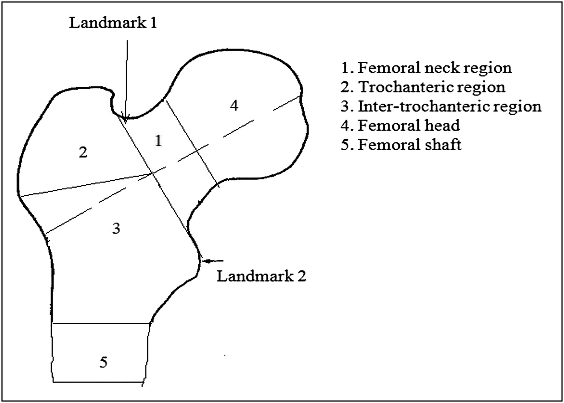
- FIGURE 1.
Two user-defined landmarks required for segmentation are shown by arrows. Different regions at femur are defined by numbers 1–5.
- FIGURE 2.
Segmented ROIs defined by semiautomatic technique. (A) Femoral-shaft ROI with cortical bone only forming hollow cylinder. (B) Femoral-neck ROI. (C) Total-hip ROI including trabecular and cortical bone of trochanter and femoral neck. ROIs are indicated by arrows. A color version of this figure is available as a supplemental file at http://tech.snmjournals.org.
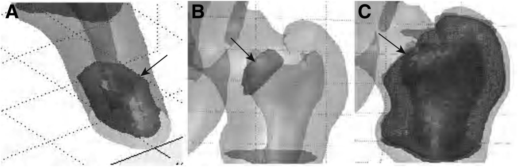
- FIGURE 3.
Two different examples of mean region (solid ovals) estimation using mean point–based distance between 2 regional boundaries (dashed ovals and dotted ovals). A color version of this figure is available as a supplemental file at http://tech.snmjournals.org.
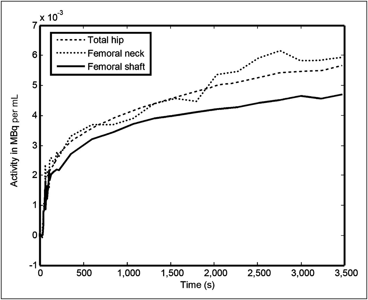
- FIGURE 4.
Mean time–activity curves generated from semiautomatically segmented ROIs. A color version of this figure is available as a supplemental file at http://tech.snmjournals.org.


Tables
- TABLE 1
Percentage Overlap and Differences Between Gold Standard and Semiautomatically Segmented ROIs
Total hip Femoral neck Femoral shaft Gold standard vs. semiautomatic Mean SD Mean SD Mean SD Percentage overlap 86.1 5.9 37.8 15.9 96.1 3.3 Percentage difference 14.5 12.5 89.7 44.2 4.7 6.0 - TABLE 2
Percentage Overlap and Differences Between Gold Standard (Mean of 8 Manually Drawn ROIs) and Manually Drawn ROIs (Each Separately Drawn Manual ROI)
Total hip Femoral neck Femoral shaft Gold standard vs. manually drawn ROI Mean SD Mean SD Mean SD Percentage overlap 85.2 42.9 39.1 24.3 95.2 47.8 Percentage difference 19.9 15 91.6 61.4 12.2 25.3 Total hip Femoral neck Femoral shaft Time (s) Mean SD Mean SD Mean SD Semiautomatic 94.5 18.6 49.0 8.0 3.28 0.6 Manual 895.3 371.9 120.1 100.1 219.5 168.9
Supplemental Data
Files in this Data Supplement:


Jump to section
Related Articles
Cited By...
- No citing articles found.