


Article Figures & Data
Figures
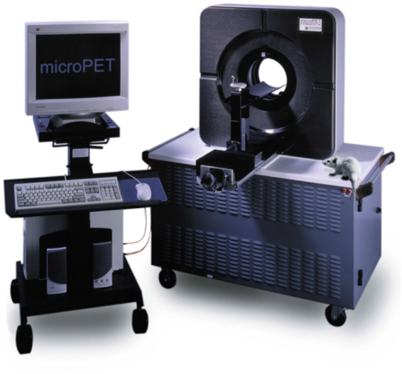
- FIGURE 1.
Photograph of microPET Focus 120 scanner (Siemens Preclinical Solutions). (Courtesy of Maurice M. Weaver.)
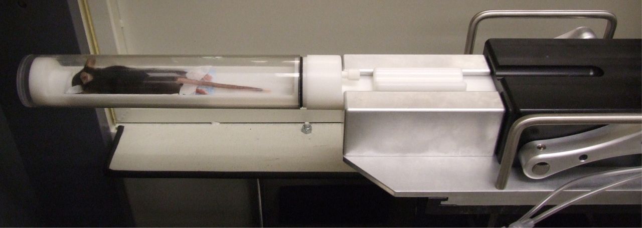
- FIGURE 2.
Mouse is placed in tube designed to facilitate anesthesia and positioning consistency. (Courtesy of David B. Stout.)
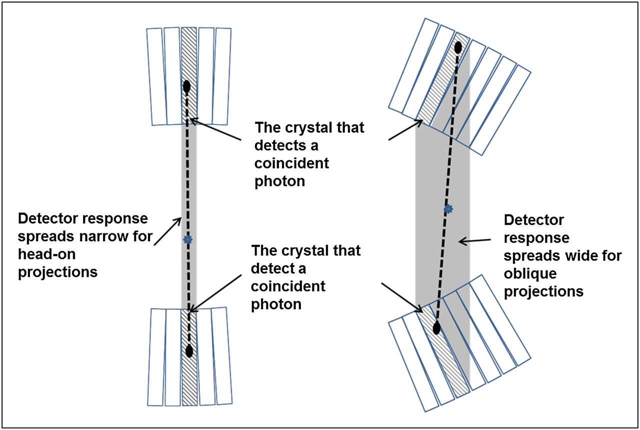
- FIGURE 3.
Diagram illustrating difference between head-on and oblique projections in terms of detector response spread (shaded area between crystals detecting coincidence event).
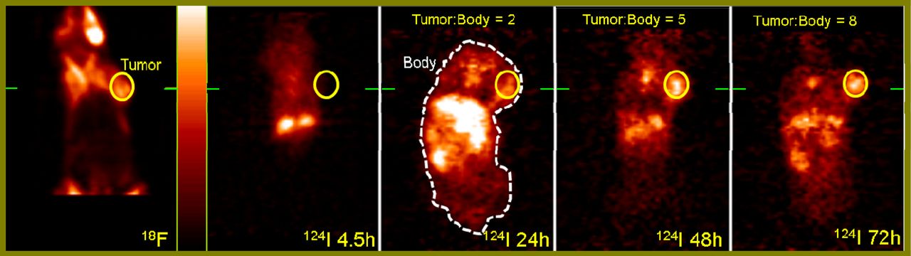
- FIGURE 4.
18F-FDG image on left (coronal view) was acquired first as reference 90 min after injection of 9.4 MBq (254 μCi) of activity via tail vein of tumor-bearing C3H mouse. Mouse was then injected with 2.7 MBq (72 μCi) of 124I-labeled derivative of pyropheophorbide-a, a bifunctional diagnostic and therapeutic agent (75). Mouse was imaged for 30 min at 4.5, 24, 48, and 72 h after injection. Concentration ratios of bifunctional agent in tumor (solid-line circle in each image) to that in animal body (dashed outline in middle image) were 2, 5, and 8 at 24, 48, and 72 h after injection, respectively, indicating that agent has desired properties to be used in therapeutic and monitoring applications. Color palette (shown to right of 18F image) was scaled to minimum/maximum of transverse slice passing through center of tumor site (indicated by green bars) in each dataset. Display scheme was same for all images. 18F = 18F-FDG; 124I = 124I-pyropheophorbide derivative.
- FIGURE 5.
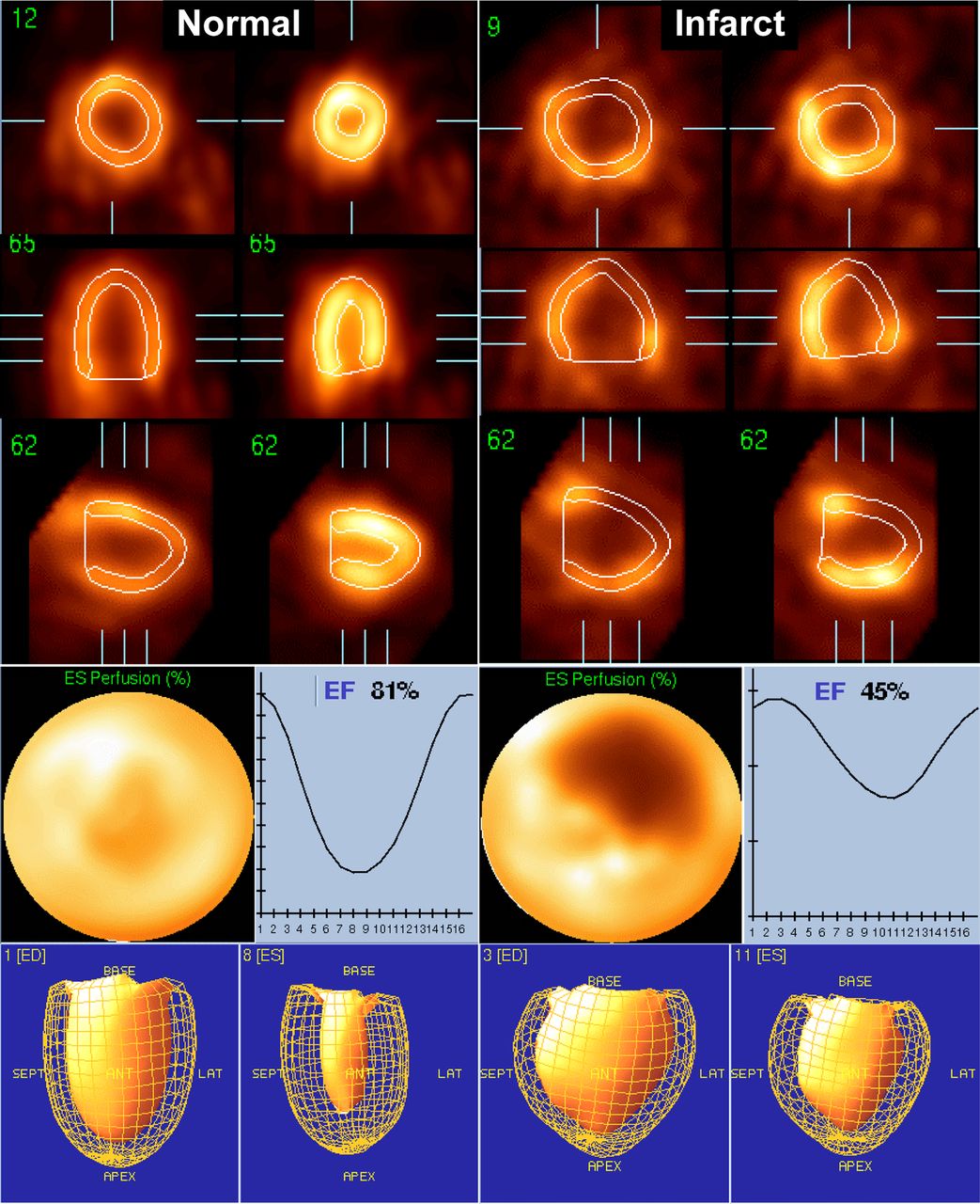
Electrocardiogram-gated 18F-FDG studies in normal and infarcted rats obtained using clinical cardiac analysis software QGS (56). Polar maps display end-systolic 18F-FDG uptake. Ejection fractions for normal and infarcted rats are 81% and 45%, respectively. ED = end-diastolic; EF = ejection fraction; ES = end-systolic. (Adapted with permission of (55).)
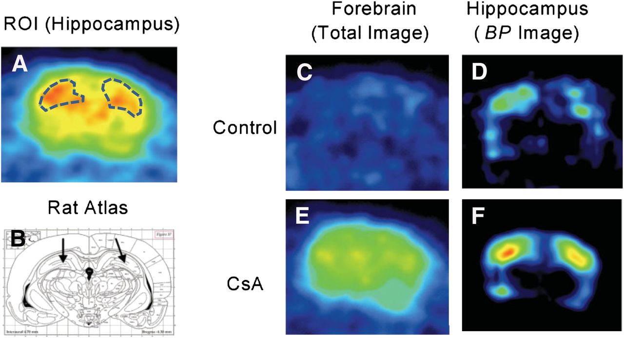
- FIGURE 6.
Uptake of 11C-(R)-(−)-RWAY in rat brain. Regions of interest were placed on left and right hippocampi (A), using coronal PET images with reference to rat brain atlas (B). Total uptake of radioactivity is shown in control rats (C) and cyclosporin A–treated rats (E). Similarly, binding potential images are shown in control rats (D) and cyclosporin A–treated rats (F). Cyclosporin A treatment significantly boosted uptake of 11C-(R)-(−)-RWAY, indicating blockade of efflux pump at blood–brain barrier. BP = binding potential; CsA = cyclosporin A; ROI = region of interest. (Reprinted with permission of (59).)


Tables
- TABLE 1
Commercially Available Small-Animal PET Scanners and Their Key System Specifications
FOV (mm) At CFOV… Manufacturer Model Transaxial Axial FWHM spatial resolution (mm) Sensitivity (%) Energy window (keV) Reference Bioscan/Mediso NanoPET 45–123 94 1.2 8.3 250–750 (69) Carestream Albira 80 40–148 <1.3 3–9 Not available (70) Gamma Medica/GE Healthcare LabPET 110 38–113 1.3 1.1–5.4 250–650 (15) Philips Mosaic HP 128 120 2.7 1.1 410–665 (71) Raytest Isotopenmessgeräte GmbH ClearPET 94 110 1.5 1.9 250–750 (72) Sedecal, S.A. rPET-1 68 47 1.5 0.5 250–650 (72) Siemens Preclinical Solutions microPET Focus 120 100 76 1.3 7.1 250–750 (73) microPET Focus 220 190 76 1.3 3.4 250–750 (74) microPET Inveon DPET 100 127 1.4 9.3 250–625 (32) CFOV = center field of view.
Target pathophysiology Tracer Working principle Metabolism (glycolysis) 18F-FDG Uptake and metabolism: tumor cells have higher rate of glucose, to which 18F-FDG is analog. Cell proliferation 3′-deoxy-3′-18F-fluorothmidine (18F-FLT) Malignant transformation increases cell proliferation, which upregulates thymidine. Gene expression 9-(4-fluoro-18F-3-hydroxymethylbutyl) guanine (18F-FHBG) Radiolabeled probe is phosphorylated by selected gene product and is trapped within cell. Thus, magnitude of probe accumulation in cell reflects level of gene expression. Tumor angiogenesis 89Zr-bevacizumab Vascular endothelial growth factor (VEGF) plays pivotal roles in regulating tumor angiogenesis. 89Zr-bevacizumab is anti-VEGF antibody and binds to VEGF. Hypoxia 18F-fluoromisonidazole (18F-FMISO) Rapid tumor growth leads to underdeveloped new vascularization, which creates hypoxia. 18F-FMISO takes advantage of increased tracer retention in hypoxic tissues with partial pressure of oxygen < 10 mm Hg. Apoptosis 18F-fluorobenzyl triphenylphosphonium cation (18F-FBnTP) Apoptosis involves permanent collapse of mitochondrial membrane electrochemical potential. 18F-FBnTP is voltage-sensitive probe.


Jump to section
Related Articles
Cited By...
- In Vivo Evaluation of Brain [18F]F-FDG Uptake Pattern Under Different Anaesthesia Protocols
- Was size of healthcare institution a factor affecting changes in healthcare utilisation during the COVID-19 pandemic in Korea? A retrospective study design analysing national healthcare big data
- Identification of pre-synaptic density networks using SV2A PET imaging and ICA in healthy and diseased mice
- Is There a Need for a Pediatric PET/CT Camera?
- Assessing PET Parameters in Oncologic 18F-FDG Studies