


Abstract
Because image fusion using 18F-FDG PET/CT allows a better localization of the pathologic uptake, this modality has a greater sensitivity than PET alone in examining the head–neck region. However, examination of this area is particularly critical because the head and neck are close to other anatomic structures and because of the high physiologic uptake of the radiocompound. The purpose of this study was to evaluate the utility of a new imaging protocol in the staging of oral carcinoma. Methods: Thirty-four consecutive patients (21 women and 13 men; age range, 20–84 y) with untreated biopsy-proven oral squamous cell carcinomas were examined using whole-body 18F-FDG PET/CT fusion imaging. All patients observed strict regulations before undergoing the PET/CT examination. At the end of the whole-body acquisition, another open-mouth scan was obtained. To compare the open- and closed-mouth methods, we analyzed features such as the feasibility of an accurate topographic localization of the tumor, evaluation of tumor extent, detection of tumor involvement with adjacent structures, and involvement of lymph nodes to which we assigned a score from 1 to 5. Results: No cases of 18F-FDG physiologic uptake in the tongue or muscles were observed. The open-mouth scan obtained a better score than did the closed-mouth scan when considering the tumor localization, tumor extent, and evaluation of adjacent anatomic structures near the clinically evident tumor. For lymph node involvement, the 2 methods showed similar results. Conclusion: The open-mouth scan improved the anatomic tumor localization and extent and detection of tumor involvement in adjacent anatomic structures achieved by the standard PET/CT procedure. In addition, time of the examination (mid morning), relaxation of muscles before the compound was administered, and an upright position while the patient waited caused a reduction of the frequent equivocal physiologic uptake in the head and neck region. The open-mouth method does not influence the nodal staging.
Correct evaluation of the extent of the lesion in oral carcinoma is essential because it represents the base on which treatment planning is built and also determines follow-up decisions (1,2). Clinical examination is limited by technical difficulties at the level of the oropharynx and tongue base, for which an anesthetic agent is required to perform a sufficient assessment (3). Moreover, clinical examination underestimates the true thickness of the tumor and of lymph node involvement (4). MRI and CT provide high-quality images, which allow for close visualization of the tumor dimension and better staging. However, even these methods present problems because of the complex anatomy of the oral cavity and the difficulty in distinguishing the lesion from surrounding structures (5).
18F-FDG PET has been reported to be a useful tool in staging primary tumors, following up treated oral squamous cell carcinomas (OSCC) (6), restaging in the case of relapse, searching for unknown primary tumors in the presence of nodal metastasis, and planning radiation therapy (7–9). PET technology with 18F-FDG shows a high sensitivity but a rather poor specificity and low spatial and anatomic resolution. Therefore, even if it is easy to differentiate a small amount of 18F-FDG uptake in malignant lesions, it is often difficult to localize these lesions with precision. Wolf et al. (10) achieved some success in improving specificity in an OSCC study, using software to fuse images independently acquired by CT and PET. Since 2001, 18F-FDG PET/CT has supplied functional and anatomic images simultaneously, improving the overimposition between areas of 18F-FDG uptake and anatomic structures (11). However, because the head–neck and other anatomic structures are close to each other and because of the high frequency of 18F-FDG physiologic uptake, the spatial resolution of 18F-FDG PET/CT examination of the oral cavity is not optimal.
Nonpathologic 18F-FDG uptake is due to a physiologic condition or function and is not directly related to a growing tumor. Tongue movement or sucking actions may increase 18F-FDG uptake in pharyngeal muscles (12). Because the genioglossus muscle prevents the tongue from falling back and obstructing the airway when the body is supine, uptake in the tongue base muscles and anterior part of the floor of the mouth is higher in patients resting supine for a long time (e.g., after a night's rest) (13). Oral activity, such as speaking during the waiting time between the injection and whole-body scan, increases 18F-FDG activity in the laryngeal muscles (14). Different uptake at the level of the orbicular muscles of the mouth, lateral pterygoid, and masseter muscles can be observed if the patient chews gum during the long wait time (15).
The aim of this study was to evaluate whether a new imaging protocol using 18F-FDG PET/CT in oral carcinomas, together with an optimal preparation of the patient and an open-mouth acquisition, may be useful in solving these problems.
MATERIALS AND METHODS
Thirty-four consecutive patients (21 women and 13 men; mean age, 67.1 y; age range, 20–84 y) with biopsy-proven previously untreated OSCC were prospectively investigated between September 2008 and November 2009. All patients were studied using whole-body 18F-FDG PET/CT fusion imaging, full clinical staging, CT, and MRI. If required, surgery was performed, an average of 15 d (range, 6–19 d) after the first imaging study. The exclusion criteria included pregnancy and breast feeding. All patients gave their informed written consent, and the study was conducted in accordance with regulations set forth by the institutional review board.
The PET/CT examinations were scheduled around mid morning to avoid the possibility of supine position–related 18F-FDG uptake in the muscles at the base of the tongue and anterior part of the mouth floor. Patients fasted for at least 6 h before examination and were instructed to remain silent and abstain from liquid intake 30 min before the injection and during the waiting time between 18F-FDG injection and whole-body scanning, to avoid 18F-FDG uptake by the tongue and vocal muscles. During this period, patients were not allowed to lie down. Before the scan, all metal objects (e.g., necklaces, earrings, and prosthesis) were removed.
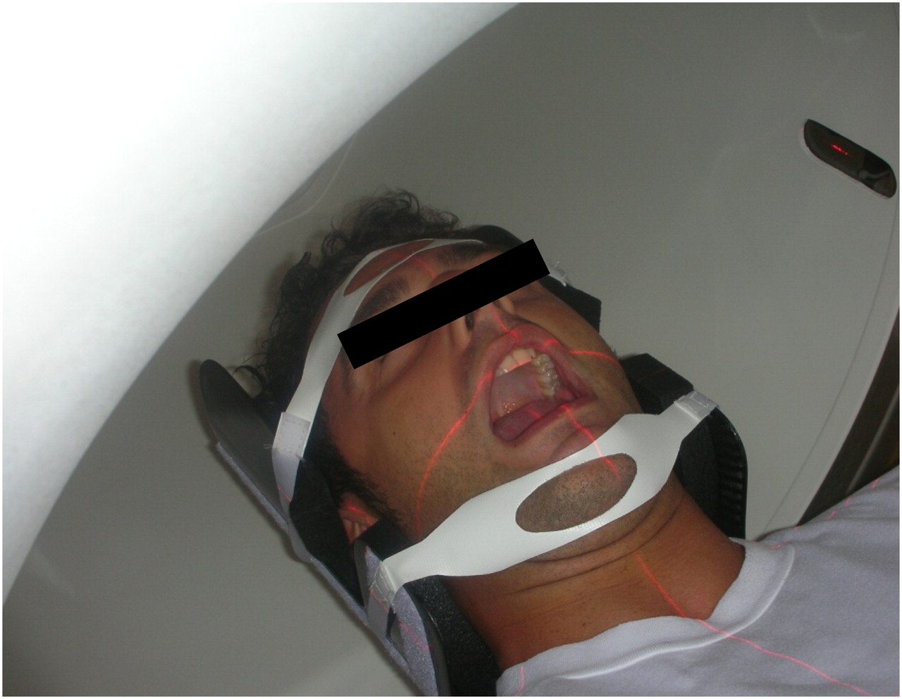
Each patient was administered a weight-related dose of 18F-FDG (277–466 MBq) by intravenous injection. The scan was obtained using a PET/CT system (Discovery ST; GE Healthcare), combining a 4-slice CT scanner and a bismuth germanate orthosilicate block detector PET tomograph, in 3 dimensions. For all patients, a whole-body PET/CT scan was acquired from the orbitomeatal line to the pelvis, with 6–7 fields of view (15 cm, 3.5 min each, 9 slices of overlap). PET was initiated immediately after the CT examination, to use CT data for the attenuation correction of PET data (CT scan thickness, 3.75 mm; 140 kV; 60–80 mA/s). Intravenous contrast material was not used for the PET/CT scans because all patients underwent PET/CT in addition to CT and MRI. Then, the laser was directed to the open mouth, and an additional 3.5-min PET/CT scan was acquired from the orbitomeatal line to the clavicular fossa, with 1 FOV (15 cm, 3.5 min). CT parameters were a scan thickness of 3.75 mm, 140 kV, and 60–80 mA/s. To optimize the visualization of the oral structures and ensure a stable open-mouth position, a bite device was used (Fig. 1). To provide radioprotection, the patient was asked to drink 500 mL of water and urinate before being dismissed from the examination.

Open-mouth scans: SCOUT from orbitomeatal line to clavicular fossa (10 mA, 120 kV), CT from orbitomeatal line to clavicular fossa (60 mA, 140 kV), and PET from clavicular fossa to orbitomeatal line (1 field of view, 15 cm for 3.5 min).
All data were evaluated by 2 experienced nuclear medicine physicians on a computer display in 3 orthogonal planes (i.e., axial, coronal, and sagittal). The observers did not have any information about the results of the CT and MRI scans, however, clinical information was available. PET scans, including anatomic information from the fused CT scan, were evaluated. Closed- and open-mouth scans were analyzed separately. Any initial disagreement between the 2 observers about the image findings was resolved by consensus. For the PET/CT scan, the invasion of bone by the cancer was suggested by 2 conditions: when 18F-FDG uptake was observed adjacent to a visible defect on CT images of the cortical bone or when 18F-FDG uptake was observed out of the cortical bone and within the bone marrow in the same region, even without a detectable cortical erosion, as described by Goerres et al. (16). The PET/CT images suggested lymph node involvement in the case of any focal 18F-FDG uptake greater than background activity and corresponding to nodular structures on CT, regardless of lymph node size. All the patients underwent surgical excision of the tumor, and all data from imaging (CT and MRI) and PET/CT scans were compared with the histologic evaluation of the excised specimens as the reference standard (pTNM) for the size of the lesions, involvement of bone or other adjacent structures, or nodal involvement. In patients without histologic N stage or M stage verification, the clinical follow-up served as the standard of reference for N and M stages.
To compare the open- and closed-mouth methods, some main characteristics were chosen (distinction of carcinoma or tumor localization, tumor extent in the primary site, evaluation of the involvement of adjacent structures, involvement of lymph nodes) and assigned a score ranging from 1 to 5, responding to the grade of each characteristic in the single examination (1: worst—when it was not possible to distinguish the characteristics; 2: bad—when an observer was not convinced that uptake correlated with the characteristics [possibly negative]; 3: sufficient—when the images were sufficiently clear to be evaluated, but the observer was undecided about the characteristics [possibly positive]; 4: good—when the observer was not completely convinced of the evaluation of the characteristics [probably positive]; and 5: best—when the observer was convinced about the evaluation of the characteristics).
Statistical Analysis
A variation of 3.2 was considered clinically relevant using an average score of 10 with an SD of 3 in a referred population studied with a standard method (17). To obtain a strength of 90%, it would have been necessary to recruit 22 patients; the available number of patients in our study (34) was a sufficient number to perform the study.
The scores of the different parameters for both closed- and open-mouth methods were reported in terms of average, SD, and 25–50–75 percentiles. For statistical analysis, the nonparametric Wilcoxon test was used for the paired data. The P value was corrected for the repeated tests with the Bonferroni method.
To evaluate the difference between the 2 methods, the results of the scores were dichotomized: a rating score of 2 or less was considered negative for tumor tissue and a rating score of 3 or greater was considered positive for tumor tissue. The results were compared using the McNemar test, and with histologic results as the reference standard.
RESULTS
Figure 1 shows patient positioning for the open-mouth technique, and Figures 2 and 3 show examples of open- and closed-mouth acquisitions. Histologic diagnoses revealed 30 squamous cell carcinomas; 3 verrucous carcinomas, which included areas of squamous cell carcinoma; and 1 recurrent leukoplakia. No patients had diabetes mellitus. All patients duly cooperated and tolerated the open-mouth position well. No cases of physiologic uptake regarding the laryngeal, tongue, and facial muscles were observed on PET/CT images.

45-y-old patient affected by squamous carcinoma of left border of tongue. (A) Closed-mouth acquisition. It is possible to identify pathologic uptake on left anterior mouth. It is difficult to evaluate involvement of the mandibular bone or mouth floor. (B) Open-mouth acquisition. Tongue is only organ involved. The tongue is also only organ involved in sagittal projection with closed-mouth (C) and open-mouth (D) procedures—clearly demonstrated in D.

Synchronous lesion in 57-y-old patient affected by carcinoma of left border of tongue. (A) Axial closed-mouth acquisition. (B and C) Axial and coronal open-mouth scan. In C, localization of lesion on right buccal mucosa is clear.
The 4 characteristics and the relative scores are shown in Table 1.
Relative Scores for 4 Characteristics Using Closed- and Open-Mouth Methods
For the 4 features, the open-mouth method resulted in either a better score than the closed mouth method or at least similar scores for each patient. The statistical analysis showed a significant difference in scores between the open-mouth and closed-mouth acquisitions for all parameters, except for lymph node involvement (Table 2).
Statistical Analysis of Different Parameters for Closed- and Open-Mouth Methods
For tumor localization, the open-mouth method always resulted in a score of at least 3 (P < 0.05), compared with scores of 1 in 4 patients and 2 in 9 patients of the 34 examined with the closed-mouth method. Thus, with the closed-mouth scan, we did not have correct tumor localization in 38% of the patients.
This same result was valid for tumor extent, for which the open-mouth scan always resulted in a score of at least 3, whereas the closed-mouth method obtained a score of 1 in 2 patients and 2 in 14 patients (P < 0.05). Thus, in 47% of closed-mouth cases, we were unable to identify the actual tumor extent.
Even in the evaluation of involved structures, the open-mouth technique was better than the closed-mouth method (P < 0.05). In fact, the open-mouth method always obtained a score of at least 3, whereas the closed-mouth method obtained a score of 1 in 4 patients and 2 in 16 patients. Consequently, this characteristic was not well evaluated in about the 59% of the analyzed patients.
In 4 patients, tumors were not detected using the closed-mouth technique but were correctly detected using the open-mouth method because of better identification of the anatomic structures.
In 3 patients, 18F-FDG PET/CT was successful in excluding a false-positive bone invasion detected by CT and MRI. In these cases, the use of the open-mouth method allowed a better separation of the anatomic structures involved.
In the evaluation of involved nodes only, the 2 methods provided similar results (P = NS). Both the 18F-FDG PET/CT open-mouth scan and the 18F-FDG PET/CT closed-mouth scan failed to correctly identify nodal involvement in some patients (3 false-positive and 2 false negative results).
Rhinopharyngeal uptake, on both closed- and open-mouth scans, suggested a secondary tumor, which was ruled out by an intraoperative histologic assessment as being an inflammatory process.
In 2 patients, the open-mouth scan allowed the identification of synchronous malignant lesions in the oral cavity, confirmed by biopsy but not identified by clinical examinations only (i.e., a lesion of the superior alveolar ridge in a patient with OSCC of the mouth floor and a lesion of the buccal mucosa in a patient with OSCC of the tongue).
DISCUSSION
The optimal examination technique for the assessment of the primary lesion of oral carcinomas is still under debate (2,18). Poor differentiation between the tumor and surrounding structures is still an important limitation and the main cause of CT understaging of the tumor. MRI overcomes some of these problems through dedicated sequences and has been shown to be superior in the detection and staging of tongue carcinomas and, in particular, in the evaluation of the base of the tongue and floor of the mouth (19).
In the literature, there are few studies that combine 18F-FDG PET and CT data or use 18F-FDG PET/CT scanners in oral cancer. Although seen to be highly sensitive in this region, PET is not able to give a precise anatomic localization of radiotracer uptake (20). 18F-FDG is also taken up by muscles and inflammatory processes and by certain metabolically active organs such as the tonsils and salivary glands. Therefore, PET may provide imprecise information on the exact location of focal pathologic uptake.
The use of 18F-FDG PET/CT, rather than PET only, seems to be particularly indicated in the head and neck region because of the anatomic complexity and high rate of physiologic 18F-FDG uptake in this area (21). The advantages of 18F-FDG PET/CT include superior lesion localization from near-perfect anatomic–functional coregistration and a better distinction between physiologic and pathologic uptake. Despite increased anatomic resolution, more improvement is needed for 18F-FDG PET/CT to differentiate the structures, because of the great complexity of the regions under examination. Improved differentiation is of great importance, in view of the reported presence of undetected multifocality in 10% of head–neck tumors and the reported risk of a synchronous or metachronous second malignancy ranging from 8% to 20% (22).
The positive results achieved in physiologic uptake suppression in this study could probably be reached simply by following a straightforward protocol consisting of mid-morning scheduling, the observation of silence, and no liquid intake for the patient half an hour before the examination. However, it is implied that these rules would be followed as a general guideline by any good team of investigators.
The open-mouth scan introduced in this study led to an increased space between the oral structures. This method allowed a much better evaluation of the palate, tongue, and alveolar ridge structures. The most promising results obtained in this study are related to the staging of the primary lesion. For each true-positive result, the site was effectively identified and extent of the primary lesion determined more easily using the open-mouth scan, as shown by the reported results in which tumor localization failed in 38% of the examined patients using the closed-mouth technique. The better results of the open-mouth scan are even more evident when considering tumor extent and involvement of adjacent structures. These data suggest that the open-mouth scan could be a great aid in detecting tumors of unknown origin in patients affected by laterocervical lymph node metastases.
The minimum tumor thickness observed in the closed-mouth scans was 4 mm, according to previously reported data regarding the low sensitivity of 18F-FDG PET in detecting superficial lesions (23). However, the 18F-FDG PET/CT false-negative results (4/34 patients) observed in our study with this method were due to misleading interpretations of nonphysiologic uptake of structures with a low anatomic definition. All of the patients with false-negative results were correctly identified using the open-mouth scan, in contrast to the previously reported data on the low sensitivity of 18F-FDG PET in identifying superficial lesions.
Furthermore, as we previously reported (24), in 3 patients 18F-FDG PET/CT was able to correctly exclude deep-bone involvement detected by CT and MRI, reducing the stage from T4 to T2. In these patients, when comparing the open- and closed-mouth scans, we observed that the open-mouth scan allowed an easier restaging of the tumor.
It could be expected that the metal artifacts would influence image quality and results differently on the open- and closed-mouth acquisitions because of the different angles of the mandible. In our study, 17 of 34 patients had metal artifacts, but in only 9 of these patients did the metal artifacts interfere with the analysis of the PET images. In 6 of 9 patients, the open-mouth method improved the image quality and final result. However, none of the patients with deep-bone involvement correctly excluded by 18F-FDG PET/CT had metal artifacts. It could be expected that some tumors would be better delineated with the mouth open, such as tumors of the buccal mucosa or tongue border, and that others would be better delineated with both methods, such as tumors of the lips.
As reported in the literature, the role of PET in N staging of oral cancer is still controversial. 18F-FDG PET combined with CT or MRI has been alternatively reported as not being significantly different from CT or MRI combined with physical findings or superior to CT or MRI itself (25,26). Moreover, it has been reported that 18F-FDG PET seems to be less useful in N staging of oral cancer than sentinel node biopsy, revealing poor sensitivity and specificity (27). In our study 18F-FDG PET/CT, using both open- and closed-mouth scans, did not show superior performance, because it was inadequate in detecting the 2 cases of nodal involvement. The results of this study reinforce what was reported by Schoder et al. in the clinically lymph nodal–negative neck (25). Despite a reasonably high overall accuracy, the clinical application of 18F-FDG PET/CT may be limited by the suboptimal sensitivity for small metastases and the relatively high number of false-positive findings (28).
Finally, given the risk of multiple lesions in the head and neck region, the open-mouth scan could be useful in localizing synchronous or metachronous tumors in the oral cavity. The identification of these conditions, as well as distant metastases, has an important implication for the management and survival of these patients (29).
CONCLUSION
Compared with PET alone, an open-mouth 18F-FDG PET/CT scan in patients with oral cavity carcinomas can improve the tumor localization, evaluation of tumor extent, and detection of tumor involvement of adjacent structures. Mid-morning scheduling of the examination, relaxation of muscles before the radiotracer administration, and a wait time with the patient upright may reduce the equivocal physiologic uptake generally frequent in this area. Finally, this imaging protocol seems to allow a better localization of potentially synchronous tumors.
Acknowledgments
We thank Glenn Richardson and Dr. Marco Pagani for assistance with the drafting of the article and Piercarlo Fania for help with the images.
REFERENCES
- Received for publication February 9, 2010.
- Accepted for publication October 29, 2010.


