
Main menu

  • Home
  • Content
    • Current
      • JNMT Supplement
    • Ahead of print
    • Past Issues
    • Continuing Education
    • JNMT Podcast
    • SNMMI Annual Meeting Abstracts
  • Subscriptions
    • Subscribers
    • Rates
    • Journal Claims
    • Institutional and Non-member
  • Authors
    • Submit to JNMT
    • Information for Authors
    • Assignment of Copyright
    • AQARA Requirements
  • Info
    • Reviewers
    • Permissions
    • Advertisers
    • Corporate & Special Sales
  • About
    • About Us
    • Editorial Board
    • Contact Information
  • More
    • Alerts
    • Feedback
    • Help
    • SNMMI Journals
  • SNMMI
    • JNMT
    • JNM
    • SNMMI Journals
    • SNMMI

User menu

  • Subscribe
  • My alerts
  • Log in
  • My Cart

Search

  • Advanced search
Journal of Nuclear Medicine Technology
  • SNMMI
    • JNMT
    • JNM
    • SNMMI Journals
    • SNMMI
  • Subscribe
  • My alerts
  • Log in
  • My Cart
Journal of Nuclear Medicine Technology

Advanced Search

  • Home
  • Content
    • Current
    • Ahead of print
    • Past Issues
    • Continuing Education
    • JNMT Podcast
    • SNMMI Annual Meeting Abstracts
  • Subscriptions
    • Subscribers
    • Rates
    • Journal Claims
    • Institutional and Non-member
  • Authors
    • Submit to JNMT
    • Information for Authors
    • Assignment of Copyright
    • AQARA Requirements
  • Info
    • Reviewers
    • Permissions
    • Advertisers
    • Corporate & Special Sales
  • About
    • About Us
    • Editorial Board
    • Contact Information
  • More
    • Alerts
    • Feedback
    • Help
    • SNMMI Journals
  • Watch or Listen to JNMT Podcast
  • Visit SNMMI on Facebook
  • Join SNMMI on LinkedIn
  • Follow SNMMI on Twitter
  • Subscribe to JNMT RSS feeds
Research ArticleIMAGING

Impact of Obesity on Nuclear Medicine Imaging

Mohammad A. Ghanem, Nafeesa A. Kazim and Abdelhamid H. Elgazzar
Journal of Nuclear Medicine Technology March 2011, 39 (1) 40-50; DOI: https://doi.org/10.2967/jnmt.110.078881
Mohammad A. Ghanem
  • Find this author on Google Scholar
  • Find this author on PubMed
  • Search for this author on this site
Nafeesa A. Kazim
  • Find this author on Google Scholar
  • Find this author on PubMed
  • Search for this author on this site
Abdelhamid H. Elgazzar
  • Find this author on Google Scholar
  • Find this author on PubMed
  • Search for this author on this site
  • Article
  • Figures & Data
  • Info & Metrics
  • PDF
Loading

Article Figures & Data

Figures

  • Tables
  • FIGURE 1.
    • Download figure
    • Open in new tab
    • Download powerpoint
    FIGURE 1.

    Whole-body PET/CT scan of 48-y-old woman (body mass index, 39; blood glucose level, 101 mg/dL, or 5.6 mmol/L) with history of breast cancer whose body contour exceeds PET and CT field of view, causing cropping and reconstruction artifact at left-breast region.
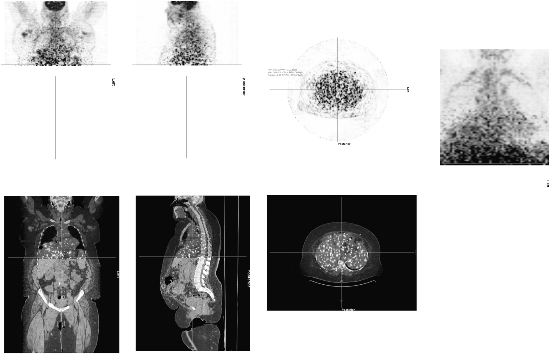
  • FIGURE 2.
    • Download figure
    • Open in new tab
    • Download powerpoint
    FIGURE 2.

    Whole-body PET/CT scan of 49-y-old woman (body mass index, 42; blood glucose level, 104 mg/dL, or 5.6 mmol/L) with history of breast cancer who was not able to complete emission part of study because of discomfort during acquisition.
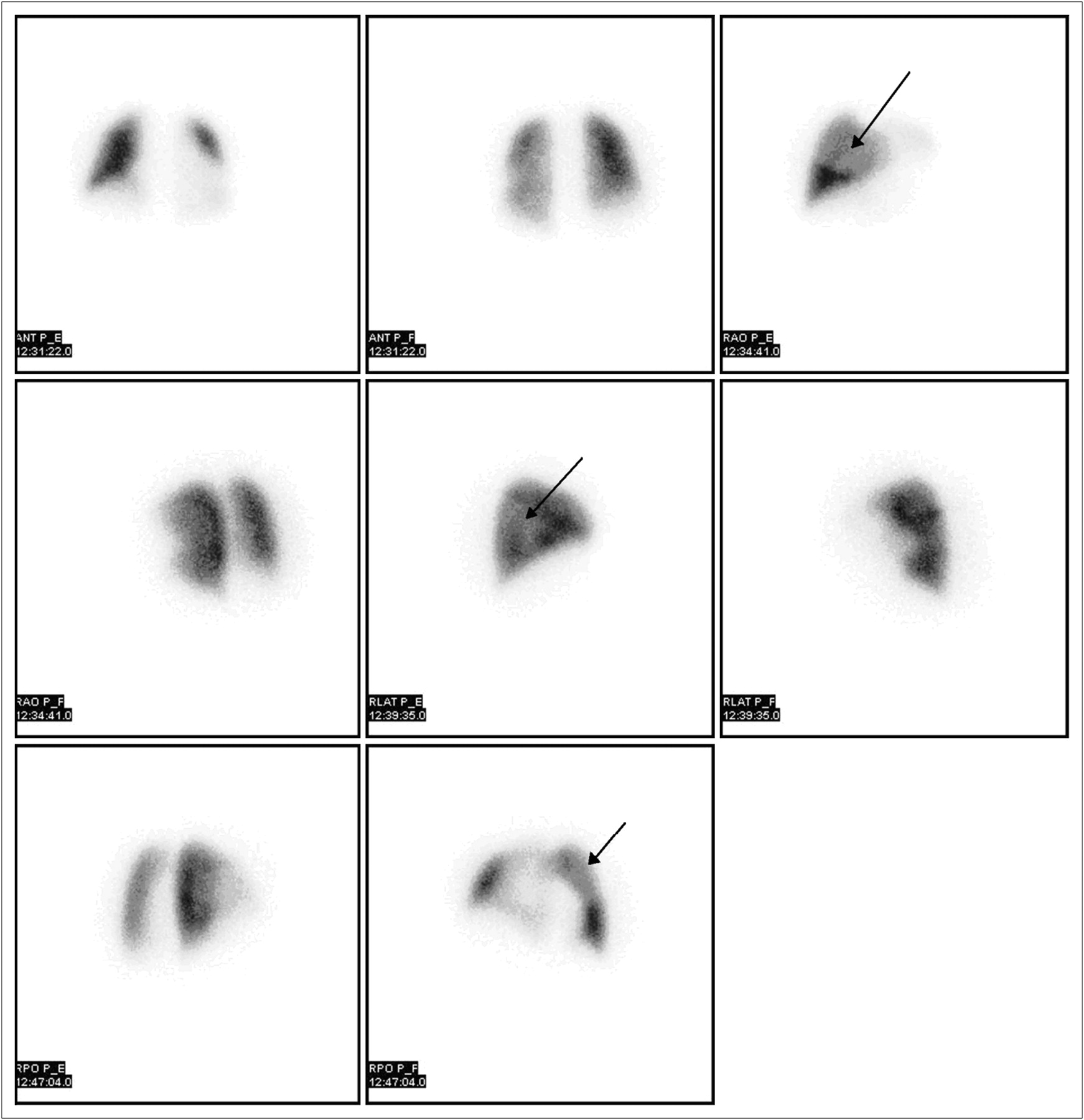
  • FIGURE 3.
    • Download figure
    • Open in new tab
    • Download powerpoint
    FIGURE 3.

    Lung perfusion images of overweight female patient who was unable to lift arms over head during acquisition, causing attenuation defects (arrows) on lateral, right anterior oblique, and left anterior oblique images.
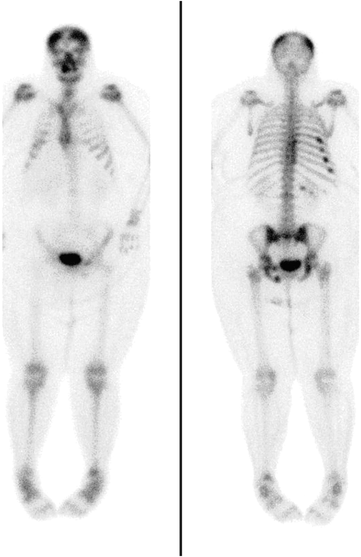
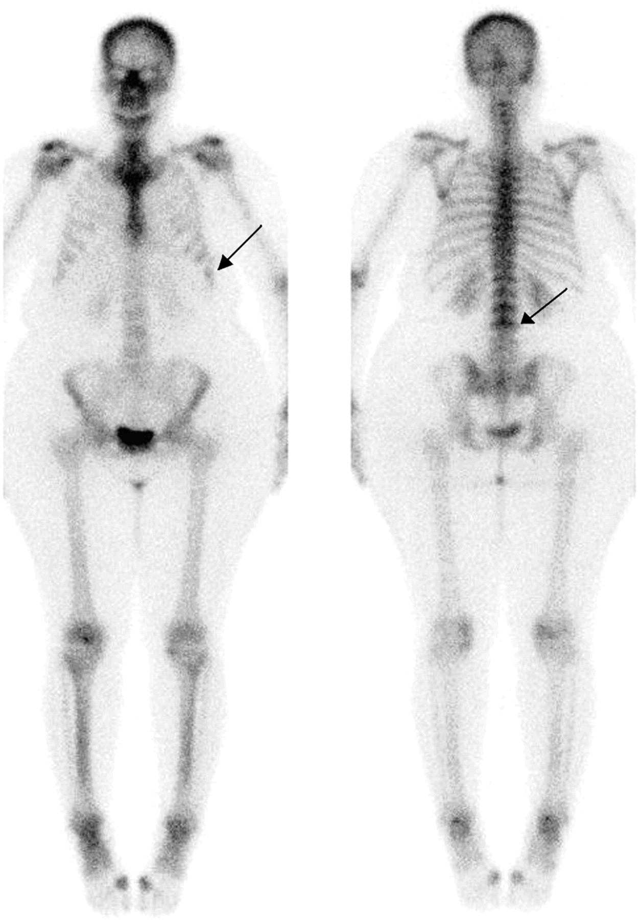
  • FIGURE 4.
    • Download figure
    • Open in new tab
    • Download powerpoint
    FIGURE 4.

    Whole-body bone scintigraphy image shows that peripheral parts of body are outside field of view of γ-camera because of patient's obesity. Most parts of upper limbs are not included in field of view.

  • FIGURE 5.
    • Download figure
    • Open in new tab
    • Download powerpoint
    FIGURE 5.

    Multislice views (A) and raw images (B) show large-breast-attenuation artifact (arrows) involving anterolateral myocardial wall of obese female patient.
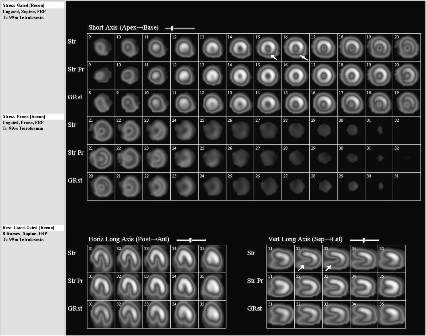
  • FIGURE 6.
    • Download figure
    • Open in new tab
    • Download powerpoint
    FIGURE 6.

    Myocardial perfusion imaging using supine and prone stress technique clearly demonstrates that inferior wall artifact (arrows) caused by diaphragmatic attenuation in obese male patient is corrected on prone images (middle row).
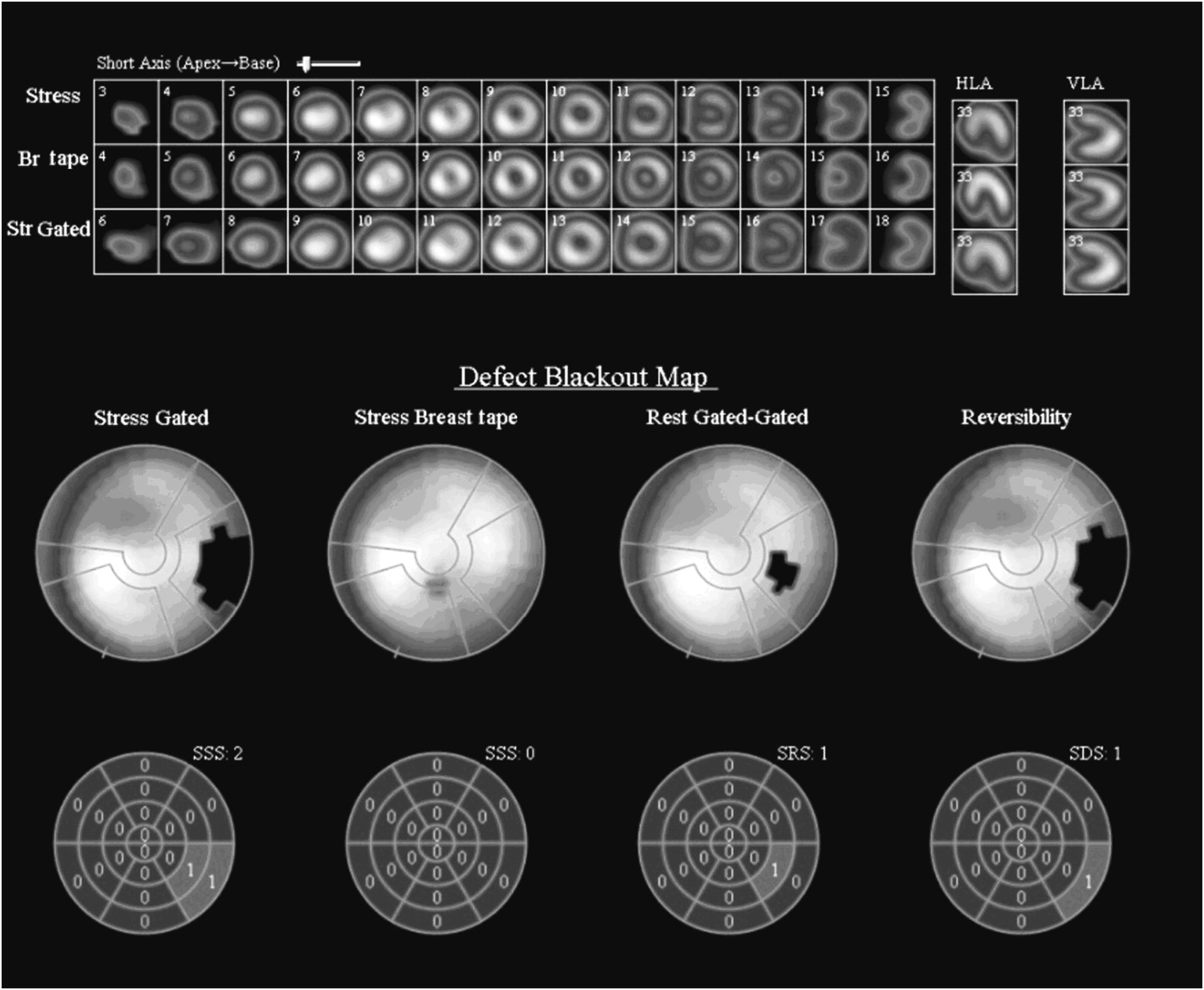
  • FIGURE 7.
    • Download figure
    • Open in new tab
    • Download powerpoint
    FIGURE 7.

    Myocardial perfusion imaging using stress technique and breast taping (middle-row images and second-from-left bull's-eye) clearly demonstrates that anterolateral wall artifact caused by large-breast attenuation in obese female patient is corrected with breast-lifting technique.
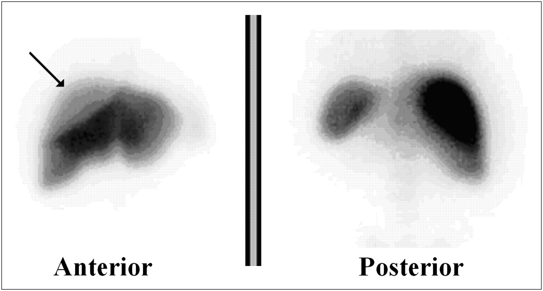
  • FIGURE 8.
    • Download figure
    • Open in new tab
    • Download powerpoint
    FIGURE 8.

    Whole-body sulfur colloid scintigraphy image shows right-breast attenuation over dome of right lobe of liver in overweight female patient.

  • FIGURE 9.
    • Download figure
    • Open in new tab
    • Download powerpoint
    FIGURE 9.

    Photon attenuation caused by massive abdominal fat, which renders interpretation of lumbar spine and pelvis difficult on anterior view of this male subject.

  • FIGURE 10.
    • Download figure
    • Open in new tab
    • Download powerpoint
    FIGURE 10.

    Fat tissue over lower abdomen and buttocks on gallium scan causes soft-tissue attenuation of detected photons on anterior and posterior views.
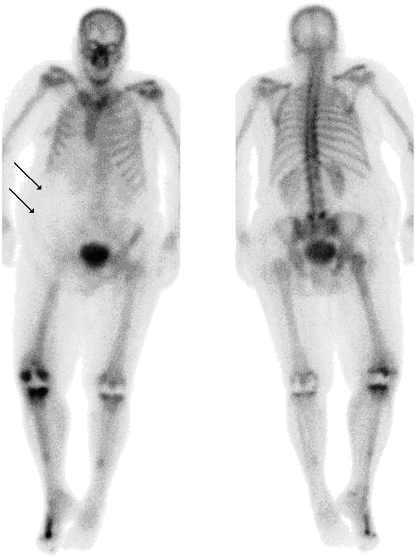
  • FIGURE 11.
    • Download figure
    • Open in new tab
    • Download powerpoint
    FIGURE 11.

    Whole-body bone scan of obese patient illustrates effect of body build on images. Attenuation is seen in region of lower lumbar spine and pelvis, in addition to edge artifact caused by fat crease in mid posterior lumbar spine and, to lesser extent, in anterior ribs.

Tables

  • Figures
    • View popup
    TABLE 1

    Main Effects of Obesity on Nuclear Medicine Imaging and Possible Remedies

    Obesity-related difficultyImpact on imagingPossible remedy
    Body configurationAttenuationProper preparation such as adequate hydration (higher amount)
    Increase of injected activity
    Delayed acquisition in some studies
    Increased time of acquisition
    Access to veinsPossible infiltrationUse of more experienced nuclear medicine technologist
    Patient discomfortUse of intravenous team
    Help from an anesthesiologist
    Use of ultrasound-guided peripheral intravenous access injection method
    Patient mobilityPossible fall injuriesUse of greater caution
    Use of accessory gadgets
    Imaging of patient on stretcher (mobile camera may be needed)
    Adequate communication with patient
    Positioning for acquisitionDifficulty in achieving proper positionsAttention to proper and secured positioning
    Use of pillow, splints, or other means to ensure patient's comfort and thus minimize motion
    Body contouringVariable distances from camera at different parts of bodyManual adjustment of camera head to obtain adequate images
    Patient girthCompton scatterUse of narrower or asymmetric energy window
    Insufficient count statisticsIncrease of acquisition time
    Beam-hardening artifact on CT.Use of caution during reading and quantification
    Reduced sensitivity to peripheral lesionsComparison with non–attenuation-corrected images
    Acquisition of additional spot image studies
    Diaphragmatic attenuation (cardiac)Masking of underlying organ activity (inferior wall artifact)Use of prone acquisition in myocardial perfusion
    Use of attenuation correction
    Breast attenuation (cardiac)Masking of underlying organ activity (anteroseptal-lateral wall defect)Breast lifting and binding
    Use of attenuation correction
    Fat crease and steatopygiaEdge effect/attenuationManipulation of crease and reimaging, or addition of SPECT
    Masking of bone details and creation of false findingsAddition of extra views or SPECT
PreviousNext
Back to top

In this issue

Journal of Nuclear Medicine Technology: 39 (1)
Journal of Nuclear Medicine Technology
Vol. 39, Issue 1
March 1, 2011
  • Table of Contents
  • About the Cover
  • Index by author
Print
Download PDF
Article Alerts
Sign In to Email Alerts with your Email Address
Email Article

Thank you for your interest in spreading the word on Journal of Nuclear Medicine Technology.

NOTE: We only request your email address so that the person you are recommending the page to knows that you wanted them to see it, and that it is not junk mail. We do not capture any email address.

Enter multiple addresses on separate lines or separate them with commas.
Impact of Obesity on Nuclear Medicine Imaging
(Your Name) has sent you a message from Journal of Nuclear Medicine Technology
(Your Name) thought you would like to see the Journal of Nuclear Medicine Technology web site.
Citation Tools
Impact of Obesity on Nuclear Medicine Imaging
Mohammad A. Ghanem, Nafeesa A. Kazim, Abdelhamid H. Elgazzar
Journal of Nuclear Medicine Technology Mar 2011, 39 (1) 40-50; DOI: 10.2967/jnmt.110.078881

Citation Manager Formats

  • BibTeX
  • Bookends
  • EasyBib
  • EndNote (tagged)
  • EndNote 8 (xml)
  • Medlars
  • Mendeley
  • Papers
  • RefWorks Tagged
  • Ref Manager
  • RIS
  • Zotero
Share
Impact of Obesity on Nuclear Medicine Imaging
Mohammad A. Ghanem, Nafeesa A. Kazim, Abdelhamid H. Elgazzar
Journal of Nuclear Medicine Technology Mar 2011, 39 (1) 40-50; DOI: 10.2967/jnmt.110.078881
Twitter logo Facebook logo LinkedIn logo Mendeley logo
  • Tweet Widget
  • Facebook Like
  • Google Plus One
Bookmark this article

Jump to section

  • Article
    • Abstract
    • PREIMAGING CONSIDERATIONS
    • IMAGING CONSIDERATIONS
    • POSTIMAGING CONSIDERATIONS
    • ROLE OF NUCLEAR PHYSICIANS AND TECHNOLOGISTS
    • CONCLUSION
    • REFERENCES
  • Figures & Data
  • Info & Metrics
  • PDF

Related Articles

  • No related articles found.
  • PubMed
  • Google Scholar

Cited By...

  • Molecular Breast Imaging: Administered Activity Does Not Require Adjustment Based on Patient Size
  • Association of Body Mass Index With Increased Cost of Care and Length of Stay for Emergency Department Patients With Chest Pain and Dyspnea
  • Cadmium-Zinc-Telluride Myocardial Perfusion Imaging in Obese Patients
  • Google Scholar

More in this TOC Section

  • Early 10-Minute Postinjection [18F]F-FAPI-42 uEXPLORER Total-Body PET/CT Scanning Protocol for Staging Lung Cancer Using HYPER Iterative Reconstruction
  • Single- Versus Dual-Time-Point Imaging for Transthyretin Cardiac Amyloid Using 99mTc-Pyrophosphate
  • Does Arthrography Improve Accuracy of SPECT/CT for Diagnosis of Aseptic Loosening in Patients with Painful Knee Arthroplasty: A Systematic Review and Metaanalysis
Show more Imaging

Similar Articles

SNMMI

© 2025 SNMMI

Powered by HighWire