


Article Figures & Data
Figures
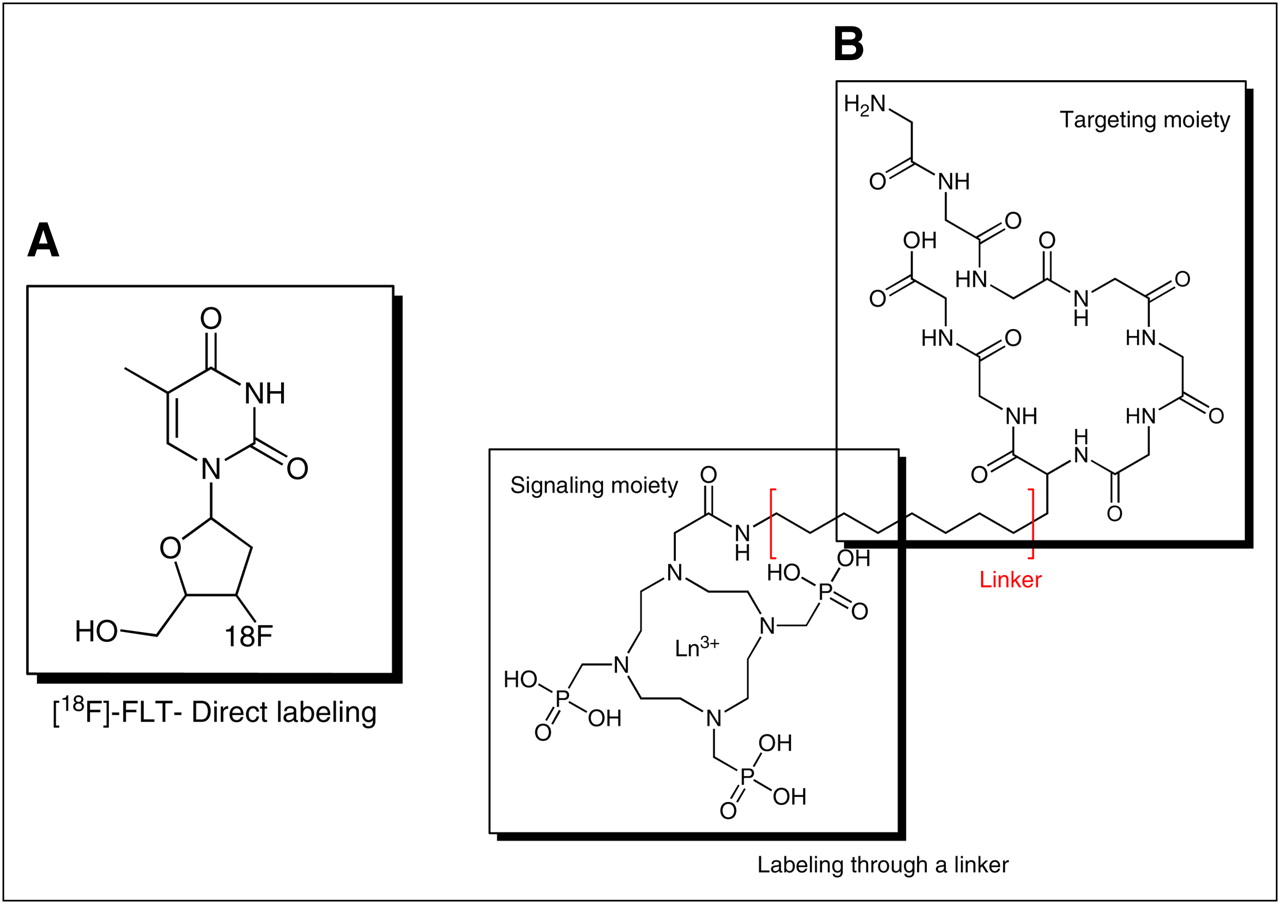
- FIGURE 1.
Examples of synthetic approaches to preparing molecular imaging agents: direct labeling (A) and labeling through linker (B).
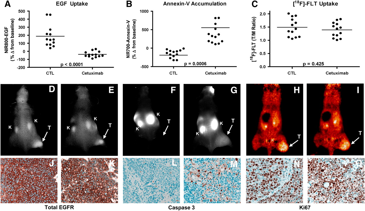
- FIGURE 2.
Noninvasive imaging assessment of response to EGFR blockade with cetuximab in CRC xenograft–bearing mice. Treated and untreated cohorts bearing DiFi xenograft tumors were simultaneously imaged with NIR800-EGF, NIR700-annexin-V, and 18F-FLT PET. After cetuximab treatment, mice bearing DiFi tumors, compared with untreated controls (CTL), exhibited significantly reduced NIR800-EGF uptake (A) and increased NIR700-annexin-V uptake (B). No statistical difference in 18F-FLT uptake was observed between treated and untreated mice (C). (D–I) Representative NIR800-EGF, NIR700-annexin-V, and 18F-FLT PET images collected from individual control (D, F, and H) and treated (E, G, and I) mice. Strong agreement between imaging metrics of response and standard immunohistochemistry was observed. Tumors from control (J) and treated (K) animals exhibited similar levels of total EGFR. Treated animals (M), compared with untreated cohorts (L), exhibited elevated caspase 3 staining. No discernible difference in Ki67 staining was observed between tumors from control (N) and treated cohorts (O). T = tumor; K = kidney. (Reprinted with permission of (24).)
Tables
Modality Signal Clinical Preclinical (rodent) Sensitivity* Quantification Acquisition time (s) PET 11C, 18F, 64Cu, 68Ga Yes Yes 1 Very good 10s−100s SPECT 99mTc, 123I, 111In, 177Lu Yes Yes 10−1−10−2 Good 100s−1,000s Fluorescence Fluorescent proteins, fluorochromes, quantum dots Potential Yes 10−2−1† Poor to fair† 1–10 BLI Luciferase No Yes 1–102† Poor to fair† 1–10 MRI Gadolinium, SPIO, USPIO, 19F Potential Yes 10−5 Fair 100s−1,000s MRS Endogenous compounds, hyperpolarized 13C Yes Yes <10−5 Fair 100s−1,000s Ultrasound Microbubbles Potential Yes ‡ Poor <1


