


Article Figures & Data
Figures
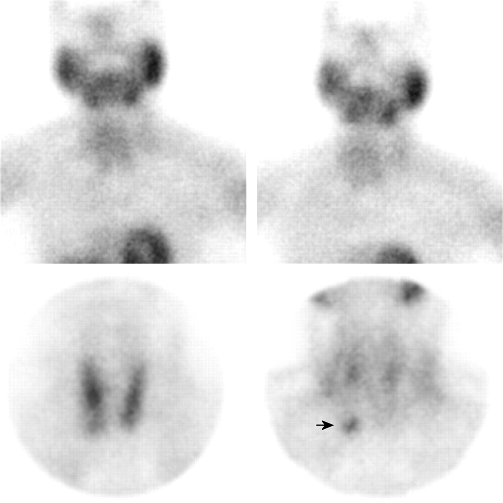
- FIGURE 1.
A 1,100-mg left inferior parathyroid lesion. Lesion is seen on both parallel-hole and pinhole imaging (arrows). (Early images are on left; late images are on right.)
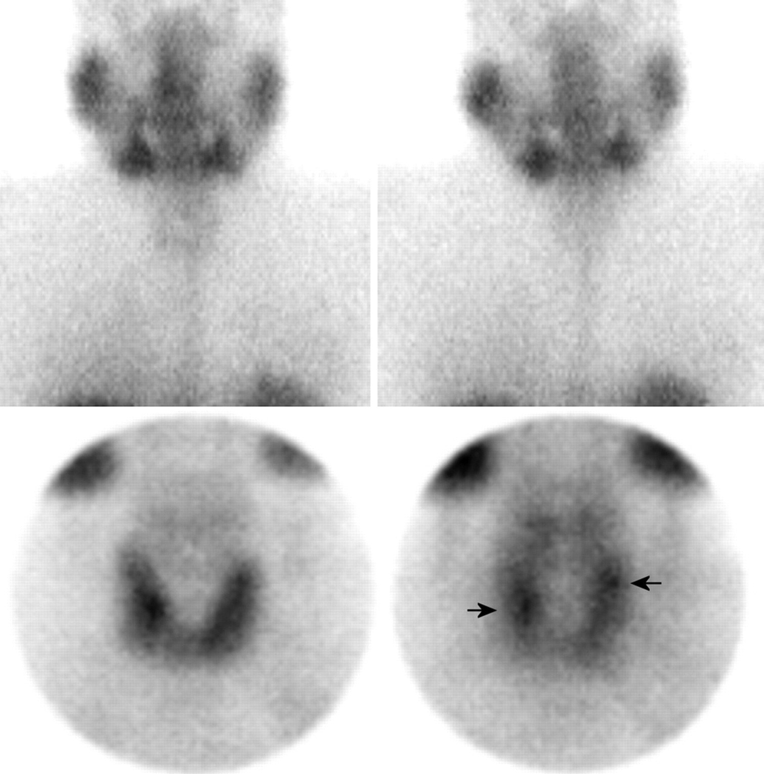
- FIGURE 2.
An 870-mg right inferior parathyroid lesion. Parallel-hole imaging (top) has negative findings. There is focally increased sestamibi accumulation, just below lower pole of right thyroid lobe (arrow) on pinhole imaging (bottom). (Early images are on left; late images are on right.)
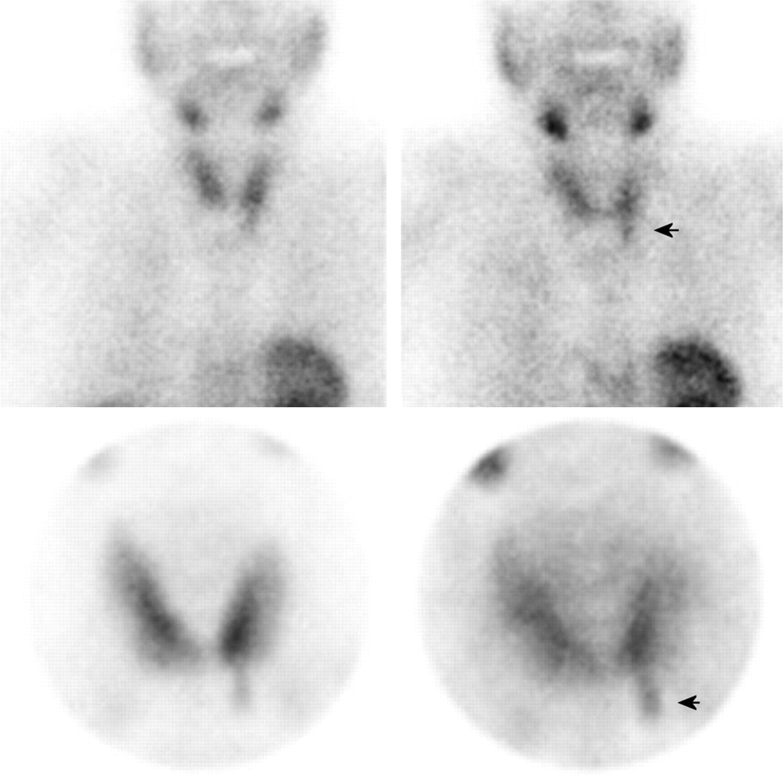
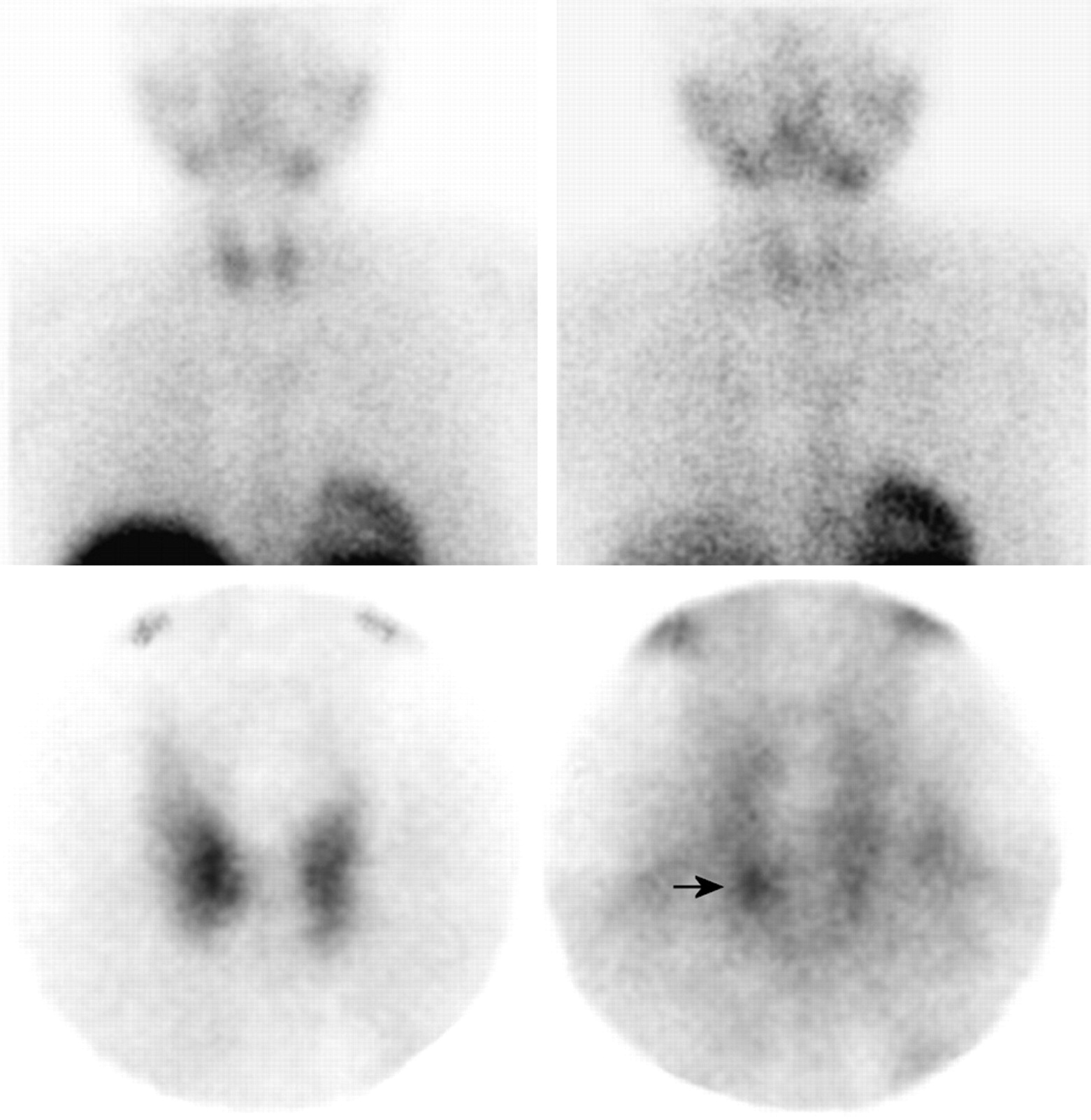
- FIGURE 3.
A 680-mg solitary right inferior parathyroid lesion. Lesion is not identified on parallel-hole imaging (top) but is clearly seen on pinhole imaging (arrow) (bottom). (Early images are on left; late images are on right.)
- FIGURE 4.
Multigland disease. Parallel-hole imaging (top) has negative findings. A 170-mg left superior lesion and 170-mg right inferior parathyroid lesion (arrows) are identified on pinhole imaging, but 90-mg left inferior parathyroid lesion is not. (Early images are on left; late images are on right.)


Tables
Index Pinhole Parallel-hole Difference (%) χ2 (df = 1) Confidence interval (%) P Sensitivity 48/54 (89%) 30/54 (56%)* 33 13.1 17 to 49 0.0003 Specificity 43/46 (93%) 44/46 (96%) 2 0.001 −7 to +11 0.29 Accuracy 91/100 (91%) 74/100 (74%)* 17 8.9 7 to 27 0.003 PPV 48/51 (94%) 30/32 (94%) 0 0.23 −11 to +11 0.78 NPV 43/49 (88%) 44/68 (65%)* 23 6.8 9 to 38 0.01 ↵* P < 0.05 for parallel-hole vs. pinhole imaging values.
df = degrees of freedom; PPV = positive predictive value; NPV = negative predictive value.
Index Pinhole Parallel-hole Difference (%) χ2 (df = 1) Confidence interval (%) P Group 1 (lesions > 840 mg) Sensitivity 28/28 (100%) 19/28 (68%)* 32 8.4 15 to 49 0.003 Specificity 30/32 (94%) 31/32 (97%) 3 0.001 −7 to +13 0.98 Accuracy 58/60 (97%) 50/60 (83%)* 14 5.1 4 to 24 0.04 PPV 28/30 (93%) 19/20 (95%) 2 0.09 −11 to +15 0.76 NPV 30/30 (100%) 31/40 (78%)* 22 5.7 9 to 35 0.01 Group 2 (lesions < 840 mg) Sensitivity 20/26 (77%)† 11/26 (42%)* 35 5.2 10 to 60 0.03 Specificity 23/24 (96%) 23/24 (96%) 0 0.54 −11 to +11 0.51 Accuracy 43/50 (86%) 34/50 (68%) 18 3.6 2 to 34 0.06 PPV 20/21 (95%) 11/12 (92%) 3 0.17 −15 to +21 0.78 NPV 23/29 (79%)† 23/38 (61%) 18 1.7 −34 to +39 0.17


