


Article Figures & Data
Figures
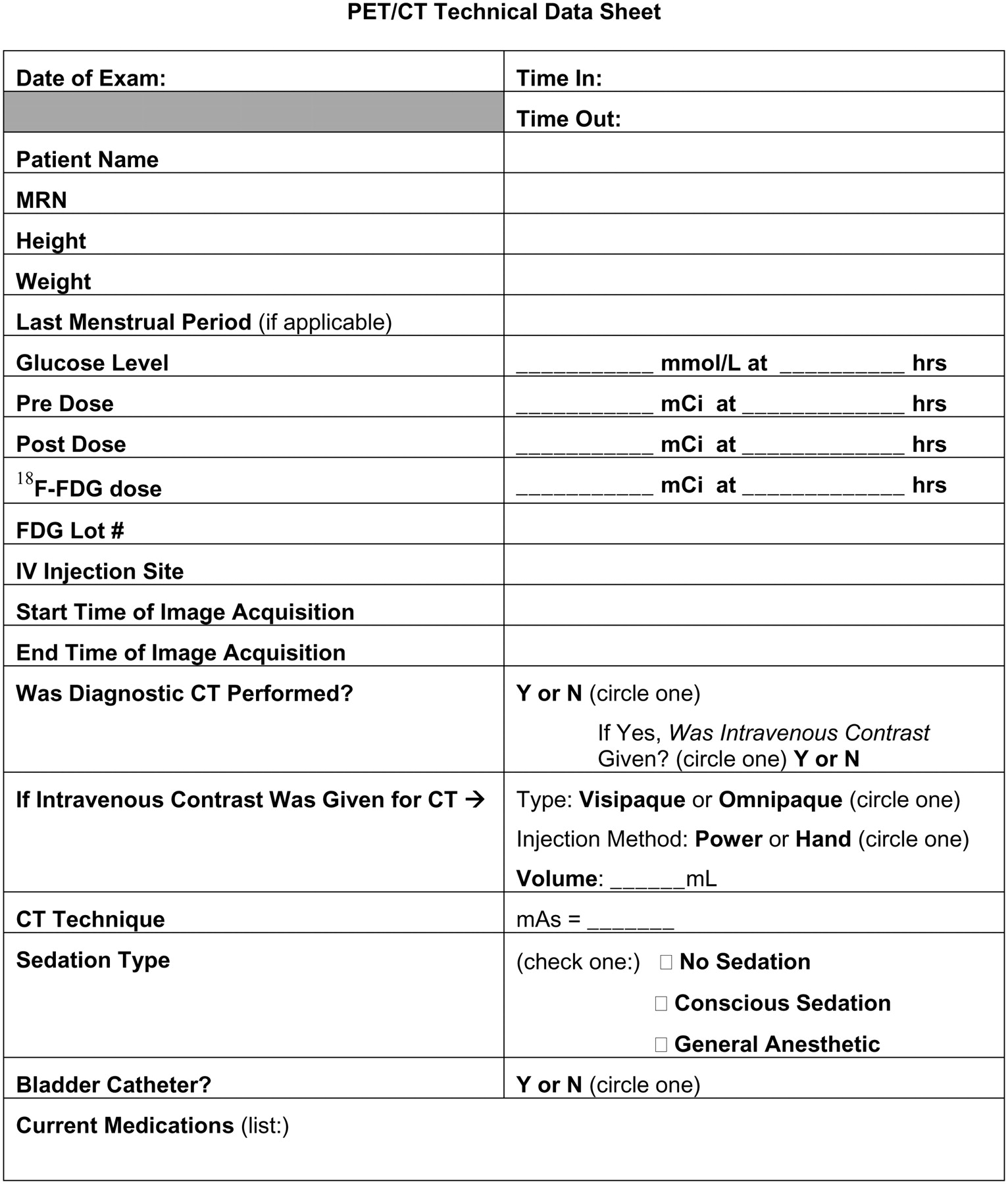
- FIGURE 1.
Example patient worksheet to help ensure that all data are recorded correctly and to help eliminate inaccuracies in standardized uptake value calculations.
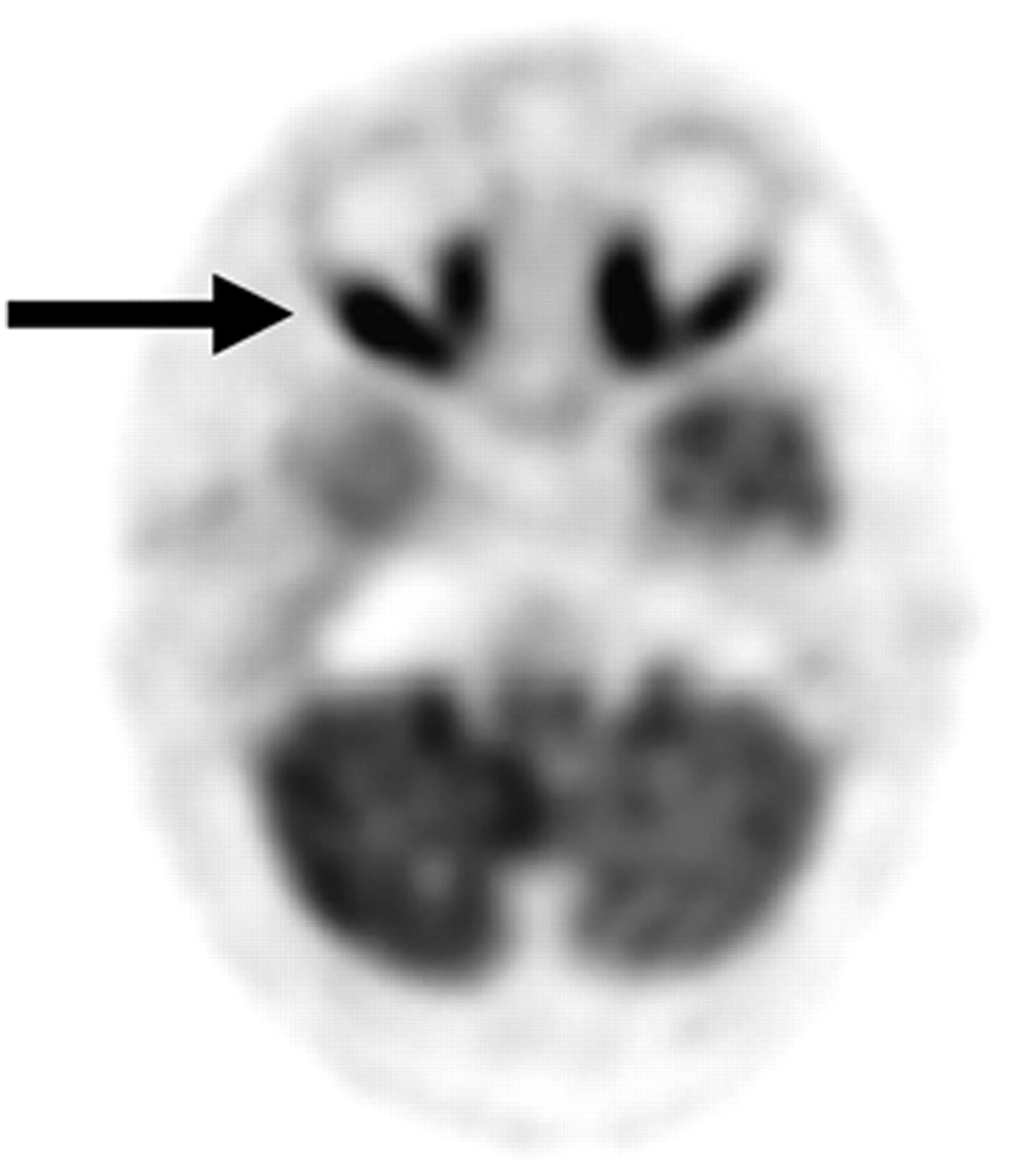
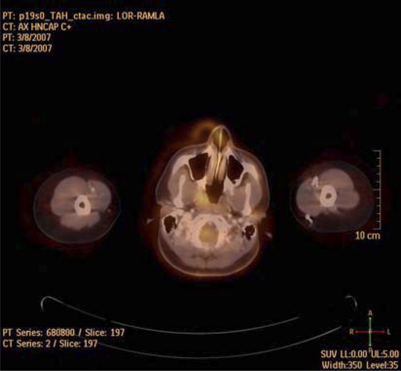
- FIGURE 2.
Transaxial projection of head revealing extraocular muscle uptake (arrow).
- FIGURE 3.
Poor registration due to motion of head between scans.
- FIGURE 4.
Misregistration artifact seen on attenuation-corrected image (left) but not on non–attenuation-corrected image (right). Artifact is result of patient motion between CT and PET acquisitions.
- FIGURE 5.
18F-FDG cardiac uptake correlated with stress MIBI imaging.
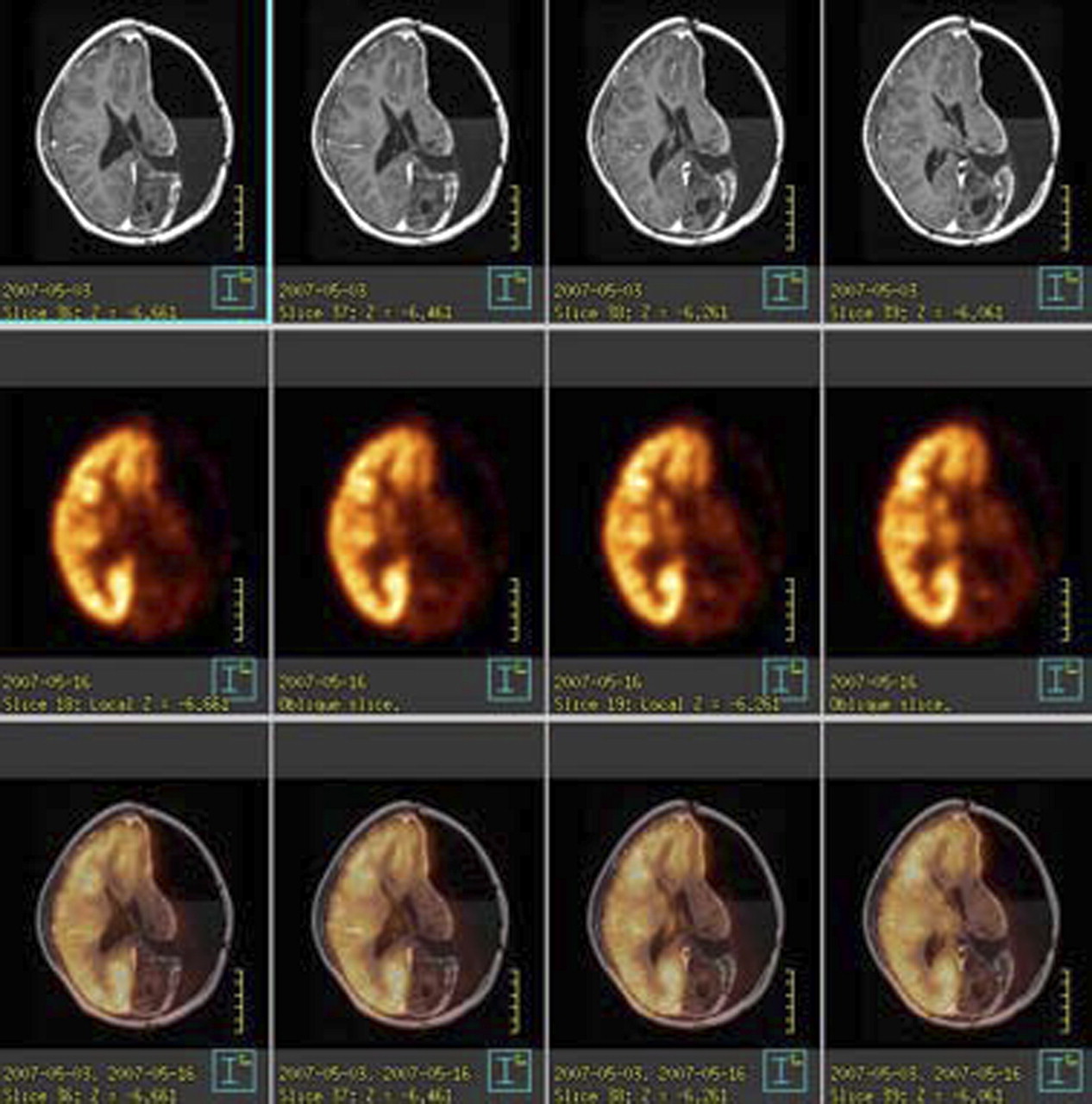
- FIGURE 6.
Transaxial views of MRI (top), PET (middle), and fused imaging (bottom) revealing matching defects in left hemisphere, with discrete focus of increased 18F-FDG uptake in occipital horn of left lateral ventricle.
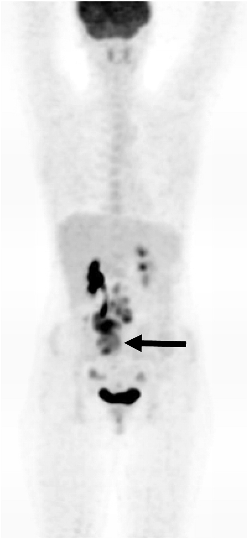
- FIGURE 7.
Uptake in obstructed kidney, ureter, ovaries, and primary neoplastic lesion (arrow).
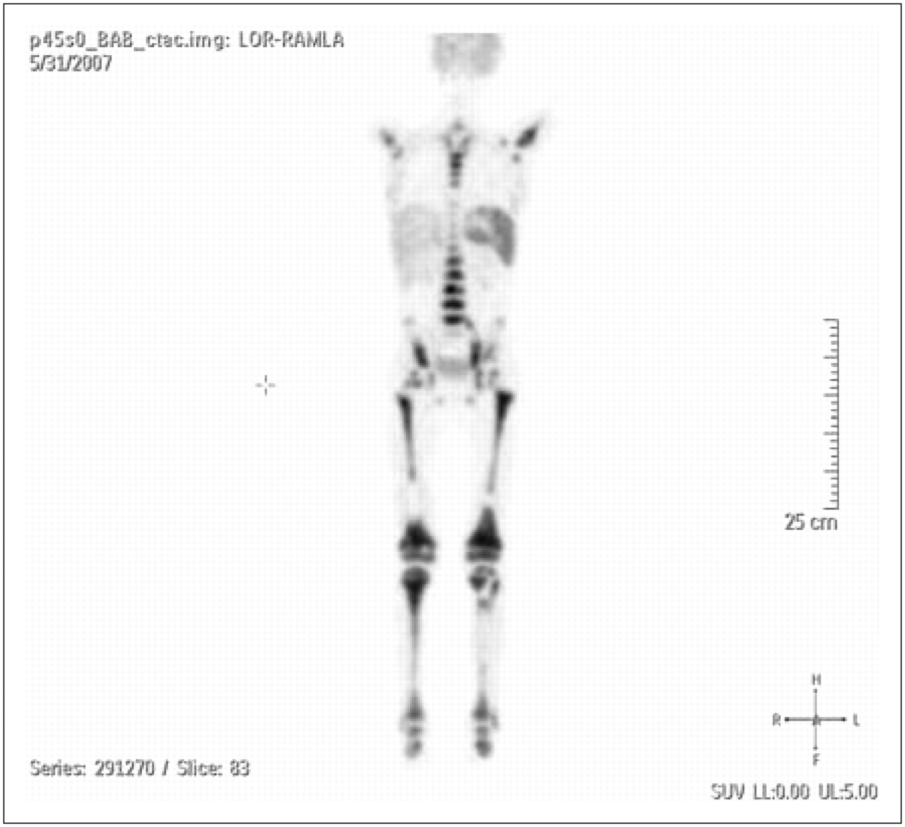
- FIGURE 8.
Image of 2-y-old child who was crying during uptake phase.
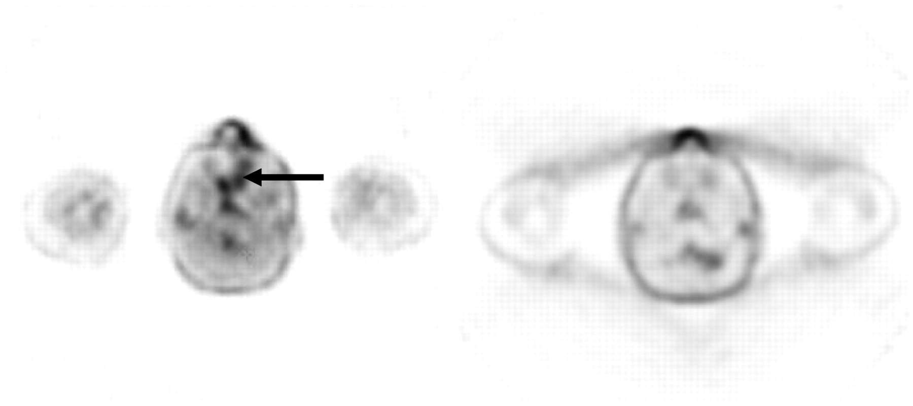
- FIGURE 9.
Asymmetric muscle uptake due to head position during uptake phase.
- FIGURE 10.
Diffuse bone marrow uptake in patient who was receiving granulocyte colony-stimulating factor.
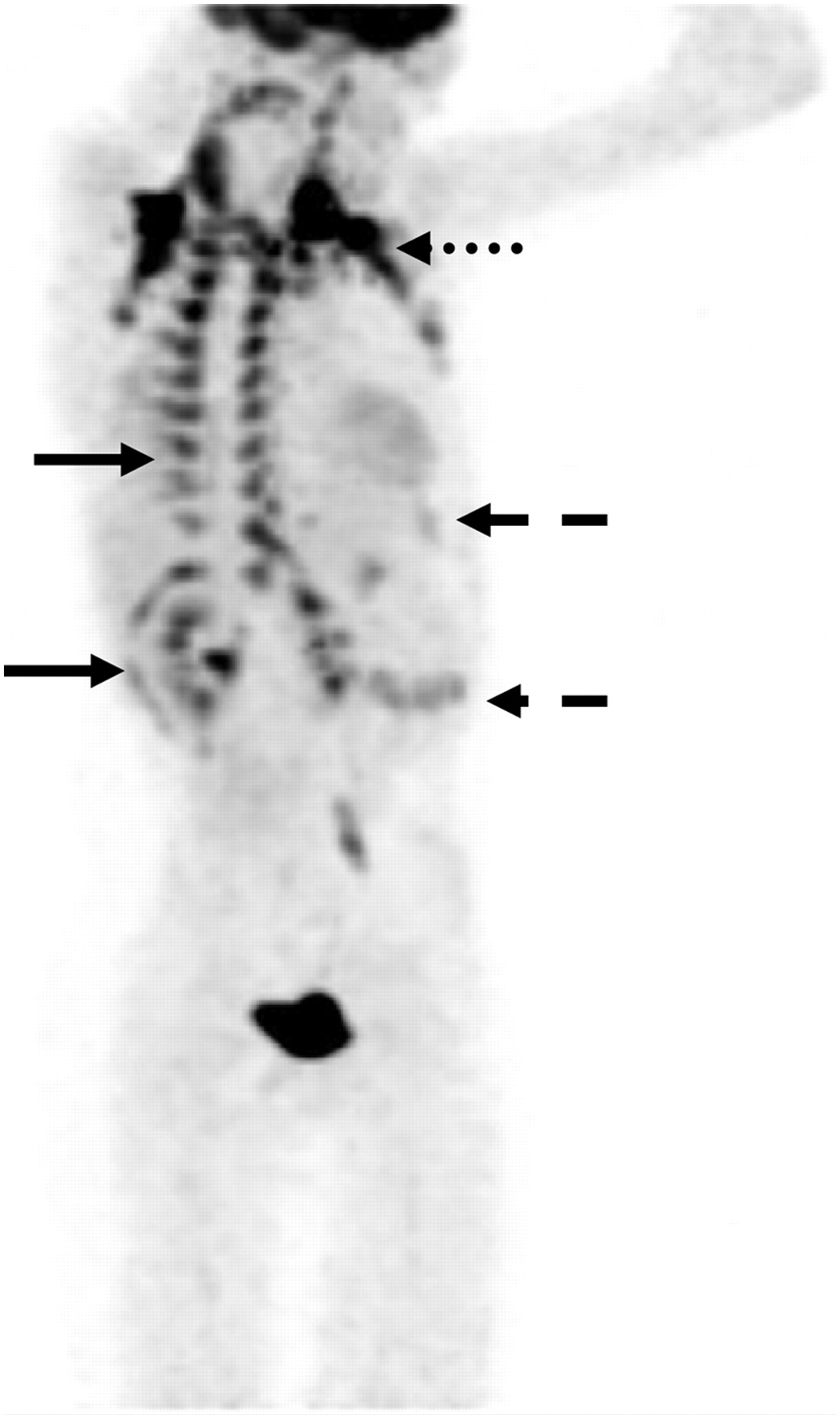
- FIGURE 11.
Brown fat uptake in costal vertebral junction and around kidney (solid arrow), clavicle (dotted arrow), abdominal wall, and intercostal region (dashed arrow).


Jump to section
Related Articles
Cited By...
- No citing articles found.