


Article Figures & Data
Figures
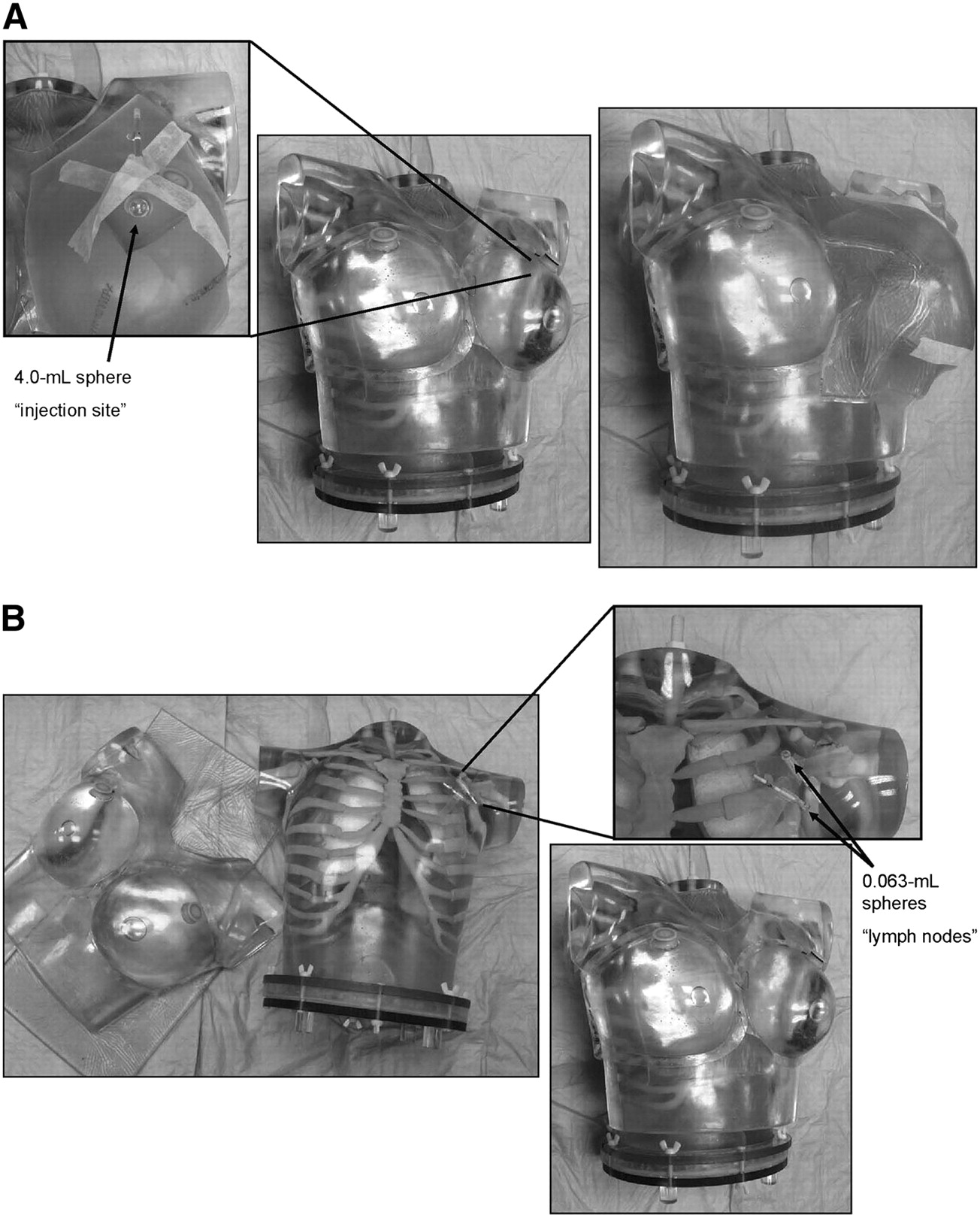
- FIGURE 1.
Modified anthropomorphic thorax breast phantom used to simulate breast lymphoscintigraphy injection-site uptake (A) and lymphatic drainage to 2 nodes (B).
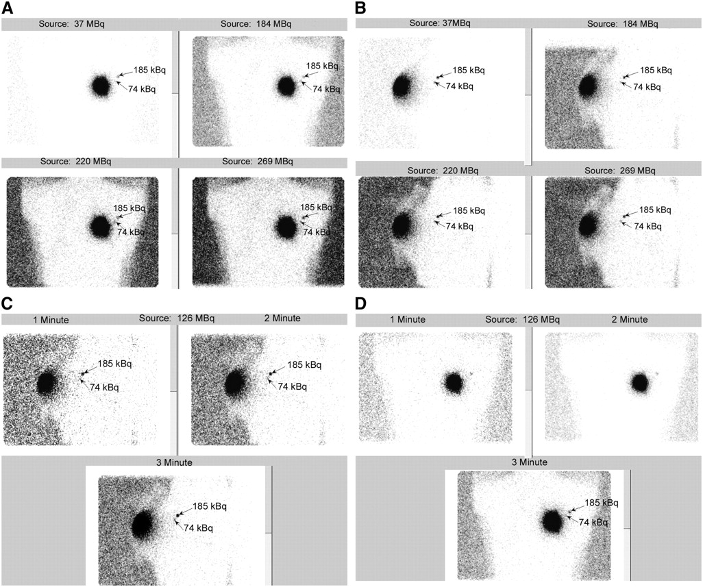
- FIGURE 2.
Different 57Co sources (activities of 37, 126, 184, 220, and 269 MBq) allowed visualization of both nodes with acquisition time of 3 min (A–D). Anterior localization images (1 and 2 min) did not show both nodes; however, both nodes were visible on lateral images (C and D).
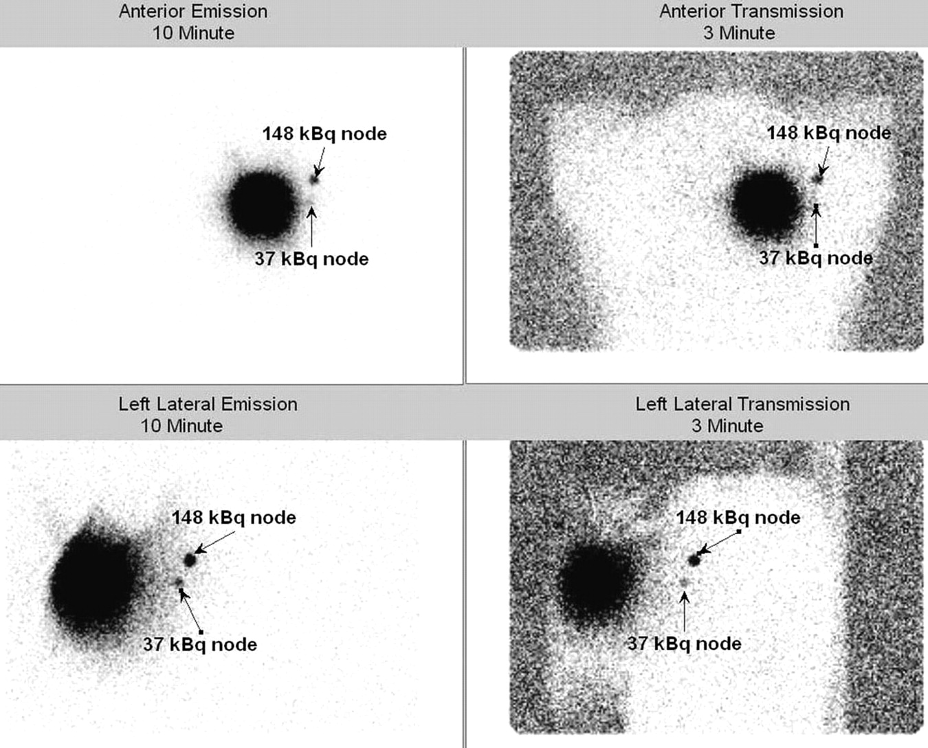
- FIGURE 3.
Node activities of 37 kBq were undetectable on anterior transmission images; however, they were visible on lateral transmission images using 126-MBq (3.41-mCi) 57Co flood source.
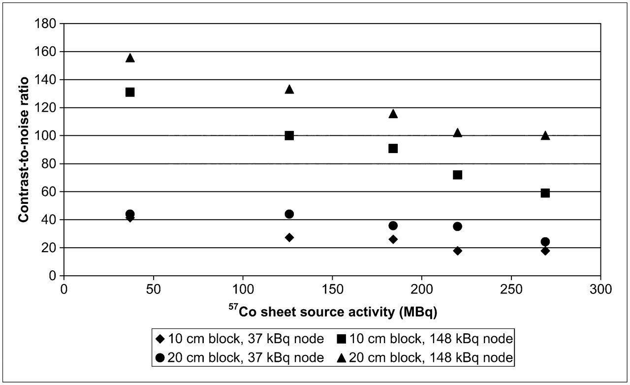
- FIGURE 4.
Plot of contrast-to-noise ratio versus source activity and block thickness for 2 node strengths.


Tables
- TABLE 1
CNR Calculations for 2 Block Thicknesses: 10 cm (Thin Patient) and 20 cm (Average Patient)
57Co source activity (MBq) Node 1 (total counts) Node 2 (total counts) Background (mean counts) No. of pixels in background ROI CNR node 1 CNR node 2 10-cm-thick acrylic 37 169 505 0.52 27 41.4 131.0 126 158 514 0.89 27 27.3 100.0 184 176 537 1.15 27 26.0 90.8 220 185 582 2.00 27 17.8 71.9 269 213 550 2.48 27 17.8 59.0 20-cm-thick acrylic 37 139 469 0.35 25 44.0 155.6 126 153 442 0.42 25 44.0 133.2 184 156 471 0.62 25 35.7 115.7 220 178 480 0.81 25 35.1 102.2 269 133 483 0.85 25 24.2 100.2 CNR = contrast-to-noise ratio.
Node detectability for transmission imaging decreases with thinner patients.
- TABLE 2
Effective Dose Estimates for 57Co Source Activities Given Standard 3-Minute Transmission Scans
57Co source activity (MBq) Measured exposure rate (μSv/h) Exposure for single view (μSv) Total for unilateral study (μSv) Total for bilateral study (μSv) 37 20 1.00 2.0 3.0 126 87 4.35 8.7 13.1 184 113 5.65 11.3 17.0 220 145 7.25 14.5 21.8 269 178 8.90 17.8 26.7


