


Article Figures & Data
Figures
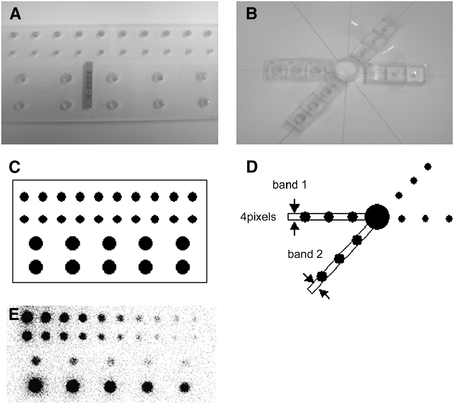
- FIGURE 1.
(A and C) Appearance and illustration of LN phantom, respectively. Holes were 8.5 mm (top row), 6.0 mm (middle row), and 12.0 mm (bottom rows) in diameter. Concentration of 99mTc-pertechnetate in 10 holes with same diameters was changed from 0.78 (right) to 400 kBq (left) per 200 μL in 1:2 increments over 10 steps. (B and D) Appearance and illustration of CB phantom, respectively. Holes containing radioactivity simulating lymph node were placed at 3, 5, and 7 cm from center of cylindric source. Band 1 and band 2 show regions for which profile curves were obtained in H and S directions, respectively. (E) Image of LN phantom taken with LEHR collimator and energy window centered at 141 keV. Note that sizes of hot spots changed with concentration of radioactivity.
- FIGURE 2.
Comparison of IS phantom images obtained with 2 cameras. (A) FORTE camera without lead shield. (B) DSX rectangular camera without lead shield. (C) FORTE camera with lead shield. (D) DSX rectangular camera with lead shield. Images were taken with LE collimator and energy window centered at 141 keV (±5%) for 250 s of data acquisition. Prominent star-shaped artifacts are shown on DSX rectangular camera images.
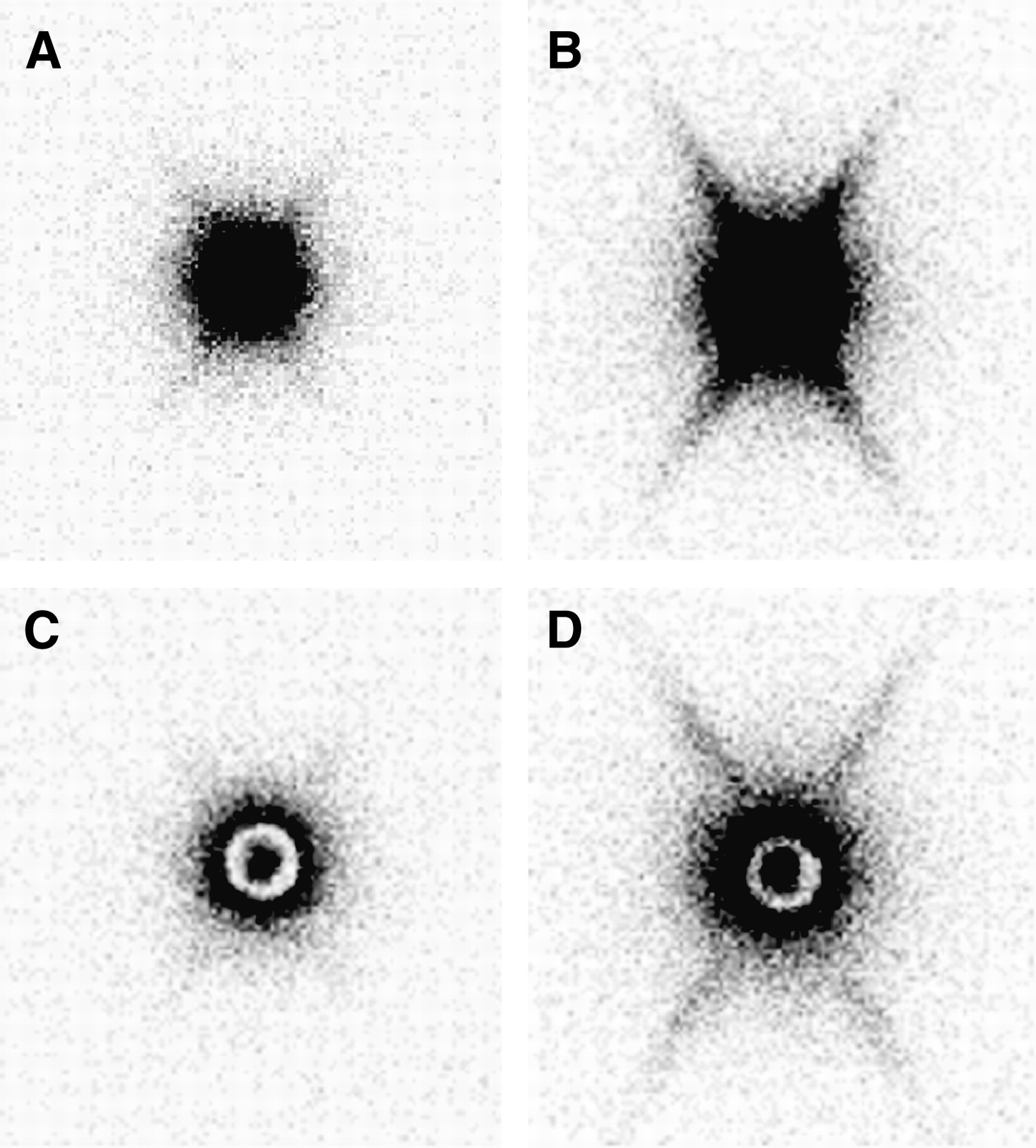
- FIGURE 3.
Comparison of CB phantom images obtained with FORTE camera (A) and DSX rectangular camera (B). (Left) 141 keV with LE collimator and lead shield—central white area shows effect of lead shield. (Middle) 141 keV with ME collimator. (Right) 146 keV with ME collimator.
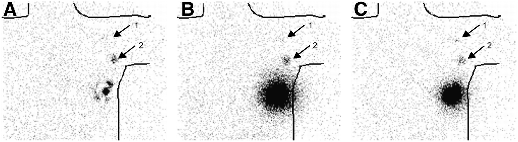
- FIGURE 4.
Static sentinel node lymphoscintigraphy (anterior views) in patient with breast cancer. (A) Image obtained with LEHR collimator and lead shield (lead shielding method). Lead shield covers imperfectly radioactivity at injection site. (B) Image obtained with ME collimator and energy window centered at 141 keV. (C) Image obtained with ME collimator and energy window centered at 146 keV (ME method). Arrows 1 and 2 show SLNs. ME method produced clear depiction of even small SLN (arrow 1).
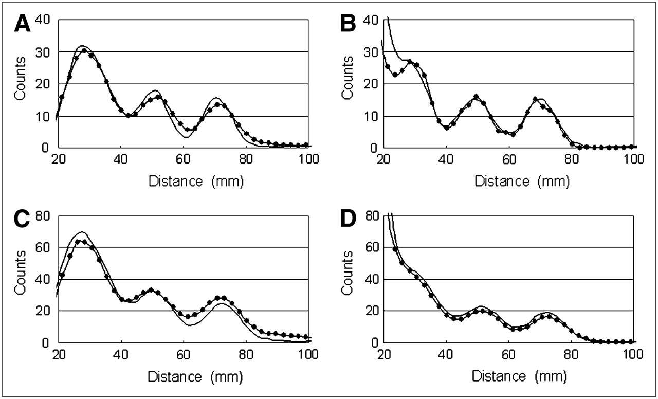
- FIGURE 5.
Profile curves acquired with 10-mm holes in CB phantom. (A and B) FORTE camera curves. (C and D) DSX rectangular camera curves. (A and C) Lead shielding method. (B and D) ME method. Note difference in longitudinal scales between FORTE and DSX rectangular camera graphs. Data obtained in H direction and data obtained in S direction are shown by plain solid line and by solid line with circles, respectively.
- FIGURE 6.
Relationship between lymph node site (distance from injection site) and image contrast with various levels of radioactivity for 5 min of data acquisition. (A) 25 kBq. (B) 100 kBq. (C) 400 kBq. Data obtained in H-direction with lead shielding method, data obtained in S-direction with lead shielding method, and data obtained with ME method are represented by plain thin line, by thin line with circles, and by thick line, respectively.
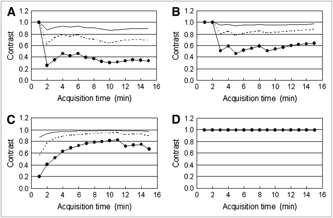
- FIGURE 7.
Relationship between image contrast and data acquisition time. (A) Contrast at 3 cm with lead shielding method. (B) Contrast at 3 cm with ME method. (C) Contrast at 5 cm with lead shielding method. (D) Contrast at 5 cm with ME method. Radioactivity concentrations of 400, 100, and 25 kBq/200 μL are represented by plain solid line, by dotted line, and by solid line with circles, respectively.


