


Article Figures & Data
Figures
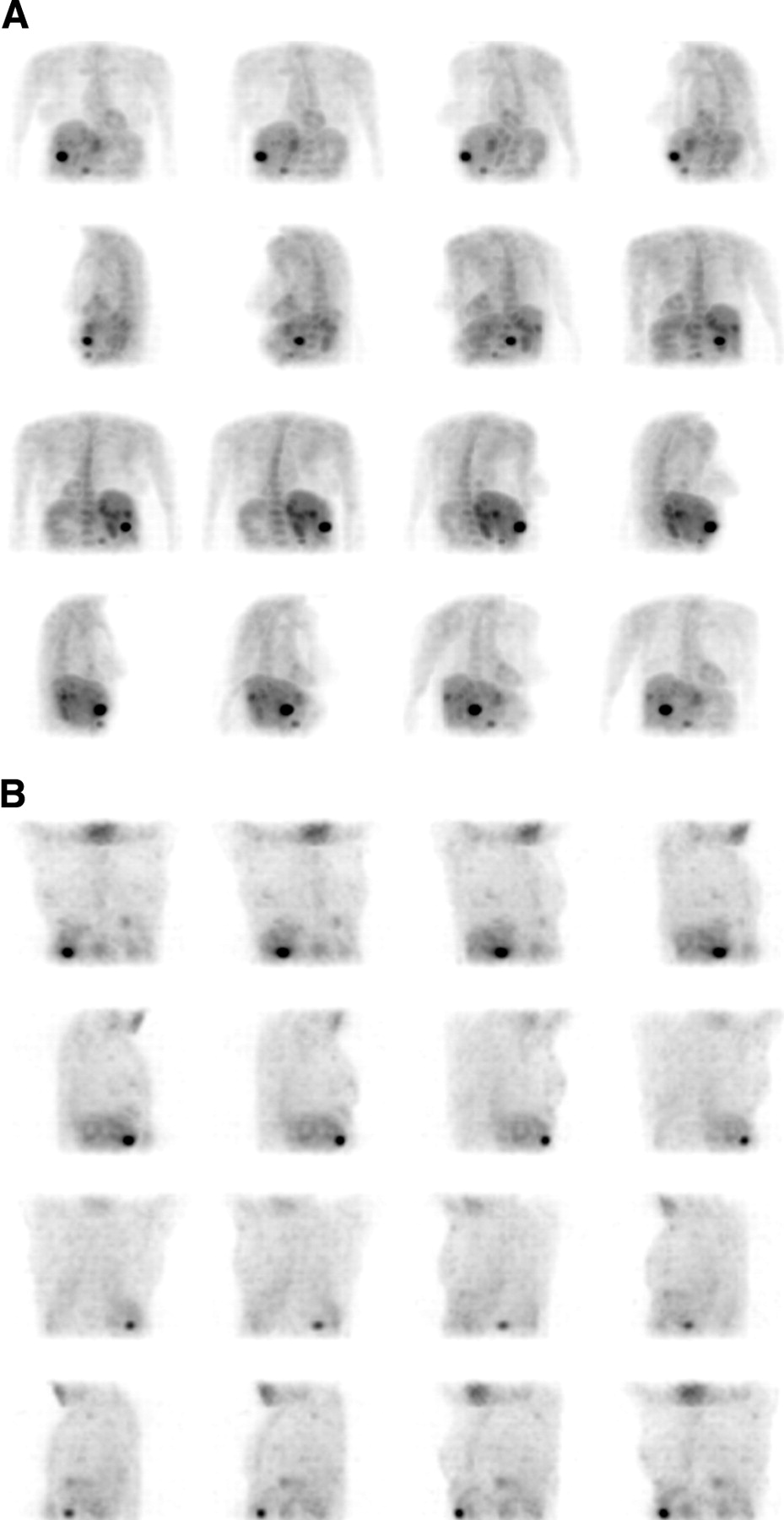
- FIGURE 1.
A 64-y-old patient with known colorectal cancer. 18F-FDG study was performed for restaging before surgical removal of solitary liver metastasis originally observed on CT. (A) 3-Di-mensional maximum-intensity-projection (MIP) images of CoDe5 without attenuation correction detected single lesion in liver. (B) 3-Dimensional MIP images of CoDe8 with attenuation correction detected multiple liver lesions in both liver lobes. Patient’s management was altered by planned surgery being canceled and patient being referred for chemotherapy and radiofrequency treatment.
- FIGURE 2.
A 57-y-old patient with known colorectal cancer was referred for 18F-FDG study because of rising carcinoembryonic antigen levels. Coincidence images detected increased uptake in anterior abdomen. Fusion of coincidence data with low-resolution CT image localized uptake in dilated collecting system of right ectopic kidney. Patient was disease free on clinical follow-up.

Tables
- TABLE 1
Added Value of Fusion of Coincidence and CT to Image Interpretation and Patient Management
Tumor type Coincidence results alone Fused image results Added value of fused images to image interpretation Added value of fused images to patient management Lung cancer Parahilar uptake Localization of uptake to lung parenchyma, no evidence of LN involvement Better anatomic localization, exclusion of nodal involvement Accurate staging, referral for surgery Lung cancer Uptake in lung tumor and in additional abdominal site Localization of abdominal site of uptake at left adrenal Better anatomic localization, detection of metastatic spread Accurate staging, referral for chemotherapy Colorectal cancer Uptake in right anterior abdomen Uptake localized in ectopic kidney Differentiation of physiologic from tumor uptake Exclusion of disease Colorectal cancer Perirectal uptake Focus of uptake in large perirectal residual mass Better anatomic localization, detection of viable tissue within fibrosis None Colorectal cancer Uptake in right upper abdomen Physiologic uptake in hepatic flexure of colon Differentiation of physiologic from tumor uptake None Colorectal cancer Presacral and perineal uptake Uptake in pelvic masses and in adjustment LNs Better anatomic localization, detection of nodal involvement None Colorectal cancer Pelvic uptake Pelvic uptake extending to sacrum Better anatomic localization, detection of adjacent bone involvement Referral for radiotherapy to involved bone Breast cancer Uptake in left pelvis Localization of uptake to iliac crest Better anatomic localization, detection of bone involvement None Breast cancer Uptake in breast and in paratracheal region Localization of cervical uptake in thyroid nodule Better anatomic localization, detection of unsuspected thyroid lesion Thyroid lesion was found to be second primary tumor; breast and thyroid tumors were removed Unknown primary Sites of uptake in cervical, oropharynx, and retrosternal regions Oropharynx uptake delineated lesion at that location on CT image; retrosternal uptake was located in postsurgical changes Better anatomic localization, detection of unknown primary tumor, exclusion of disease in region of previous surgery Referral for surgery and radiotherapy LN = lymph node.
Parameter CoDe5NC CoDe8NC P* 95% CI All tumor lesions (n = 54) T/NT ratio 4.2 ± 3.5 6.3 ± 5.2 <0.0005 1.3, 3.0 T/Bg ratio 4.9 ± 3.9 6.4 ± 5 <0.0005 0.8, 2.3 Assessment of lesions according to location Above diaphragm (n = 31) T/NT ratio 4.5 ± 4 7.1 ± 5.7 <0.0005 1.3, 3.9 T/Bg ratio 5.25 ± 4.6 7.5 ± 5.7 <0.0005 1.1, 3.4 Below diaphragm (n = 23) T/NT ratio 3.7 ± 2.6 5.2 ± 4.3 <0.001 0.6, 2.4 T/Bg ratio 4.3 ± 2.7 4.8 ± 3.3 NS −0.3, 1.4 Assessment of lesion of different sizes† Lesions <7.2 mL (n = 14) T/NT ratio 3.6 ± 2.8 6.2 ± 4.7 <0.001 1.2, 4.0 T/Bg ratio 4 ± 3 6.6 ± 4.3 <0.0005 1.4, 3.6 Lesions 7.2–12.5 mL (n = 13) T/NT ratio 4.2 ± 3.7 6 ± 2.7 <0.01 0.3, 3.4 T/Bg ratio 4.6 ± 3 6.5 ± 3.3 <0.03 −0.02, 3.9 Lesions 12.5–17.5 mL (n = 14) T/NT ratio 4.4 ± 4 7 ± 7.7 <0.01 0.4, 4.9 T/Bg ratio 5 ± 3.8 6.4 ± 6.4 <0.05 −0.3, 3.2 Lesions >17.5 mL (n = 13) T/NT ratio 4.5 ± 3.5 6 ± 4.9 NS −0.5, 3.3 T/Bg ratio 6 ± 5.4 6 ± 5.7 NS −1.3, 1.5 ↵* P < 0.05 was considered significant. NS = not significant.
↵† Parameters evaluated: T/NT ratio and T/Bg ratio. Lesion size was measured by functional volumes. Descriptive statistics were used to define 4 subgroup limits.
Image sets compared were CoDe5NC, 5/8-in. NaI(Tl) crystals without attention correction; and CoDe8NC, 1-in. NaI(Tl) crystals without attention correction.
Parameter CoDe8NC CoDe8AC P* 95% CI All tumor lesions (n = 54) T/NT ratio 6.3 ± 5.2 7.5 ± 5.8 <0.05 −0.2, 2.8 T/Bg ratio 6.4 ± 5 8.9 ± 5.6 <0.0005 1.4, 3.8 Assessment of lesions according to location Above diaphragm (n = 31) T/NT ratio 7.1 ± 5.7 9.0 ± 6.4 <0.04 −0.1, 4.1 T/Bg ratio 7.5 ± 5.7 10.4 ± 6.2 <0.001 1.2, 4.6 Below diaphragm (n = 23) T/NT ratio 5.2 ± 4.3 5.6 ± 4.2 NS −1.8, 2.5 T/Bg ratio 4.8 ± 3.3 7 ± 4.1 <0.003 0.4, 3.9 Assessment of lesion n of different sizes† Lesions <7.2 mL (n = 14) T/NT ratio 6.2 ± 4.7 8.7 ± 5.3 NS −0.2, 5.6 T/Bg ratio 6.6 ± 4.3 9.9 ± 4.4 <0.01 1.2, 5.7 Lesions 7.2–12.5 mL (n = 13) T/NT ratio 6 ± 2.7 8.3 ± 7.6 NS −2.0, 6.5 T/Bg ratio 6.5 ± 3.3 10.1 ± 6.9 <0.03 −0.1, 7.3 Lesions 12.5–17.5 mL (n = 14) T/NT ratio 7 ± 7.7 7.4 ± 6.3 NS −2.6, 3.3 T/Bg ratio 6.4 ± 6.4 7.94 ± 4.6 <0.03 −0.8, 3.9 Lesions >17.5 mL (n = 13) T/NT ratio 6 ± 4.9 5.7 ± 3.5 NS −3.1, 2.5 T/Bg ratio 6 ± 5.7 7.8 ± 6.6 <0.03 −0.04, 3.7 ↵* P < 0.05 was considered significant. NS = not significant.
↵† Parameters evaluated: T/NT ratio and T/Bg ratio. Lesion size was measured by functional volumes. Descriptive statistics were used to define 4 subgroup limits.
Image sets compared were 1-in. NaI(Tl) crystals with and without attenuation correction (CoDe8NC and CoDe8AC).


Jump to section
Related Articles
Cited By...
- No citing articles found.