
Main menu

  • Home
  • Content
    • Current
      • JNMT Supplement
    • Ahead of print
    • Past Issues
    • Continuing Education
    • JNMT Podcast
    • SNMMI Annual Meeting Abstracts
  • Subscriptions
    • Subscribers
    • Rates
    • Journal Claims
    • Institutional and Non-member
  • Authors
    • Submit to JNMT
    • Information for Authors
    • Assignment of Copyright
    • AQARA Requirements
  • Info
    • Reviewers
    • Permissions
    • Advertisers
    • Corporate & Special Sales
  • About
    • About Us
    • Editorial Board
    • Contact Information
  • More
    • Alerts
    • Feedback
    • Help
    • SNMMI Journals
  • SNMMI
    • JNMT
    • JNM
    • SNMMI Journals
    • SNMMI

User menu

  • Subscribe
  • My alerts
  • Log in
  • My Cart

Search

  • Advanced search
Journal of Nuclear Medicine Technology
  • SNMMI
    • JNMT
    • JNM
    • SNMMI Journals
    • SNMMI
  • Subscribe
  • My alerts
  • Log in
  • My Cart
Journal of Nuclear Medicine Technology

Advanced Search

  • Home
  • Content
    • Current
    • Ahead of print
    • Past Issues
    • Continuing Education
    • JNMT Podcast
    • SNMMI Annual Meeting Abstracts
  • Subscriptions
    • Subscribers
    • Rates
    • Journal Claims
    • Institutional and Non-member
  • Authors
    • Submit to JNMT
    • Information for Authors
    • Assignment of Copyright
    • AQARA Requirements
  • Info
    • Reviewers
    • Permissions
    • Advertisers
    • Corporate & Special Sales
  • About
    • About Us
    • Editorial Board
    • Contact Information
  • More
    • Alerts
    • Feedback
    • Help
    • SNMMI Journals
  • Watch or Listen to JNMT Podcast
  • Visit SNMMI on Facebook
  • Join SNMMI on LinkedIn
  • Follow SNMMI on Twitter
  • Subscribe to JNMT RSS feeds
OtherCONTINUING EDUCATION

Gated Myocardial Perfusion SPECT: Basic Principles, Technical Aspects, and Clinical Applications

Asit K. Paul and Hani A. Nabi
Journal of Nuclear Medicine Technology December 2004, 32 (4) 179-187;
Asit K. Paul
  • Find this author on Google Scholar
  • Find this author on PubMed
  • Search for this author on this site
Hani A. Nabi
  • Find this author on Google Scholar
  • Find this author on PubMed
  • Search for this author on this site
  • Article
  • Figures & Data
  • Info & Metrics
  • PDF
Loading

Article Figures & Data

Figures

  • Tables
  • FIGURE 1.
    • Download figure
    • Open in new tab
    • Download powerpoint
    FIGURE 1.

    Principle of ECG-gated acquisition. R–R interval on ECG, representing 1 cardiac cycle, is typically divided into 8 frames of equal duration (A). Image data from each frame are acquired over multiple cardiac cycles and stored separately in specific locations (“bin”) of computer memory (B). When all data in a bin are added together, image represents a specific phase of cardiac cycle. Typically, a volume curve is obtained, which represents endocardial volume for each of 8 frames (C). ED = end-diastole; ES = end-systole.
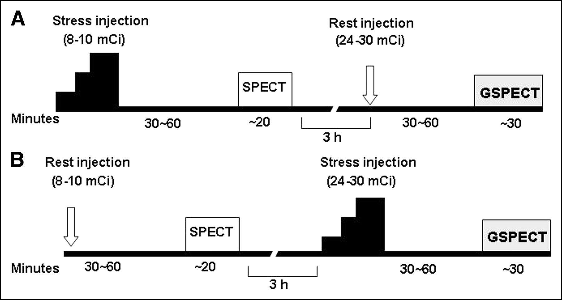
  • FIGURE 2.
    • Download figure
    • Open in new tab
    • Download powerpoint
    FIGURE 2.

    Same-day GSPECT protocols with 99mTc-sestamibi or 99mTc-tetrofosmin. Myocardial perfusion imaging can be gated either at rest or after exercise–stress in stress–rest (A) or rest–stress (B) sequence.
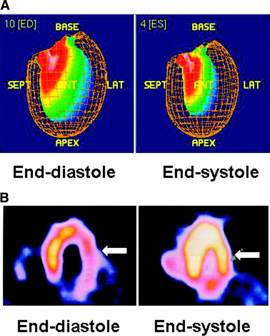
  • FIGURE 3.
    • Download figure
    • Open in new tab
    • Download powerpoint
    FIGURE 3.

    Assessment of LV regional function by GSPECT. RWM is assessed by inward excursion of endocardial wall (A) and SWT is assessed by myocardial brightening (arrow) from end-diastole to end-systole (B). ED = end-diastole; ES = end-systole; SEPT = septal; ANT = anterior; LAT = lateral.

  • FIGURE 4.
    • Download figure
    • Open in new tab
    • Download powerpoint
    FIGURE 4.

    Comparison of LVEF between GSPECT and contrast left ventrioculography (LVG) in a patient. ISE = base; ANT = anterior; SEPT = septal; INF = inferior.
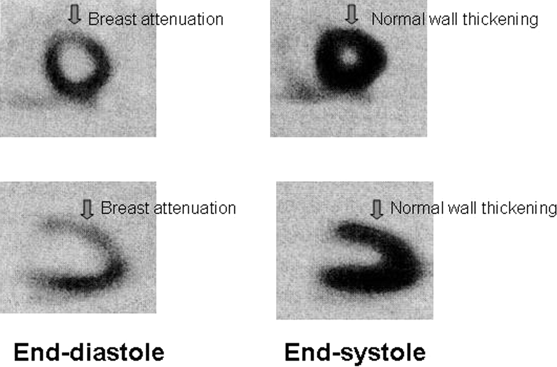
  • FIGURE 5.
    • Download figure
    • Open in new tab
    • Download powerpoint
    FIGURE 5.

    GSPECT shows preserved SWT of anterior wall of LV, suggesting presence of attenuation artifact rather than true infarct.

Tables

  • Figures
    • View popup
    TABLE 1

    Acquisition Parameters for GSPECT with 99mTc-Perfusion Tracers

    ParameterCharacteristic
    γ-CameraDual or triple detector
    CollimatorLow energy, high resolution
    Orbit180° (dual detector) or 360° (triple detector)
    Projections32∼64
    Acquisition time/projection>25 s
    Matrix64 × 64
    Framing8∼16/R–R interval
    Beat acceptance±20%
    Total acquisition time∼20–30 min
    • View popup
    TABLE 2

    Repeatability of Gated SPECT Measurements

    ReferenceParameterNo. of patientsMeasurementAgreement
    YearAuthor
    1997Johnson et al. (23)LVEF15Consecutive daysr = 0.98, SD = 2.6%
    1998Berman et al. (24)LVEF180Proner = 0.93, SD = 3.2%
    EDV180Supiner = 0.97, SD = 2.6 mL
    ESV180Same-day consecutive scansr = 0.98, SD = 4.8 mL
    2001Paeng et al. (25)RWM31Same-day consecutive scansr = 0.95 (quantitative); 79%*, κ = 0.81 (visual)
    SWT31Same-day consecutive scansr = 0.88 (quantitative); 71%*, κ = 0.71 (visual)
    • ↵* Exact segmental agreement.

    • κ = Cohen’s κ.

    • Measurements were performed using QGS software (18,19).

    • View popup
    TABLE 3

    Validation of LV Volumes and EF Measurements Obtained by GSPECT

    ReferenceStudy patientsNo. of patientsModality of comparisonr with GSPECT measurements
    YearAuthorEDVESVLVEF
    1999Vaduganathan et al. (26)Recent MI25MRI0.810.920.93
    1999Tadamura et al. (27)CAD20MRI0.920.970.94
    2000Bavelaar-Croon et al. (28)CAD21MRI0.940.950.85
    2000Bax et al. (29)CAD with LV dysfunction25MRI0.840.870.90
    1998Nichols et al. (30)CAD58LVG0.870.910.86
    1999Yoshioka et al. (31)Mixed21LVG0.730.830.87
    1999Cwajg et al. (32)Mixed109Echo0.870.860.72
    2001Vourvouri et al. (33)CAD with LV dysfunction32Echo0.940.960.83
    2000Paul et al. (34)Mixed15ERNA (tomographic)0.990.990.97
    2000Chua et al. (35)CAD with large defect62ERNA (planar)0.880.950.94
    • MI = myocardial infarction; LVG = contrast left ventriculography; Echo = echocardiography.

    • View popup
    TABLE 4

    Validation of LV Regional Function Obtained by GSPECT

    ReferencePatientsNo. of patientsNo. of segmentsGSPECT evaluationModality of comparisonExact agreement, κ value
    YearAuthorRWMSWT
    1997Gunning et al. (38)Mixed28252Visual, SQMRI78%, 0.6678%, 0.62
    1999Tadamura et al. (27)CAD20180Visual, SQMRI84%, 0.7387%, 0.76
    1999Tadamura et al. (39)After CABG16128Visual, SQMRINA76%, 0.62
    2001Wahba et al. (40)CAD21273Visual, SQMRI84%, 0.7286%, 0.77
    1994Chua et al. (41)CAD40640Visual, SQEcho91%, 0.6890%, 0.62
    1997Germano et al. (19)Mixed791,580QuantitativeVisual, SQ GSPECT73%, 0.4375%, 0.41
    2000Sharir et al. (58)Mixed1002,000QuantitativeVisual, SQ GSPECT80%, 0.7186%, 0.68
    • κ = Cohen’s κ; SQ = semiquantitative; CABG = coronary artery bypass graft; NA = not available; Echo = echocardiography.

PreviousNext
Back to top

In this issue

Journal of Nuclear Medicine Technology: 32 (4)
Journal of Nuclear Medicine Technology
Vol. 32, Issue 4
December 1, 2004
  • Table of Contents
  • About the Cover
  • Index by author
Print
Download PDF
Article Alerts
Sign In to Email Alerts with your Email Address
Email Article

Thank you for your interest in spreading the word on Journal of Nuclear Medicine Technology.

NOTE: We only request your email address so that the person you are recommending the page to knows that you wanted them to see it, and that it is not junk mail. We do not capture any email address.

Enter multiple addresses on separate lines or separate them with commas.
Gated Myocardial Perfusion SPECT: Basic Principles, Technical Aspects, and Clinical Applications
(Your Name) has sent you a message from Journal of Nuclear Medicine Technology
(Your Name) thought you would like to see the Journal of Nuclear Medicine Technology web site.
Citation Tools
Gated Myocardial Perfusion SPECT: Basic Principles, Technical Aspects, and Clinical Applications
Asit K. Paul, Hani A. Nabi
Journal of Nuclear Medicine Technology Dec 2004, 32 (4) 179-187;

Citation Manager Formats

  • BibTeX
  • Bookends
  • EasyBib
  • EndNote (tagged)
  • EndNote 8 (xml)
  • Medlars
  • Mendeley
  • Papers
  • RefWorks Tagged
  • Ref Manager
  • RIS
  • Zotero
Share
Gated Myocardial Perfusion SPECT: Basic Principles, Technical Aspects, and Clinical Applications
Asit K. Paul, Hani A. Nabi
Journal of Nuclear Medicine Technology Dec 2004, 32 (4) 179-187;
Twitter logo Facebook logo LinkedIn logo Mendeley logo
  • Tweet Widget
  • Facebook Like
  • Google Plus One
Bookmark this article

Jump to section

  • Article
    • Abstract
    • GENERAL PRINCIPLES
    • ACQUISITION AND ANALYSIS
    • VALIDATION
    • CLINICAL APPLICATIONS
    • CONCLUSION
    • Acknowledgments
    • Footnotes
    • REFERENCES
  • Figures & Data
  • Info & Metrics
  • PDF

Related Articles

  • No related articles found.
  • PubMed
  • Google Scholar

Cited By...

  • Gated Myocardial Perfusion Imaging
  • Gating: Keep It Regular
  • Validation of Left Ventricular Ejection Fraction with the IQ*SPECT System in Small-Heart Patients
  • Clinical Utility of Enhanced Relative Activity Recovery on Systolic Myocardial Perfusion SPECT: Lessons from PET
  • Artifacts and Pitfalls in Myocardial Perfusion Imaging
  • Gated Myocardial Perfusion SPECT
  • REPLY:
  • Google Scholar

More in this TOC Section

  • Illuminating the Hidden: Standardizing Cardiac MIBG Imaging for Sympathetic Dysfunction
  • PET/CT Case Series: Unmasking the Mystery of Cardiac Sarcoidosis
  • Delivery Methods of Radiopharmaceuticals: Exploring Global Strategies to Minimize Occupational Radiation Exposure
Show more Continuing Education

Similar Articles

SNMMI

© 2025 SNMMI

Powered by HighWire