


Article Figures & Data
Figures
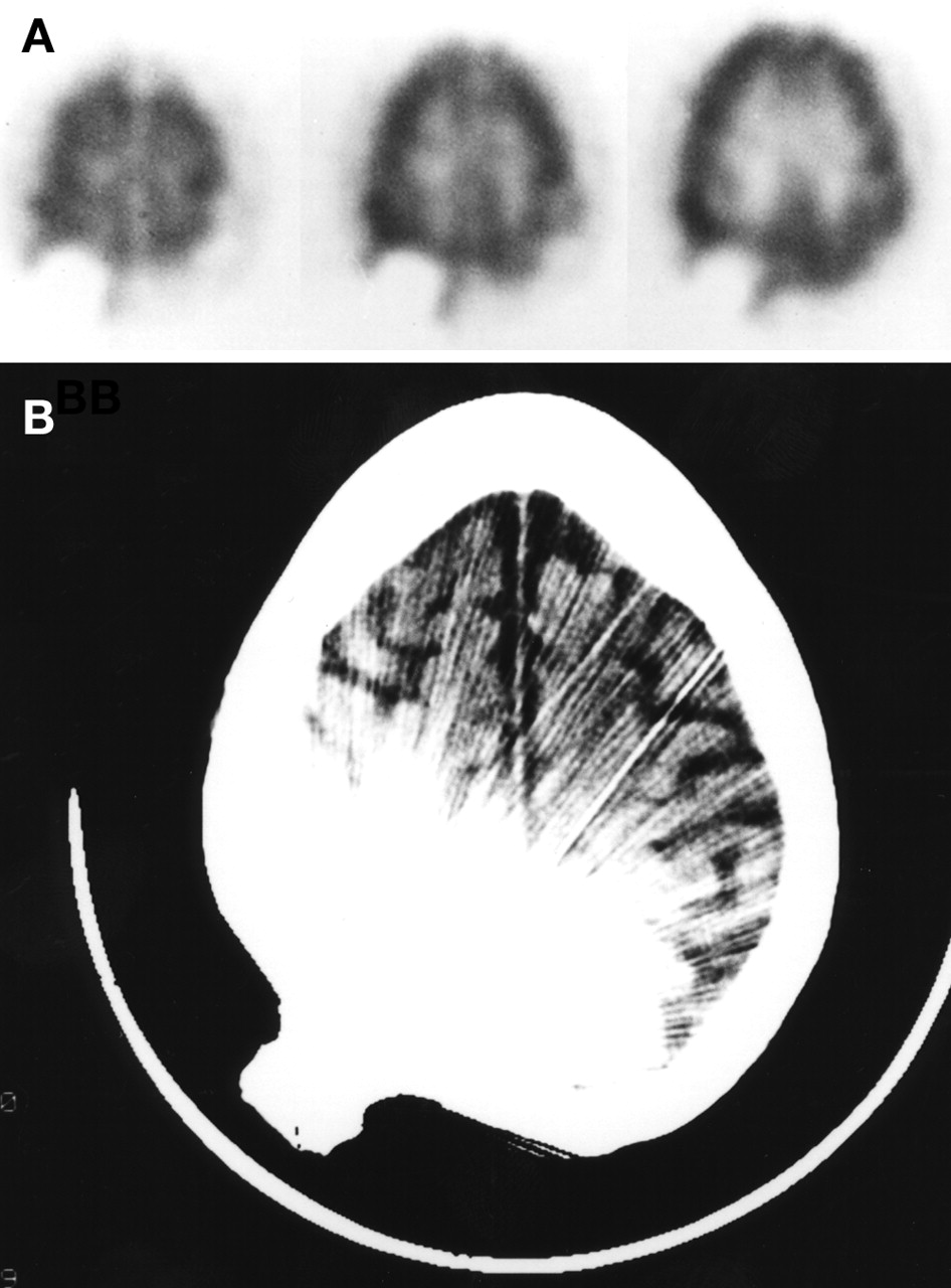
- FIGURE 1.
Patient 1, with head trauma in the 1940s that required a metallic plate in the left frontal area. (A) Transaxial images from brain SPECT study show photon-deficient area in the left frontal region with faint increased tracer activity extending into scalp area at periphery. (B) CT shows significant streak artifact caused by the metallic plate.
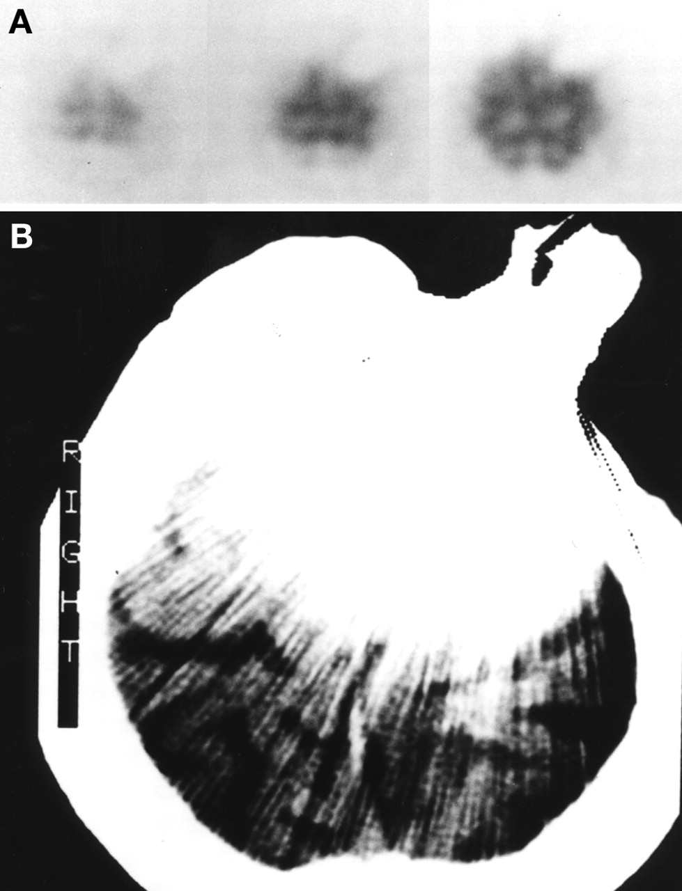
- FIGURE 2.
Patient 2, with head trauma in the 1970s that required a metallic plate in the right occipital area. (A) Transaxial images from brain SPECT study show photon-deficient area in the right occipital region with prominent increased tracer activity extending into scalp area at periphery. (B) CT shows significant streak artifact caused by the metallic plate.
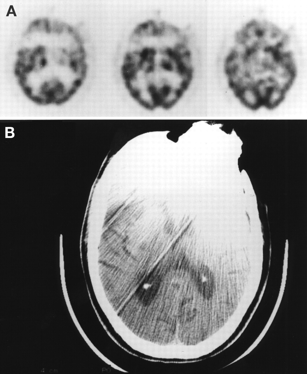
- FIGURE 3.
Patient 3, with head trauma in the 1940s that required a metallic plate in the left frontal area. (A) Transaxial images from brain SPECT study show photon-deficient area in the left frontal region with very faint increased tracer activity extending into scalp area at periphery. (B) CT shows significant streak artifact caused by the metallic plate.
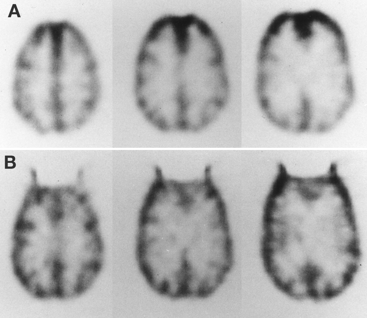
- FIGURE 4.
Transaxial images of SPECT brain phantom with no metallic sheets in place (A) and with 0.076-cm lead sheet placed over midline of the frontal region (B). The 0.038-cm lead sheet produced a similar artifact, though not as prominent.


Tables
Type of Metallic Plate/Sheet Aluminum Stainless Steel Tantalum Lead Mass attenuation coefficient for 140 keV photon (cm2/g) 0.143 NA 1.825 2.393 Density (g/cm3) 2.70 NA 16.65 11.35 Reported metallic plate thickness used for cranioplasty (cm) 0.064 0.046 0.038, 0.051 NA Theoretical attenuation for reported metallic plate (%) 2 NA 68, 79 NA Experimental metallic sheet thickness used for phantom study (cm) 0.064 0.061 NA 0.038, 0.076 Theoretical attenuation for experimental metallic sheet (%) 2 NA NA 64, 87 Calculated attenuation from planar image (%) 4 9 NA NA, 83 Mass attenuation coefficient and density information obtained from www.physics.NIST.gov Web site’s reference tables of x-ray mass attenuation coefficients.


Jump to section
Related Articles
Cited By...
- No citing articles found.