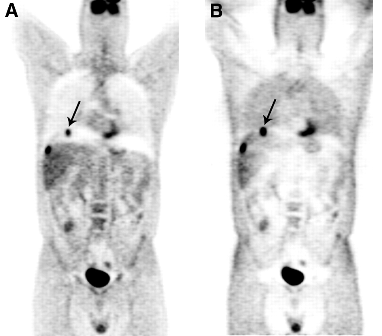
Main menu

  • Home
  • Content
    • Current
      • JNMT Supplement
    • Ahead of print
    • Past Issues
    • Continuing Education
    • JNMT Podcast
    • SNMMI Annual Meeting Abstracts
  • Subscriptions
    • Subscribers
    • Rates
    • Journal Claims
    • Institutional and Non-member
  • Authors
    • Submit to JNMT
    • Information for Authors
    • Assignment of Copyright
    • AQARA Requirements
  • Info
    • Reviewers
    • Permissions
    • Advertisers
    • Corporate & Special Sales
  • About
    • About Us
    • Editorial Board
    • Contact Information
  • More
    • Alerts
    • Feedback
    • Help
    • SNMMI Journals
  • SNMMI
    • JNMT
    • JNM
    • SNMMI Journals
    • SNMMI

User menu

  • Subscribe
  • My alerts
  • Log in
  • Log out
  • My Cart

Search

  • Advanced search
Journal of Nuclear Medicine Technology
  • SNMMI
    • JNMT
    • JNM
    • SNMMI Journals
    • SNMMI
  • Subscribe
  • My alerts
  • Log in
  • Log out
  • My Cart
Journal of Nuclear Medicine Technology

Advanced Search

  • Home
  • Content
    • Current
    • Ahead of print
    • Past Issues
    • Continuing Education
    • JNMT Podcast
    • SNMMI Annual Meeting Abstracts
  • Subscriptions
    • Subscribers
    • Rates
    • Journal Claims
    • Institutional and Non-member
  • Authors
    • Submit to JNMT
    • Information for Authors
    • Assignment of Copyright
    • AQARA Requirements
  • Info
    • Reviewers
    • Permissions
    • Advertisers
    • Corporate & Special Sales
  • About
    • About Us
    • Editorial Board
    • Contact Information
  • More
    • Alerts
    • Feedback
    • Help
    • SNMMI Journals
  • Watch or Listen to JNMT Podcast
  • Visit SNMMI on Facebook
  • Join SNMMI on LinkedIn
  • Follow SNMMI on Twitter
  • Subscribe to JNMT RSS feeds
OtherCONTINUING EDUCATION

PET/CT Imaging Artifacts

Waheeda Sureshbabu and Osama Mawlawi
Journal of Nuclear Medicine Technology September 2005, 33 (3) 156-161;
Waheeda Sureshbabu
  • Find this author on Google Scholar
  • Find this author on PubMed
  • Search for this author on this site
Osama Mawlawi
  • Find this author on Google Scholar
  • Find this author on PubMed
  • Search for this author on this site

FIGURE 7.

FIGURE 7.
  • Download figure
  • Open in new tab
  • Download powerpoint
FIGURE 7.

(A) 58-y-old man with colon cancer. Lesion at dome of liver is mislocalized to right lung (arrow) because of respiratory motion. (B) Image without attenuation correction shows that all lesions are confined to liver.

  View article

In this issue

Journal of Nuclear Medicine Technology: 53 (1)
Journal of Nuclear Medicine Technology
Vol. 53, Issue 1
March 1, 2025
  • Table of Contents
  • About the Cover
  • Index by author
  • Complete Issue (PDF)
Print
Download PDF
Article Alerts
Sign In to Email Alerts with your Email Address
Email Article

Thank you for your interest in spreading the word on Journal of Nuclear Medicine Technology.

NOTE: We only request your email address so that the person you are recommending the page to knows that you wanted them to see it, and that it is not junk mail. We do not capture any email address.

Enter multiple addresses on separate lines or separate them with commas.
FIGURE 7.
(Your Name) has sent you a message from Journal of Nuclear Medicine Technology
(Your Name) thought you would like to see the Journal of Nuclear Medicine Technology web site.
Citation Tools
PET/CT Imaging Artifacts
Waheeda Sureshbabu, Osama Mawlawi
Journal of Nuclear Medicine Technology Sep 2005, 33 (3) 156-161;

Citation Manager Formats

  • BibTeX
  • Bookends
  • EasyBib
  • EndNote (tagged)
  • EndNote 8 (xml)
  • Medlars
  • Mendeley
  • Papers
  • RefWorks Tagged
  • Ref Manager
  • RIS
  • Zotero
Share
PET/CT Imaging Artifacts
Waheeda Sureshbabu, Osama Mawlawi
Journal of Nuclear Medicine Technology Sep 2005, 33 (3) 156-161;
Twitter logo Facebook logo LinkedIn logo Mendeley logo
  • Tweet Widget
  • Facebook Like
  • Google Plus One
Bookmark this article

Related Articles

  • Technologists News
  • PubMed
  • Google Scholar

Cited By...

  • Diagnostic Accuracy of FDG PET/CT in Suspected LVAD Infections: A Case Series, Systematic Review, and Meta-Analysis
  • Measuring PET Spatial Resolution Using a Cylinder Phantom Positioned at an Oblique Angle
  • Clinical Impact of Respiratory Motion Correction in Simultaneous PET/MR, Using a Joint PET/MR Predictive Motion Model
  • Pitfalls and Pearls of Wisdom in 18F-FDG PET Imaging of Tumors
  • PET artefact masquerading as a PET positive lung mass
  • Technical Considerations in Brain Amyloid PET Imaging with 18F-Florbetapir
  • Whole-Body 18F-FDG PET/CT: The Need for a Standardized Field of View--A Referring-Physician Aid
  • Google Scholar

More in this TOC Section

  • Illuminating the Hidden: Standardizing Cardiac MIBG Imaging for Sympathetic Dysfunction
  • PET/CT Case Series: Unmasking the Mystery of Cardiac Sarcoidosis
  • Technegas at Last! Implementing Technegas into Clinical Practice in the United States: Considerations, Challenges, and Recommendations
Show more Continuing Education

Similar Articles

SNMMI

© 2025 SNMMI

Powered by HighWire