Abstract
A strong relationship exists between the severity of carotid stenosis and early stroke risk. Inflammation is believed to be an important event for atherosclerotic plaque destabilization and subsequent thromboembolism. 18F-FDG can image atherosclerotic inflammation, providing information about plaque biology, which may serve as a useful biomarker for the assessment of early stroke risk.
A strong relationship exists between the severity of carotid stenosis and early stroke risk that is independent of other prognostic indicators and neuroimaging findings (1,2). Inflammation may lead to atherosclerotic plaque destabilization and thromboembolism (3). Increased 18F-FDG uptake represents carotid plaque inflammation on PET and predicts early recurrent stroke, independent of age and degree of stenosis (4).
CASE REPORT
A 58-y-old man presented with transient right hemiparesis and aphasia. His cardiovascular risk factors included hypertension, ischemic heart disease, and dyslipidemia. He had been diagnosed 2 y previously with asymptomatic severe (>70%) stenosis of the left internal carotid artery. The findings on clinical examination and CT scanning of the brain were unremarkable. CT angiography (Fig. 1A) confirmed the preexisting severe focal stenosis of the left internal carotid artery.
Diffusion-weighted MR imaging (A) shows ischemic infarcts in left internal carotid artery territory with high-grade stenosis (arrow) on CT angiography (B). Transcranial Doppler monitoring shows spontaneous microembolic signals (arrow) (C).
Clopidogrel, aspirin, and atorvastatin were started with a plan for early carotid endarterectomy. However, on day 3 he developed right hemiparesis. Brain MR imaging revealed multiple acute ischemic infarctions in the left middle and anterior cerebral artery territories (Fig. 1B). Transcranial Doppler monitoring of the left middle cerebral artery revealed spontaneous microemboli (Fig. 1C). Carotid PET/CT fused with CT angiography showed increased 18F-FDG uptake in the left carotid plaque (Figs. 2A and 2B), representing acute inflammation. His family members refused an urgent carotid endarterectomy. On day 5, he developed another severe ischemic stroke in the left middle cerebral artery, with clinical worsening. His further stay in the hospital was uneventful until discharge to a rehabilitation unit.
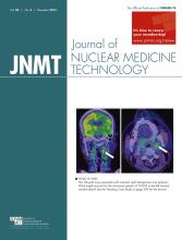
PET/CT imaging shows increased uptake of 18F-FDG (arrows) in left internal carotid artery plaque on coronal plane (A) and axial plane (B), suggestive of acute inflammation.
DISCUSSION
Our case demonstrates the role of 18F-FDG PET imaging of carotid plaque inflammation as a reliable marker for risk stratification. To date, risk stratification for carotid atherosclerosis has been based mainly on arterial lumen stenosis, and understanding of plaque biology remains poor (1).
Limited data are available on the prognostic utility of metabolic imaging with PET to predict short-term stroke risk in patients with recently symptomatic carotid atherosclerosis. The addition of plaque 18F-FDG data to the Framingham Risk Score significantly improved long-term risk prediction (5). In a study in Ireland, the predictive utility of 18F-FDG plaque uptake was independent of age and degree of lumen stenosis (categorized as 50%–69% and 70%–99%; adjusted hazard ratio, 6.1; confidence interval, 1.3–28.8; P = 0.02) (4). In addition to risk stratification, molecular imaging with 18F-FDG PET may have potential to monitor the response of atherosclerotic plaque to therapeutic intervention.
CONCLUSION
Inflammation in a previously stable carotid plaque is an important event that leads to local thrombosis and cerebral embolization. This phenomenon can be reliably imaged with 18F-FDG PET for better risk stratification and therapeutic strategizing in carotid stenosis.
DISCLOSURE
No potential conflict of interest relevant to this article was reported.
Footnotes
Published online Apr. 9, 2015.
REFERENCES
- Received for publication December 29, 2014.
- Accepted for publication February 4, 2015.


