Main menu

  • Home
  • Content
    • Current
      • JNMT Supplement
    • Ahead of print
    • Past Issues
    • Continuing Education
    • JNMT Podcast
    • SNMMI Annual Meeting Abstracts
  • Subscriptions
    • Subscribers
    • Rates
    • Journal Claims
    • Institutional and Non-member
  • Authors
    • Submit to JNMT
    • Information for Authors
    • Assignment of Copyright
    • AQARA Requirements
  • Info
    • Reviewers
    • Permissions
    • Advertisers
    • Corporate & Special Sales
  • About
    • About Us
    • Editorial Board
    • Contact Information
  • More
    • Alerts
    • Feedback
    • Help
    • SNMMI Journals
  • SNMMI
    • JNMT
    • JNM
    • SNMMI Journals
    • SNMMI

User menu

  • Subscribe
  • My alerts
  • Log in
  • Log out
  • My Cart

Search

  • Advanced search
Journal of Nuclear Medicine Technology
  • SNMMI
    • JNMT
    • JNM
    • SNMMI Journals
    • SNMMI
  • Subscribe
  • My alerts
  • Log in
  • Log out
  • My Cart
Journal of Nuclear Medicine Technology

Advanced Search

  • Home
  • Content
    • Current
    • Ahead of print
    • Past Issues
    • Continuing Education
    • JNMT Podcast
    • SNMMI Annual Meeting Abstracts
  • Subscriptions
    • Subscribers
    • Rates
    • Journal Claims
    • Institutional and Non-member
  • Authors
    • Submit to JNMT
    • Information for Authors
    • Assignment of Copyright
    • AQARA Requirements
  • Info
    • Reviewers
    • Permissions
    • Advertisers
    • Corporate & Special Sales
  • About
    • About Us
    • Editorial Board
    • Contact Information
  • More
    • Alerts
    • Feedback
    • Help
    • SNMMI Journals
  • Watch or Listen to JNMT Podcast
  • Visit SNMMI on Facebook
  • Join SNMMI on LinkedIn
  • Follow SNMMI on Twitter
  • Subscribe to JNMT RSS feeds
Review ArticleTeaching Case Studies

Plaque Inflammation Imaging in Severe Carotid Stenosis and Recurrent Cerebral Ischemia

Vijay K. Sharma, Prakash R. Paliwal and Arvind K. Sinha
Journal of Nuclear Medicine Technology December 2015, 43 (4) 299-300; DOI: https://doi.org/10.2967/jnmt.114.153627
Vijay K. Sharma
1Division of Neurology, National University Hospital, Singapore
2YLL School of Medicine, National University of Singapore, Singapore; and
  • Find this author on Google Scholar
  • Find this author on PubMed
  • Search for this author on this site
Prakash R. Paliwal
1Division of Neurology, National University Hospital, Singapore
  • Find this author on Google Scholar
  • Find this author on PubMed
  • Search for this author on this site
Arvind K. Sinha
3Department of Diagnostic Imaging, National University Hospital, Singapore
  • Find this author on Google Scholar
  • Find this author on PubMed
  • Search for this author on this site
  • Article
  • Figures & Data
  • Info & Metrics
  • PDF
Loading

Abstract

A strong relationship exists between the severity of carotid stenosis and early stroke risk. Inflammation is believed to be an important event for atherosclerotic plaque destabilization and subsequent thromboembolism. 18F-FDG can image atherosclerotic inflammation, providing information about plaque biology, which may serve as a useful biomarker for the assessment of early stroke risk.

  • acute ischemic stroke
  • carotid stenosis
  • plaque inflammation
  • PET/CT

A strong relationship exists between the severity of carotid stenosis and early stroke risk that is independent of other prognostic indicators and neuroimaging findings (1,2). Inflammation may lead to atherosclerotic plaque destabilization and thromboembolism (3). Increased 18F-FDG uptake represents carotid plaque inflammation on PET and predicts early recurrent stroke, independent of age and degree of stenosis (4).

CASE REPORT

A 58-y-old man presented with transient right hemiparesis and aphasia. His cardiovascular risk factors included hypertension, ischemic heart disease, and dyslipidemia. He had been diagnosed 2 y previously with asymptomatic severe (>70%) stenosis of the left internal carotid artery. The findings on clinical examination and CT scanning of the brain were unremarkable. CT angiography (Fig. 1A) confirmed the preexisting severe focal stenosis of the left internal carotid artery.

FIGURE 1.
  • Download figure
  • Open in new tab
  • Download powerpoint
FIGURE 1.

Diffusion-weighted MR imaging (A) shows ischemic infarcts in left internal carotid artery territory with high-grade stenosis (arrow) on CT angiography (B). Transcranial Doppler monitoring shows spontaneous microembolic signals (arrow) (C).

Clopidogrel, aspirin, and atorvastatin were started with a plan for early carotid endarterectomy. However, on day 3 he developed right hemiparesis. Brain MR imaging revealed multiple acute ischemic infarctions in the left middle and anterior cerebral artery territories (Fig. 1B). Transcranial Doppler monitoring of the left middle cerebral artery revealed spontaneous microemboli (Fig. 1C). Carotid PET/CT fused with CT angiography showed increased 18F-FDG uptake in the left carotid plaque (Figs. 2A and 2B), representing acute inflammation. His family members refused an urgent carotid endarterectomy. On day 5, he developed another severe ischemic stroke in the left middle cerebral artery, with clinical worsening. His further stay in the hospital was uneventful until discharge to a rehabilitation unit.

FIGURE 2.
  • Download figure
  • Open in new tab
  • Download powerpoint
FIGURE 2.

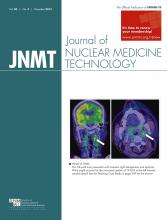
PET/CT imaging shows increased uptake of 18F-FDG (arrows) in left internal carotid artery plaque on coronal plane (A) and axial plane (B), suggestive of acute inflammation.

DISCUSSION

Our case demonstrates the role of 18F-FDG PET imaging of carotid plaque inflammation as a reliable marker for risk stratification. To date, risk stratification for carotid atherosclerosis has been based mainly on arterial lumen stenosis, and understanding of plaque biology remains poor (1).

Limited data are available on the prognostic utility of metabolic imaging with PET to predict short-term stroke risk in patients with recently symptomatic carotid atherosclerosis. The addition of plaque 18F-FDG data to the Framingham Risk Score significantly improved long-term risk prediction (5). In a study in Ireland, the predictive utility of 18F-FDG plaque uptake was independent of age and degree of lumen stenosis (categorized as 50%–69% and 70%–99%; adjusted hazard ratio, 6.1; confidence interval, 1.3–28.8; P = 0.02) (4). In addition to risk stratification, molecular imaging with 18F-FDG PET may have potential to monitor the response of atherosclerotic plaque to therapeutic intervention.

CONCLUSION

Inflammation in a previously stable carotid plaque is an important event that leads to local thrombosis and cerebral embolization. This phenomenon can be reliably imaged with 18F-FDG PET for better risk stratification and therapeutic strategizing in carotid stenosis.

DISCLOSURE

No potential conflict of interest relevant to this article was reported.

Footnotes

  • Published online Apr. 9, 2015.

REFERENCES

  1. 1.↵
    1. Sheehan OC,
    2. Kyne L,
    3. Kelly LA,
    4. et al
    . A population based comparison of ABCD2 score, atrial fibrillation and carotid stenosis for prediction of early stroke recurrence after transient ischaemic attack. The North Dublin TIA Study. Stroke. 2010;41:844–850.
    OpenUrlAbstract/FREE Full Text
  2. 2.↵
    1. Merwick A,
    2. Albers GW,
    3. Amarenco P,
    4. et al
    . Addition of brain and carotid imaging to the ABCD2 score to improve identification of patients at high early stroke risk after transient ischaemic attack. Lancet Neurol. 2010;9:1060–1069.
    OpenUrlCrossRefPubMed
  3. 3.↵
    1. Libby P,
    2. Ridker P,
    3. Hansson GK
    . Progress and challenges in translating the biology of atherosclerosis. Nature. 2011;473:317–325.
    OpenUrlCrossRefPubMed
  4. 4.↵
    1. Marnane M,
    2. Merwick A,
    3. Sheehan OC,
    4. et al
    . Carotid plaque inflammation on 18FDG-PET predicts early stroke recurrence: the Dublin Carotid Atherosclerosis Stroke Study. Ann Neurol. 2012;71:709–718.
    OpenUrlCrossRefPubMed
  5. 5.↵
    1. Tawakol A,
    2. Finn AV
    . Imaging inflammatory changes in atherosclerosis multimodal imaging hitting stride. JACC Cardiovasc Imaging. 2011;4:1119–1122.
    OpenUrlCrossRefPubMed
  • Received for publication December 29, 2014.
  • Accepted for publication February 4, 2015.
PreviousNext
Back to top

In this issue

Journal of Nuclear Medicine Technology: 43 (4)
Journal of Nuclear Medicine Technology
Vol. 43, Issue 4
December 1, 2015
  • Table of Contents
  • About the Cover
  • Index by author
Print
Download PDF
Article Alerts
Sign In to Email Alerts with your Email Address
Email Article

Thank you for your interest in spreading the word on Journal of Nuclear Medicine Technology.

NOTE: We only request your email address so that the person you are recommending the page to knows that you wanted them to see it, and that it is not junk mail. We do not capture any email address.

Enter multiple addresses on separate lines or separate them with commas.
Plaque Inflammation Imaging in Severe Carotid Stenosis and Recurrent Cerebral Ischemia
(Your Name) has sent you a message from Journal of Nuclear Medicine Technology
(Your Name) thought you would like to see the Journal of Nuclear Medicine Technology web site.
Citation Tools
Plaque Inflammation Imaging in Severe Carotid Stenosis and Recurrent Cerebral Ischemia
Vijay K. Sharma, Prakash R. Paliwal, Arvind K. Sinha
Journal of Nuclear Medicine Technology Dec 2015, 43 (4) 299-300; DOI: 10.2967/jnmt.114.153627

Citation Manager Formats

  • BibTeX
  • Bookends
  • EasyBib
  • EndNote (tagged)
  • EndNote 8 (xml)
  • Medlars
  • Mendeley
  • Papers
  • RefWorks Tagged
  • Ref Manager
  • RIS
  • Zotero
Share
Plaque Inflammation Imaging in Severe Carotid Stenosis and Recurrent Cerebral Ischemia
Vijay K. Sharma, Prakash R. Paliwal, Arvind K. Sinha
Journal of Nuclear Medicine Technology Dec 2015, 43 (4) 299-300; DOI: 10.2967/jnmt.114.153627
Twitter logo Facebook logo LinkedIn logo Mendeley logo
  • Tweet Widget
  • Facebook Like
  • Google Plus One
Bookmark this article

Jump to section

  • Article
    • Abstract
    • CASE REPORT
    • DISCUSSION
    • CONCLUSION
    • DISCLOSURE
    • Footnotes
    • REFERENCES
  • Figures & Data
  • Info & Metrics
  • PDF

Related Articles

  • No related articles found.
  • PubMed
  • Google Scholar

Cited By...

  • No citing articles found.
  • Google Scholar

More in this TOC Section

  • High-Sensitivity Troponin Elevation in a Young Woman with Typical Chest Pain: The Heart of the Matter
  • Pulmonary Adenocarcinoma Revealed by Parathyroid Scintigraphy: An Incidental Case to Remember
  • Prominent Right Ventricular Tracer Uptake: A Harbinger of Multivessel Coronary Artery Disease
Show more Teaching Case Studies

Similar Articles

Keywords

  • acute ischemic stroke
  • carotid stenosis
  • plaque inflammation
  • PET/CT
SNMMI

© 2025 SNMMI

Powered by HighWire