  • Main menu
  • User menu
  • Search
  • English ▼
    • English
    • Afrikaans
    • Albanian
    • Amharic
    • Arabic
    • Armenian
    • Azerbaijani
    • Basque
    • Belarusian
    • Bengali
    • Bosnian
    • Bulgarian
    • Catalan
    • Cebuano
    • Chichewa
    • Chinese (Simplified)
    • Chinese (Traditional)
    • Corsican
    • Croatian
    • Czech
    • Danish
    • Dutch
    • Esperanto
    • Estonian
    • Filipino
    • Finnish
    • French
    • Frisian
    • Galician
    • Georgian
    • German
    • Greek
    • Gujarati
    • Haitian Creole
    • Hausa
    • Hawaiian
    • Hebrew
    • Hindi
    • Hmong
    • Hungarian
    • Icelandic
    • Igbo
    • Indonesian
    • Irish
    • Italian
    • Japanese
    • Javanese
    • Kannada
    • Kazakh
    • Khmer
    • Korean
    • Kurdish (Kurmanji)
    • Kyrgyz
    • Lao
    • Latin
    • Latvian
    • Lithuanian
    • Luxembourgish
    • Macedonian
    • Malagasy
    • Malay
    • Malayalam
    • Maltese
    • Maori
    • Marathi
    • Mongolian
    • Myanmar (Burmese)
    • Nepali
    • Norwegian
    • Pashto
    • Persian
    • Polish
    • Portuguese
    • Punjabi
    • Romanian
    • Russian
    • Samoan
    • Scottish Gaelic
    • Serbian
    • Sesotho
    • Shona
    • Sindhi
    • Sinhala
    • Slovak
    • Slovenian
    • Somali
    • Spanish
    • Sudanese
    • Swahili
    • Swedish
    • Tajik
    • Tamil
    • Telugu
    • Thai
    • Turkish
    • Ukrainian
    • Urdu
    • Uzbek
    • Vietnamese
    • Welsh
    • Xhosa
    • Yiddish
    • Yoruba
    • Zulu

Main menu

  • Home
  • Content
    • Current
    • Ahead of print
    • Past Issues
    • JNM Supplement
    • SNMMI Annual Meeting Abstracts
    • Continuing Education
    • JNM Podcasts
  • Subscriptions
    • Subscribers
    • Institutional and Non-member
    • Rates
    • Journal Claims
    • Corporate & Special Sales
  • Authors
    • Submit to JNM
    • Information for Authors
    • Assignment of Copyright
    • AQARA requirements
  • Info
    • Reviewers
    • Permissions
    • Advertisers
  • About
    • About Us
    • Editorial Board
    • Contact Information
  • More
    • Alerts
    • Feedback
    • Help
    • SNMMI Journals
  • SNMMI
    • JNM
    • JNMT
    • SNMMI Journals
    • SNMMI

User menu

  • Subscribe
  • My alerts
  • Log in
  • Log out
  • My Cart
Institution: Mass Inst of Tech Libraries

Search

  • Advanced search
Journal of Nuclear Medicine
  • SNMMI
    • JNM
    • JNMT
    • SNMMI Journals
    • SNMMI
Institution: Mass Inst of Tech Libraries
  • Subscribe
  • My alerts
  • Log in
  • Log out
  • My Cart
Journal of Nuclear Medicine

Advanced Search

English ▼
  • English
  • Afrikaans
  • Albanian
  • Amharic
  • Arabic
  • Armenian
  • Azerbaijani
  • Basque
  • Belarusian
  • Bengali
  • Bosnian
  • Bulgarian
  • Catalan
  • Cebuano
  • Chichewa
  • Chinese (Simplified)
  • Chinese (Traditional)
  • Corsican
  • Croatian
  • Czech
  • Danish
  • Dutch
  • Esperanto
  • Estonian
  • Filipino
  • Finnish
  • French
  • Frisian
  • Galician
  • Georgian
  • German
  • Greek
  • Gujarati
  • Haitian Creole
  • Hausa
  • Hawaiian
  • Hebrew
  • Hindi
  • Hmong
  • Hungarian
  • Icelandic
  • Igbo
  • Indonesian
  • Irish
  • Italian
  • Japanese
  • Javanese
  • Kannada
  • Kazakh
  • Khmer
  • Korean
  • Kurdish (Kurmanji)
  • Kyrgyz
  • Lao
  • Latin
  • Latvian
  • Lithuanian
  • Luxembourgish
  • Macedonian
  • Malagasy
  • Malay
  • Malayalam
  • Maltese
  • Maori
  • Marathi
  • Mongolian
  • Myanmar (Burmese)
  • Nepali
  • Norwegian
  • Pashto
  • Persian
  • Polish
  • Portuguese
  • Punjabi
  • Romanian
  • Russian
  • Samoan
  • Scottish Gaelic
  • Serbian
  • Sesotho
  • Shona
  • Sindhi
  • Sinhala
  • Slovak
  • Slovenian
  • Somali
  • Spanish
  • Sudanese
  • Swahili
  • Swedish
  • Tajik
  • Tamil
  • Telugu
  • Thai
  • Turkish
  • Ukrainian
  • Urdu
  • Uzbek
  • Vietnamese
  • Welsh
  • Xhosa
  • Yiddish
  • Yoruba
  • Zulu
  • Home
  • Content
    • Current
    • Ahead of print
    • Past Issues
    • JNM Supplement
    • SNMMI Annual Meeting Abstracts
    • Continuing Education
    • JNM Podcasts
  • Subscriptions
    • Subscribers
    • Institutional and Non-member
    • Rates
    • Journal Claims
    • Corporate & Special Sales
  • Authors
    • Submit to JNM
    • Information for Authors
    • Assignment of Copyright
    • AQARA requirements
  • Info
    • Reviewers
    • Permissions
    • Advertisers
  • About
    • About Us
    • Editorial Board
    • Contact Information
  • More
    • Alerts
    • Feedback
    • Help
    • SNMMI Journals
  • View or Listen to JNM Podcast
  • Visit JNM on Facebook
  • Join JNM on LinkedIn
  • Follow JNM on Twitter
  • Subscribe to our RSS feeds
Research ArticleThe State of the Art

FAPI PET/CT: Will It End the Hegemony of 18F-FDG in Oncology?

Rodney J. Hicks, Peter J. Roselt, Kumarswamy G. Kallur, Richard W. Tothill and Linda Mileshkin
Journal of Nuclear Medicine March 2021, 62 (3) 296-302; DOI: https://doi.org/10.2967/jnumed.120.256271
Rodney J. Hicks
1Sir Peter MacCallum Department of Oncology, University of Melbourne, Melbourne, Victoria, Australia
  • Find this author on Google Scholar
  • Find this author on PubMed
  • Search for this author on this site
Peter J. Roselt
2Cancer Imaging, Peter MacCallum Cancer Centre, Melbourne, Victoria, Australia
  • Find this author on Google Scholar
  • Find this author on PubMed
  • Search for this author on this site
Kumarswamy G. Kallur
3Healthcare Global Enterprises Ltd., Bangalore, Karnataka, India; and
  • Find this author on Google Scholar
  • Find this author on PubMed
  • Search for this author on this site
Richard W. Tothill
4Department of Clinical Pathology and Centre for Cancer Research, University of Melbourne, Melbourne, Australia
  • Find this author on Google Scholar
  • Find this author on PubMed
  • Search for this author on this site
Linda Mileshkin
1Sir Peter MacCallum Department of Oncology, University of Melbourne, Melbourne, Victoria, Australia
  • Find this author on Google Scholar
  • Find this author on PubMed
  • Search for this author on this site
  • Article
  • Figures & Data
  • Info & Metrics
  • PDF
Loading

Abstract

For over 40 years, 18F-FDG has been the dominant PET tracer in neurology, cardiology, inflammatory diseases, and, most particularly, oncology. Combined with the ability to perform whole-body scanning, 18F-FDG has revolutionized the evaluation of cancer and has stifled the adoption of other tracers, except in situations where low avidity or high background activity limits diagnostic performance. The strength of 18F-FDG has generally been its ability to detect disease in the absence of structural abnormality, thereby enhancing diagnostic sensitivity, but its simultaneous weakness has been a lack of specificity due to diverse pathologies with enhanced glycolysis. Radiotracers that leverage other hallmarks of cancer or specific cell-surface targets are gradually finding a niche in the diagnostic armamentarium. However, none have had sufficient sensitivity to realistically compete with 18F-FDG for evaluation of the broad spectrum of malignancies. Perhaps, this situation is about to change with development of a class of tracers targeting fibroblast activation protein that have low uptake in almost all normal tissues but high uptake in most cancer types. In this review, the development and exciting preliminary clinical data relating to various fibroblast activation protein–specific small-molecule inhibitor tracers in oncology will be discussed along with potential nononcologic applications.

  • PET
  • theranostics
  • fibroblast activation protein
  • oncology
  • radiochemistry

Inspired by Sokoloff’s autoradiographic assessment of cerebral glucose metabolism, David Kuhl approached the Brookhaven National Laboratory to develop a positron-emitting tracer of deoxyglucose for his new tomographic scanner. As a consequence, 18F-FDG, which Dr. Henry Wagner once described as being “the molecule of the 20th century,” was conceived. Roles in epilepsy (1), neurodegeneration (2), and ischemic heart disease (3) were initially described, but it was in oncology that 18F-FDG has most profoundly impacted the current practice of molecular imaging. This was accelerated by the development of whole-body scanning capability (4) and particularly PET/CT (5).

In the developed world, PET/CT is now widely used to diagnose, stage, and monitor treatment response with more accuracy, sparing inappropriate use of expensive and toxic therapies (6). 18F-FDG is by far the dominant tracer in oncology, despite increasing use of more specific tracers in certain malignancies, such as 68Ga-DOTA-octreotate in neuroendocrine neoplasia (7) and various prostate-specific membrane antigen tracers for prostate cancer evaluation (8,9). These two have become particularly important as part of theranostic paradigms including peptide receptor radionuclide therapy (10) and prostate-specific membrane antigen–based radioligand therapy (11), respectively. Similarly, high uptake of 18F-FDG in the brain has opened the way for amino acid analogs in the evaluation of brain tumors (12).

Despite the attraction of such tracers in advancing precision medicine and replacing standard nuclear medicine procedures (13), none have the broadly based appeal of 18F-FDG across the spectrum of malignancies in which it currently holds hegemony. For a tracer to compete, it would need to vie with 18F-FDG for sensitivity and surpass it with respect to specificity or, alternatively, offer the opportunity for theranostic application. Although data are preliminary, emerging evidence suggests that small-molecule fibroblast activation protein inhibitor (FAPI) tracers may be candidates for widespread oncologic application. In this review, the development and performance of FAPI PET/CT will be discussed along with possible therapeutic use. As with 18F-FDG, FAPI agents may also find an important diagnostic niche in nononcologic conditions.

THE ROLE OF CAFs IN CANCER

Although neoplastic cells have traditionally been thought to be primary determinants of cancer behavior, nonmalignant cells within the tumor microenvironment are increasingly recognized as modulators of both tumor progression and therapeutic response. Accordingly, the stromal compartment, including immune cells, vascular elements, and cancer-associated fibroblasts (CAFs), has been a focus of investigation. Key functions of CAFs are deposition and remodeling of extracellular matrix as part of a fibrotic reaction, which, when prominent in tumors such as pancreatic adenocarcinoma (14), is termed desmoplasia. However, CAFs are commonly found in solid tumor types without desmoplasia and therefore represent an attractive diagnostic and therapeutic target (15). Indeed, there is a concerted international effort to develop therapeutic agents targeting CAFs (16). Recently, it has become clear that the complex interplay of CAFs and immune cells can both suppress and accelerate tumor growth. This is likely to be driven by the significant biologic heterogeneity that has been identified in CAF subtypes both within and between cancers (17,18). The origins of CAFs are postulated to include activation of quiescent tissue fibroblasts, transdifferentiation of other stromal or epithelial cells, recruitment of circulating mesenchymal stem cells, or differentiation from tissue-resident stem cells (19). Any or all of these mechanisms may coexist, but how this ontogeny influences their biologic behavior remains to be fully elucidated. Epigenetic processes are thought to be involved in the development of protumorigenic CAFs (20).

One of the major impacts of CAFs is the development of an immunosuppressive tumor microenvironment (21) by processes including desmoplastic stroma excluding T cells from tumor deposits and secretion of various chemokines that recruit myeloid-derived suppressor cells and regulatory T cells. Chief among these is transforming growth factor β (22). The desmoplasia initiated by transforming growth factor β has recently been shown to be mediated through downregulation of signaling through the type I interferon receptor (23). CAFs also interact to alter the behavior of epithelial cancer cells through cell–cell interactions mediated by cadherins, cytokine signaling, and secretion of exosomes (24). Further, by modifying the extracellular matrix, CAFs are permissive for cancer invasion and tumor cell survival (25,26).

The biology and nomenclature of subsets of CAFs in different cancer types are still evolving (18), but recent advances, including single-cell technologies, have helped to define specialized subsets of CAFs (21). In pancreatic cancer, for instance, CAFs can be classified as myofibroblastic, inflammatory, and antigen-presenting (27).

FIBROBLAST ACTIVATION PROTEIN (FAP) AND CANCER BIOLOGY

FAP is a type II transmembrane glycoprotein belonging to the dipeptidyl peptidase IV–like family of postprolyl cleaving serine proteases that are expressed in dimeric form on the surface of CAFs (28); as such, FAP is an attractive diagnostic and therapeutic target. It plays a complex role in modulating the extracellular matrix (29). FAP differs from other members of the dipeptidyl peptidase family in acting as a gelatinase on denatured collagen, especially when cleaved by matrix metallopeptidases (30). Although the role of specific subtypes of CAFs in promoting or suppressing tumorigenesis remains controversial, it is becoming increasing clear that CAFs with high FAP expression are associated with an adverse prognosis by promoting invasion, angiogenesis, microenvironmental immune suppression, and metastasis (15). At least in part, this may reflect production of an immunosuppressive tumor microenvironment (31). Interestingly, in addition to expression on CAFs, FAP is also expressed on tumor-associated macrophages, which are important mediators of immune suppression (32). FAP has been shown to be important in recruitment of immunosuppressive myeloid-derived suppressor cells in cholangiocarcinoma (33). One of the potential effector molecules secreted by FAP-expressing CAFs is chemokine (C-X-C motif) ligand 12, which leads to polarization of macrophages to the immunosuppressive M2 phenotype (34) and also promotes angiogenesis in both breast (35) and colorectal carcinoma (36).

There is still much to learn about the biologic and clinical significance of FAP expression in different tumor types. For example, in the brain, where CAFs do not exist in the microenvironment, FAP overexpression has been found both in nonmalignant mesenchymal cells in the stroma and in transformed cells but had no association with prognosis (37). FAP expression was particularly associated with dysplastic blood vessels and highest in the mesenchymal subtype of glioblastoma. In a subsequent study of 13 patients with glioblastoma (38), no correlation was found with the apparent diffusion coefficient, which is generally associated with variation in prognosis. There was, however, significant intratumoral heterogeneity, with a relationship to better-perfused regions, possibly reflecting FAP expression in the neovasculature (39). Additionally, FAP expression is also seen in certain sarcomas and some benign mesenchymal tumors, including desmoid, giant cell tumors, and chondroblastoma (40), suggesting that FAP expression is not limited to CAFs.

Besides the opportunity that FAP provides as a diagnostic target, its high expression in several cancers with a poor response to current therapeutic approaches, including pancreatic (41) and ovarian carcinoma (42,43), makes it also an attractive therapeutic target. Even in more common and generally chemoresponsive tumors such as colorectal cancer, high FAP expression has also been found to be associated with poor prognosis (36,44). At least in preclinical models, use of chimeric antigen receptor T cells directed against FAP-expressing CAFs has been a successful therapeutic strategy (45). FAP is also being investigated as a target for antibody–drug conjugates (46). Accordingly, companion diagnostics would be valuable for selecting patients for such treatments or even for selecting patients for radioligand therapy. Importantly, FAP appears to be overexpressed across a diverse range of cancers (Fig. 1).

FIGURE 1.
  • Download figure
  • Open in new tab
  • Download powerpoint
FIGURE 1.

Cancer FAP expression results based on data generated by Cancer Genome Atlas Research Network (99). mRNA = messenger RNA; SCC = squamous cell carcinoma.

FAP AS A TARGET FOR IMAGING CANCER

Early attempts to develop FAP imaging probes involved radiolabeled antibodies. The 19F probe that had been used for immunohistochemical identification of FAP expression (47) was labeled with 131I and evaluated in patients with metastatic colorectal cancer (48). However, the long circulation time of intact antibodies poses logistic challenges and stimulated investigation of smaller molecules that may be suitable for imaging. It had long been known that novel dipeptides containing the boronic acid analog of proline could form potent yet nonselective inhibitors of the dipeptidyl peptidase family of peptidases (49). It was the introduction of the structurally similar 2-cyanopyrollidine that led to the discovery of FAPIs with increased selectivity and nanomolar affinity (50,51). By building on this work, the Heidelberg group based its first FAP-targeted tracers, FAPI-01 and FAPI-02, radiolabeled with 131I, and 68Ga, respectively, through chelation to DOTA (52).

Initial reports primarily involved preclinical validation (52,53). Compared with FAPI-01, FAPI-02 was found to specifically bind to FAP-expressing cells and have greater cellular retention. Internalization was confirmed using a fluorescent FAPI-02 with colocalization with endosomes. Small-animal PET demonstrated high and specific tumor uptake. Encouraged by these results, a preliminary human evaluation was performed on 3 patients, 1 each with metastatic breast, lung, and pancreatic cancer. All 3 patients had visualization of primary and metastatic disease but declining tumor uptake over 3 h. The primary route of excretion was renal, with little parenchymal retention. A series of variants based on FAPI-02 was designed to increase cellular retention. The most promising agent in preclinical models, FAPI-04, was tested in 2 women with metastatic breast cancer, demonstrating encouraging tumor visualization and a favorable biodistribution (54).

A further report expanded these preliminary clinical results in 50 patients equally split between 68Ga-FAPI-02 or 68Ga-FAPI-04 PET/CT (55). As suggested by the earlier reports, there was significantly greater washout of 68Ga-FAPI-02 than of 68Ga-FAPI-04 from tumor deposits at 1–3 h, but both performed comparably at 1 h, with clinically acceptable dosimetry. In 6 patients with contemporaneous 18F-FDG PET/CT available for direct correlation, comparable tumor SUV results were observed, except for 1 patient with iodine-negative thyroid cancer in whom 18F-FDG visualized several sites of disease not identified on FAPI PET/CT.

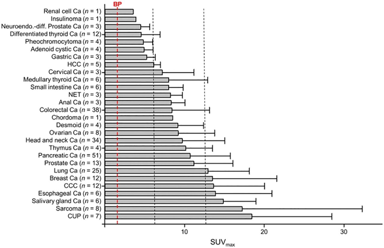
The Heidelberg experience with 68Ga-FAPI-04 was subsequently expanded to 80 patients with a range of cancers (56). Of 28 cancer types evaluated, the highest uptake (average SUVmax > 12) was observed in carcinoma of unknown primary, sarcoma, cholangiocarcinoma, and cancers of the esophagus, breast, and lung, but most had uptake significantly higher than that in the blood pool (Fig. 2). However, a case report from another group suggested that inflammatory conditions, such as IgG4-related disease, can also be positive (57). Similarly, another group evaluating 68Ga-FAPI-04 in 68 patients with equivocal 18F-FDG PET/CT findings found a small number of false-positive results related to inflammatory conditions despite clarifying the extent of disease in most patients (58). Another study evaluated 75 patients with FAPI-04, including 54 with 18F-FDG PET/CT correlation (59). Across 12 different cancer groupings, FAPI PET/CT identified more primary, nodal, and distant metastatic sites than did 18F-FDG PET/CT, with a significantly higher SUV in lung, pancreatic, and gastric cancer. The high tumor-to-background ratios provided by 68Ga-FAPI have been leveraged to guide radiotherapy planning in 14 patients with head and neck cancer (60), a site of complex anatomy and where muscular and brown fat activity can make 18F-FDG PET/CT difficult to interpret. Given that FAP seems to invoke an immune-excluded tumor microenvironment, FAPI imaging may potentially play a role in identifying patients who are less likely to respond to immune checkpoint inhibitor therapy (61).

FIGURE 2.
  • Download figure
  • Open in new tab
  • Download powerpoint
FIGURE 2.

Average SUVmax of 68Ga-FAPI PET/CT in various tumor entities in comparison to blood pool. Ca = cancer; CCC = cholangiocellular carcinoma; CUP = carcinoma of unknown primary; HCC = hepatocellular carcinoma; NET = neuroendocrine tumor; neuroendo.-diff. = neuroendocrine differentiation. (Reprinted with permission of (56).)

The Heidelberg group continued to experiment with variations on FAPI-04 using chemical adaptations, including alteration of lipophilicity and modification of the DOTA/linker attachment at the quinoline moiety to improve tumor retention (53). On the basis of cell culture and preclinical imaging, 2 lead compounds entered clinical evaluation in 8 patients. These were designated FAPI-21 and FAPI-46. Although both had high tumor uptake, FAPI-21 inexplicably had higher uptake in the major salivary glands, mouth, and thyroid, rendering 68Ga-FAPI-46 the leading candidate for clinical evaluation. Formal dosimetry studies performed on 6 patients were favorable (62). The main off-target organs to receive radiation were, as expected, the urinary bladder and kidneys. This suggests that adequate hydration and frequent voiding would be protective in the therapeutic setting. From a combination of scans with 68Ga-FAPI-04 (n = 16) and 68Ga-FAPI-46 (n = 6), promising results were reported in tumors of the lower gastrointestinal tract (63). Stage was changed in 50% of patients without prior treatment, with significantly more metastases being identified in those with already known metastasis, and management was changed in 17 of 21 evaluable patients. However, relatively few of these patients had been staged with 18F-FDG PET/CT. The study included 6 patients with anal cancer, which can be difficult to assess because of physiologic 18F-FDG uptake in sphincteric muscle. Low bowel activity is a potential advantage of FAPI in delineating peritoneal disease (Fig. 3).

FIGURE 3.
  • Download figure
  • Open in new tab
  • Download powerpoint
FIGURE 3.

Peritoneal carcinomatosis. (A) 18F-FDG PET maximum-intensity-projection image demonstrates focal uptake in region of cecum and transverse colon but impression of diffuse peritoneal disease. (B) Corresponding FAPI-04 PET maximum-intensity projection clearly demonstrates diffuse peritoneal disease including involvement of subphrenic spaces.

In a further advance, an 18F-labeled agent, FAPI-74, was recently described that uses an aluminum-fluoride labeling method on a precursor that can also be labeled as a cold kit using 68Ga (64). Evaluation in 10 patients with lung cancer revealed somewhat higher vascular visualization than that obtained with FAPI-02 or FAPI-04 but high tumor uptake enabling radiotherapy planning. Radiosynthesis and preclinical evaluation of another fluorinated FAPI tracer, 18FGlc‐FAPI, has also been recently described (65). This agent had higher hepatobiliary clearance than 68Ga-FAPI-04. There is currently limited information on the clinical performance of this tracer.

Low uptake of FAPI agents in the brain is a potential advantage over 18F-FDG for evaluation of both primary and secondary malignancies (Fig. 4). Recognition of cerebral metastasis (66) and leptomeningeal disease (67) has been reported in individual patients with adenocarcinoma of the lung. However, uptake may not necessarily be specific for malignancy, with infectious etiologies such as tuberculosis also demonstrating increased activity (68). In a series of patients with various primary brain tumors, uptake of 68Ga-FAPI-02 or 68Ga-FAPI-04 was observed in all isocitrate dehydrogenase wild-type glioblastomas and in higher-grade isocitrate dehydrogenase mutant gliomas (38). In a preliminary evaluation, the potential application of FAPI PET/CT or PET/MRI for radiotherapy planning has been described (69).

FIGURE 4.
  • Download figure
  • Open in new tab
  • Download powerpoint
FIGURE 4.

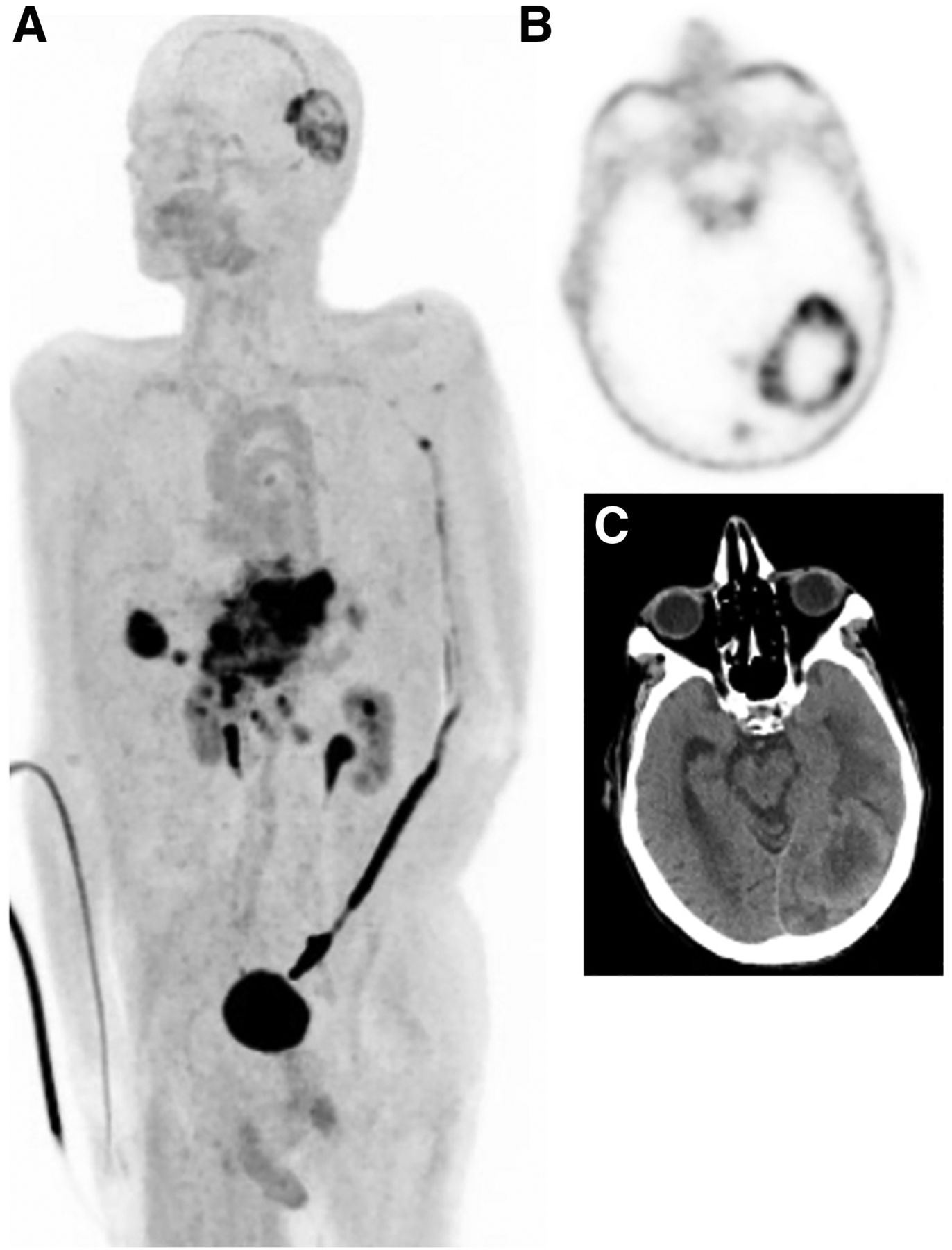
Gastric cancer with node, liver, and brain metastases. (A) FAPI PET maximum-intensity projection. (B) Necrotic left occipital metastasis on transaxial FAPI PET. (C) Correlative CT.

Beside the diagnostic utility of FAPI agents, the potential ability to select patients for radionuclide therapy is an attractive prospect. Preclinical studies of 177Lu-labeled internalizing antibodies directed against FAP have been tested in preclinical models and demonstrated to have efficacy (70). For agents with very high uptake but a short biologic half-life, a radionuclide with a short physical half-life may be more appropriate than a long-lived radionuclide. With these factors in mind, FAPI analogs were evaluated preclinically using 64Cu (71). The pairing of 64Cu and 67Cu represents an exciting theranostic combination. 64Cu is a positron-emitting radioisotope with a 12.7-h half-life, allowing centralized manufacturing and distribution of diagnostic tracers as well as the ability to perform prospective dosimetry (72), whereas 67Cu is a β-emitting radiometal with physical characteristics similar to 177Lu but a shorter half-life. Compared with 68Ga-FAPI-04, 64Cu-FAPI-04 showed higher liver and intestinal activity. Although it is known that the radiometal component of otherwise identical targeting agents can alter biodistribution (73), for copper, there is a possibility that relatively poor chelation by DOTA may increase free-64Cu uptake in the liver. For radiopeptides, alternative chelating agents can significantly reduce liver activity (74). However, unless related to free copper, altering clearance from rapid renal clearance to slower hepatobiliary excretion may benefit therapeutic applications by increasing the bioavailability of the radioligand.

THERAPEUTIC TARGETING OF FAP IN CANCER

One of the first attempts to leverage high FAP expression as a therapeutic target was a trial in 26 patients, primarily with metastatic colorectal cancer, using an antibody called sibrotuzumab (75). Unfortunately, no objective responses were observed. Further antibodies developed against FAP identified by phage display have been labeled with 177Lu and tested in preclinical models (70). These agents demonstrated encouraging efficacy. Other approaches have included development of FAP-targeting prodrugs, vaccines, and nanoparticles (76), although these have largely been limited to preclinical evaluation as yet.

The therapeutic application of FAPI agents has thus far been relatively limited. The original FAPI-04 report included a single patient treated with 2.9 GBq of 90Y-FAPI-04 for metastatic breast cancer (54). Bremsstrahlung imaging after treatment demonstrated significant tumor retention, and the patient experienced a symptomatic improvement in bone pain, thereby demonstrating the theranostic potential of these agents.

FAP IMAGING IN OTHER PATHOLOGIES

There is a wide range of conditions that involve deposition of collagen. In normal-tissue homeostasis, wound healing is the obvious example (77,78). However, an excessive fibrotic reaction characterizes many pathologic states, including keloid formation in the skin, pulmonary fibrosis, asbestosis in the lungs, postmyocardial healing, restrictive pericarditis in the heart, interstitial nephritis in the kidney, and sclerosing cholangitis and cirrhosis in the liver, to name but a few. Additionally, increased FAP expression has be reported in synoviocytes in refractory rheumatoid arthritis (79). In preclinical models, an antibody targeting FAP, 111In-28H1, had impressive uptake but the 89Zn version for PET was less impressive because of higher bone uptake (80).

Benign processes characterized by fibrosis can, of course, coexist with cancer or be caused by cancer treatments, particularly radiotherapy and bleomycin chemotherapy. For example, radiotherapy can induce radiation pneumonitis that can be identified by a geographic increase in uptake of 18F-FDG (81), which often precedes the visualization of abnormality on CT (82). In a preclinical model of bleomycin lung injury, FAP levels were increased and fibrosis correspondingly reduced by use of a FAPI (83). Nonmalignant fibrosis of the mesentery and cardiac valves is a characteristic feature of carcinoid syndrome. Serotonin and bradykinins secreted by enterochromaffinlike cells have been implicated in this process through stimulating fibroblasts (84). In the heart, transforming growth factor β, which is also a key cytokine in cancer-associated desmoplasia, has been shown to be involved in the development of cardiac valvular thickening that characterizes carcinoid heart disease (85). Under the influence of transforming growth factor β, FAP is also instrumental in remodeling in the periinfarct zone after ischemic injury to the myocardium (86,87). The potential for FAPI PET to image this process has been demonstrated clinically (88,89).

Thus far, only limited clinical studies have been performed in benign fibrotic conditions using newer FAPI agents. Diffusely increased uptake in a cirrhotic liver but not in associated nodules, one of which was a hepatic adenoma on biopsy, has been reported (90). High uptake in hepatic cirrhosis could potentially impair the detection of hepatocellular carcinoma arising in this setting. However, in a recent series of 17 patients, who included 11 patients with intrahepatic hepatocellular carcinoma, all 15 identified lesions were positive on FAPI-04 PET/CT despite the fact that the liver-parenchyma SUVmean was significantly higher in patients with cirrhosis than in patients without cirrhosis (91).

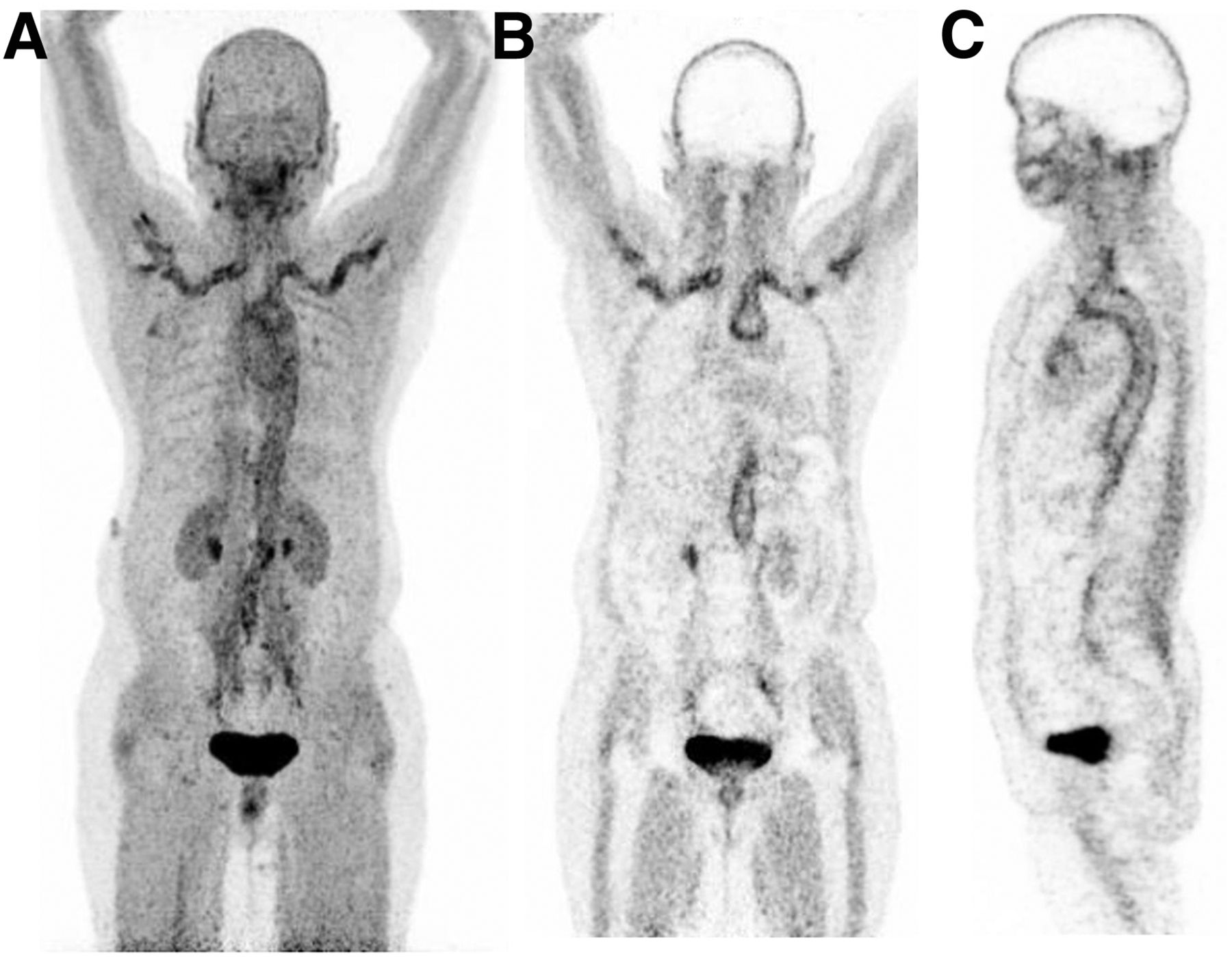
IgG4-related disease seems to have high FAPI avidity, which may be focal or diffuse in the pancreas and involve other organs, particularly the lungs, salivary glands, and biliary tract (57,92,93). In the largest series published as yet, 26 patients were evaluated with both 18F-FDG and FAPI-04, with the latter demonstrating active disease in all cases and additional sites of involvement in 50% of cases (93). Activation of myofibroblasts by polarized CD4-positive T lymphocytes is the likely explanation for this finding (94). Large-vessel vasculitis is another potential application because of the rapid blood clearance and high uptake in vasculitis lesions (Fig. 5). The ability to differentiate vasculitis from active atherosclerotic plaques may, however, be limited, with an autoradiographic using an 125I-labeled FAPI agent demonstrating high binding to plaques in an animal model (95).

FIGURE 5.
  • Download figure
  • Open in new tab
  • Download powerpoint
FIGURE 5.

Giant cell arteritis. (A) FAPI PET maximum-intensity projection. (B) Coronal slice through aortic arch and subclavian arteries. (C) Sagittal slice through thoracic aorta.

FUTURE DIRECTIONS

There are a large number of ongoing or planned clinical trials using various FAPI tracers suitable for PET imaging, primarily in cancer (96). The need for agents suitable for use in parts of the world where PET is not routinely available is potentially addressed by the development of FAPI agents labeled with 99mTc (97). 99mTc-FAPI-34 has been tested in preclinical models and evaluated in one patient with ovarian cancer and another with pancreatic cancer, with the biodistribution in each case being similar to that for prior 68Ga-FAPI PET/CT and also posttreatment 90Y-FAPI bremsstrahlung imaging. A further attraction of this agent is the potential for therapeutic use with 188Re. This generator-produced β-emitting radionuclide has a relatively short half-life (16.9 h), which may be advantageous given the clearance kinetics of FAPI-34, and an imageable γ-emission (155 keV), allowing posttreatment dosimetry (98).

CONCLUSION

Although preliminary, there are several reasons to be hopeful that FAPI agents will play an important role in the molecular imaging armamentarium. These include high expression across a wide range of cancer types (including several with typically low 18F-FDG avidity), low uptake in almost all normal tissues (including the brain and bowel, where high physiologic uptake can obscure primary or metastatic disease), and the ability to provide prognostic information that might guide therapeutic options. Although it remains unclear whether targeting FAP will be an effective therapeutic strategy, the possibility of theranostics remains intriguing. As with 18F-FDG, the causes of false-positive cancer diagnoses open possibilities for a broader use of these agents, particularly in a range of fibrotic diseases that are difficult to monitor noninvasively.

DISCLOSURE

No potential conflict of interest relevant to this article was reported.

Footnotes

  • Published online Dec. 4, 2020.

  • © 2021 by the Society of Nuclear Medicine and Molecular Imaging.

REFERENCES

  1. 1.↵
    1. Kuhl DE,
    2. Engel J Jr.,
    3. Phelps ME,
    4. Kowell AP
    . Epileptic patterns of local cerebral metabolism and perfusion in man: investigation by emission computed tomography of 18F-fluorodeoxyglucose and 13N-ammonia. Trans Am Neurol Assoc. 1978;103:52–53.
    OpenUrlPubMedGoogle Scholar
  2. 2.↵
    1. Kuhl DE,
    2. Metter EJ,
    3. Riege WH,
    4. Phelps ME
    . Effects of human aging on patterns of local cerebral glucose utilization determined by the [18F]fluorodeoxyglucose method. J Cereb Blood Flow Metab. 1982;2:163–171.
    OpenUrlCrossRefPubMedGoogle Scholar
  3. 3.↵
    1. Phelps ME,
    2. Schelbert HR,
    3. Hoffman EJ,
    4. Huang SC,
    5. Kuhl DE
    . Positron tomography of the heart. Prog Nucl Med. 1980;6:183–209.
    OpenUrlPubMedGoogle Scholar
  4. 4.↵
    1. Hoh CK,
    2. Schiepers C,
    3. Seltzer MA,
    4. et al
    . PET in oncology: will it replace the other modalities? Semin Nucl Med. 1997;27:94–106.
    OpenUrlCrossRefPubMedGoogle Scholar
  5. 5.↵
    1. Beyer T,
    2. Townsend DW,
    3. Brun T,
    4. et al
    . A combined PET/CT scanner for clinical oncology. J Nucl Med. 2000;41:1369–1379.
    OpenUrlAbstract/FREE Full TextGoogle Scholar
  6. 6.↵
    1. Sullivan R,
    2. Peppercorn J,
    3. Sikora K,
    4. et al
    . Delivering affordable cancer care in high-income countries. Lancet Oncol. 2011;12:933–980.
    OpenUrlCrossRefPubMedGoogle Scholar
  7. 7.↵
    1. Kwekkeboom DJ,
    2. Kam BL,
    3. van Essen M,
    4. et al
    . Somatostatin-receptor-based imaging and therapy of gastroenteropancreatic neuroendocrine tumors. Endocr Relat Cancer. 2010;17:R53–R73.
    OpenUrlAbstract/FREE Full TextGoogle Scholar
  8. 8.↵
    1. Hofman MS,
    2. Lawrentschuk N,
    3. Francis RJ,
    4. et al
    . Prostate-specific membrane antigen PET-CT in patients with high-risk prostate cancer before curative-intent surgery or radiotherapy (proPSMA): a prospective, randomised, multicentre study. Lancet. 2020;395:1208–1216.
    OpenUrlGoogle Scholar
  9. 9.↵
    1. Fendler WP,
    2. Eiber M,
    3. Beheshti M,
    4. et al
    . Ga-PSMA PET/CT: joint EANM and SNMMI procedure guideline for prostate cancer imaging: version 1.0. Eur J Nucl Med Mol Imaging. 2017;44:1014–1024.
    OpenUrlGoogle Scholar
  10. 10.↵
    1. Hicks RJ,
    2. Kwekkeboom DJ,
    3. Krenning E,
    4. et al
    . ENETS consensus guidelines for the standards of care in neuroendocrine neoplasia: peptide receptor radionuclide therapy with radiolabeled somatostatin analogues. Neuroendocrinology. 2017;105:295–309.
    OpenUrlGoogle Scholar
  11. 11.↵
    1. Kratochwil C,
    2. Fendler WP,
    3. Eiber M,
    4. et al
    . EANM procedure guidelines for radionuclide therapy with 177Lu-labeled PSMA-ligands (177Lu-PSMA-RLT). Eur J Nucl Med Mol Imaging. 2019;46:2536–2544.
    OpenUrlGoogle Scholar
  12. 12.↵
    1. Galldiks N,
    2. Lohmann P,
    3. Albert NL,
    4. Tonn JC,
    5. Langen KJ
    . Current status of PET imaging in neuro-oncology. Neurooncol Adv. 2019;1:vdz010.
    OpenUrlGoogle Scholar
  13. 13.↵
    1. Hicks RJ,
    2. Hofman MS
    . Is there still a role for SPECT-CT in oncology in the PET-CT era? Nat Rev Clin Oncol. 2012;9:712–720.
    OpenUrlCrossRefPubMedGoogle Scholar
  14. 14.↵
    1. Ho WJ,
    2. Jaffee EM,
    3. Zheng L
    . The tumour microenvironment in pancreatic cancer: clinical challenges and opportunities. Nat Rev Clin Oncol. 2020;17:527–540.
    OpenUrlGoogle Scholar
  15. 15.↵
    1. Kalluri R
    . The biology and function of fibroblasts in cancer. Nat Rev Cancer. 2016;16:582–598.
    OpenUrlCrossRefPubMedGoogle Scholar
  16. 16.↵
    1. Chen X,
    2. Song E
    . Turning foes to friends: targeting cancer-associated fibroblasts. Nat Rev Drug Discov. 2019;18:99–115.
    OpenUrlCrossRefPubMedGoogle Scholar
  17. 17.↵
    1. Helms E,
    2. Onate MK,
    3. Sherman MH
    . Fibroblast heterogeneity in the pancreatic tumor microenvironment. Cancer Discov. 2020;10:648–656.
    OpenUrlAbstract/FREE Full TextGoogle Scholar
  18. 18.↵
    1. Sahai E,
    2. Astsaturov I,
    3. Cukierman E,
    4. et al
    . A framework for advancing our understanding of cancer-associated fibroblasts. Nat Rev Cancer. 2020;20:174–186.
    OpenUrlCrossRefPubMedGoogle Scholar
  19. 19.↵
    1. Kobayashi H,
    2. Enomoto A,
    3. Woods SL,
    4. Burt AD,
    5. Takahashi M,
    6. Worthley DL
    . Cancer associated fibroblasts in gastrointestinal cancer. Nat Rev Gastroenterol Hepatol. 2019;16:282–295.
    OpenUrlGoogle Scholar
  20. 20.↵
    1. Fiori ME,
    2. Di Franco S,
    3. Villanova L,
    4. Bianca P,
    5. Stassi G,
    6. De Maria R
    . Cancer-associated fibroblasts as abettors of tumor progression at the crossroads of EMT and therapy resistance. Mol Cancer. 2019;18:70.
    OpenUrlGoogle Scholar
  21. 21.↵
    1. Kieffer Y,
    2. Hocine HR,
    3. Gentric G,
    4. et al
    . Single-cell analysis reveals fibroblast clusters linked to immunotherapy resistance in cancer. Cancer Discov. 2020;10:1330–1351.
    OpenUrlAbstract/FREE Full TextGoogle Scholar
  22. 22.↵
    1. Flavell RA,
    2. Sanjabi S,
    3. Wrzesinski SH,
    4. Licona-Limón P
    . The polarization of immune cells in the tumour environment by TGFβ. Nat Rev Immunol. 2010;10:554–567.
    OpenUrlCrossRefPubMedGoogle Scholar
  23. 23.↵
    1. Cho C,
    2. Mukherjee R,
    3. Peck AR,
    4. et al
    . Cancer-associated fibroblasts downregulate type I interferon receptor to stimulate intratumoral stromagenesis. Oncogene. 2020;39:6129–6137.
    OpenUrlGoogle Scholar
  24. 24.↵
    1. Ireland AS,
    2. Micinski AM,
    3. Kastner DW,
    4. et al
    . MYC drives temporal evolution of small cell lung cancer subtypes by reprogramming neuroendocrine fate. Cancer Cell. 2020;38:60–78.e12.
    OpenUrlGoogle Scholar
  25. 25.↵
    1. Goetz JG,
    2. Minguet S,
    3. Navarro-Lérida I,
    4. et al
    . Biomechanical remodeling of the microenvironment by stromal caveolin-1 favors tumor invasion and metastasis. Cell. 2011;146:148–163.
    OpenUrlCrossRefPubMedGoogle Scholar
  26. 26.↵
    1. Riching KM,
    2. Cox BL,
    3. Salick MR,
    4. et al
    . 3D collagen alignment limits protrusions to enhance breast cancer cell persistence. Biophys J. 2014;107:2546–2558.
    OpenUrlCrossRefPubMedGoogle Scholar
  27. 27.↵
    1. Elyada E,
    2. Bolisetty M,
    3. Laise P,
    4. et al
    . Cross-species single-cell analysis of pancreatic ductal adenocarcinoma reveals antigen-presenting cancer-associated fibroblasts. Cancer Discov. 2019;9:1102–1123.
    OpenUrlAbstract/FREE Full TextGoogle Scholar
  28. 28.↵
    1. Scanlan MJ,
    2. Raj BK,
    3. Calvo B,
    4. et al
    . Molecular cloning of fibroblast activation protein alpha, a member of the serine protease family selectively expressed in stromal fibroblasts of epithelial cancers. Proc Natl Acad Sci USA. 1994;91:5657–5661.
    OpenUrlAbstract/FREE Full TextGoogle Scholar
  29. 29.↵
    1. Šimková A,
    2. Busek P,
    3. Sedo A,
    4. Konvalinka J
    . Molecular recognition of fibroblast activation protein for diagnostic and therapeutic applications. Biochim Biophys Acta Proteins Proteomics. 2020;1868:140409.
    OpenUrlGoogle Scholar
  30. 30.↵
    1. Park JE,
    2. Lenter MC,
    3. Zimmermann RN,
    4. Garin-Chesa P,
    5. Old LJ,
    6. Rettig WJ
    . Fibroblast activation protein, a dual specificity serine protease expressed in reactive human tumor stromal fibroblasts. J Biol Chem. 1999;274:36505–36512.
    OpenUrlAbstract/FREE Full TextGoogle Scholar
  31. 31.↵
    1. Kraman M,
    2. Bambrough PJ,
    3. Arnold JN,
    4. et al
    . Suppression of antitumor immunity by stromal cells expressing fibroblast activation protein-alpha. Science. 2010;330:827–830.
    OpenUrlAbstract/FREE Full TextGoogle Scholar
  32. 32.↵
    1. Arnold JN,
    2. Magiera L,
    3. Kraman M,
    4. Fearon DT
    . Tumoral immune suppression by macrophages expressing fibroblast activation protein- and heme oxygenase-1. Cancer Immunol Res. 2014;2:121–126.
    OpenUrlAbstract/FREE Full TextGoogle Scholar
  33. 33.↵
    1. Lin Y,
    2. Li B,
    3. Yang X,
    4. et al
    . Fibroblastic FAP promotes intrahepatic cholangiocarcinoma growth via MDSCs recruitment. Neoplasia. 2019;21:1133–1142.
    OpenUrlGoogle Scholar
  34. 34.↵
    1. Feig C,
    2. Jones JO,
    3. Kraman M,
    4. et al
    . Targeting CXCL12 from FAP-expressing carcinoma associated fibroblasts synergizes with anti-PD-L1 immunotherapy in pancreatic cancer. Proc Natl Acad Sci USA. 2013;110:20212–20217.
    OpenUrlAbstract/FREE Full TextGoogle Scholar
  35. 35.↵
    1. Orimo A,
    2. Gupta PB,
    3. Sgroi DC,
    4. et al
    . Stromal fibroblasts present in invasive human breast carcinomas promote tumor growth and angiogenesis through elevated SDF-1/CXCL12 secretion. Cell. 2005;121:335–348.
    OpenUrlCrossRefPubMedGoogle Scholar
  36. 36.↵
    1. Coto-Llerena M,
    2. Ercan C,
    3. Kancherla V,
    4. et al
    . High expression of FAP in colorectal cancer is associated with angiogenesis and immunoregulation processes. Front Oncol. 2020;10:979.
    OpenUrlGoogle Scholar
  37. 37.↵
    1. Busek P,
    2. Balaziova E,
    3. Matrasova I,
    4. et al
    . Fibroblast activation protein alpha is expressed by transformed and stromal cells and is associated with mesenchymal features in glioblastoma. Tumour Biol. 2016;37:13961–13971.
    OpenUrlGoogle Scholar
  38. 38.↵
    1. Röhrich M,
    2. Floca R,
    3. Loi L,
    4. et al
    . FAP-specific PET signaling shows a moderately positive correlation with relative CBV and no correlation with ADC in 13 IDH wildtype glioblastomas. Eur J Radiol. 2020;127:109021.
    OpenUrlGoogle Scholar
  39. 39.↵
    1. Aimes RT,
    2. Zijlstra A,
    3. Hooper JD,
    4. et al
    . Endothelial cell serine proteases expressed during vascular morphogenesis and angiogenesis. Thromb Haemost. 2003;89:561–572.
    OpenUrlPubMedGoogle Scholar
  40. 40.↵
    1. Dohi O,
    2. Ohtani H,
    3. Hatori M,
    4. et al
    . Histogenesis-specific expression of fibroblast activation protein and dipeptidylpeptidase-IV in human bone and soft tissue tumours. Histopathology. 2009;55:432–440.
    OpenUrlCrossRefPubMedGoogle Scholar
  41. 41.↵
    1. Cohen SJ,
    2. Alpaugh RK,
    3. Palazzo I,
    4. et al
    . Fibroblast activation protein and its relationship to clinical outcome in pancreatic adenocarcinoma. Pancreas. 2008;37:154–158.
    OpenUrlCrossRefPubMedGoogle Scholar
  42. 42.↵
    1. Zhang Y,
    2. Tang H,
    3. Cai J,
    4. et al
    . Ovarian cancer-associated fibroblasts contribute to epithelial ovarian carcinoma metastasis by promoting angiogenesis, lymphangiogenesis and tumor cell invasion. Cancer Lett. 2011;303:47–55.
    OpenUrlCrossRefPubMedGoogle Scholar
  43. 43.↵
    1. Mhawech-Fauceglia P,
    2. Yan L,
    3. Sharifian M,
    4. et al
    . Stromal expression of fibroblast activation protein alpha (FAP) predicts platinum resistance and shorter recurrence in patients with epithelial ovarian cancer. Cancer Microenviron. 2015;8:23–31.
    OpenUrlGoogle Scholar
  44. 44.↵
    1. Wikberg ML,
    2. Edin S,
    3. Lundberg IV,
    4. et al
    . High intratumoral expression of fibroblast activation protein (FAP) in colon cancer is associated with poorer patient prognosis. Tumour Biol. 2013;34:1013–1020.
    OpenUrlCrossRefPubMedGoogle Scholar
  45. 45.↵
    1. Lo A,
    2. Wang LS,
    3. Scholler J,
    4. et al
    . Tumor-promoting desmoplasia is disrupted by depleting FAP-expressing stromal cells. Cancer Res. 2015;75:2800–2810.
    OpenUrlAbstract/FREE Full TextGoogle Scholar
  46. 46.↵
    1. Ostermann E,
    2. Garin-Chesa P,
    3. Heider KH,
    4. et al
    . Effective immunoconjugate therapy in cancer models targeting a serine protease of tumor fibroblasts. Clin Cancer Res. 2008;14:4584–4592.
    OpenUrlAbstract/FREE Full TextGoogle Scholar
  47. 47.↵
    1. Garin-Chesa P,
    2. Old LJ,
    3. Rettig WJ
    . Cell surface glycoprotein of reactive stromal fibroblasts as a potential antibody target in human epithelial cancers. Proc Natl Acad Sci USA. 1990;87:7235–7239.
    OpenUrlAbstract/FREE Full TextGoogle Scholar
  48. 48.↵
    1. Welt S,
    2. Divgi CR,
    3. Scott AM,
    4. et al
    . Antibody targeting in metastatic colon cancer: a phase I study of monoclonal antibody F19 against a cell-surface protein of reactive tumor stromal fibroblasts. J Clin Oncol. 1994;12:1193–1203.
    OpenUrlAbstract/FREE Full TextGoogle Scholar
  49. 49.↵
    1. Flentke GR,
    2. Munoz E,
    3. Huber BT,
    4. Plaut AG,
    5. Kettner CA,
    6. Bachovchin WW
    . Inhibition of dipeptidyl aminopeptidase IV (DP-IV) by Xaa-boroPro dipeptides and use of these inhibitors to examine the role of DP-IV in T-cell function. Proc Natl Acad Sci USA. 1991;88:1556–1559.
    OpenUrlAbstract/FREE Full TextGoogle Scholar
  50. 50.↵
    1. Tsai TY,
    2. Yeh TK,
    3. Chen X,
    4. et al
    . Substituted 4-carboxymethylpyroglutamic acid diamides as potent and selective inhibitors of fibroblast activation protein. J Med Chem. 2010;53:6572–6583.
    OpenUrlCrossRefPubMedGoogle Scholar
  51. 51.↵
    1. Jansen K,
    2. Heirbaut L,
    3. Cheng JD,
    4. et al
    . Selective inhibitors of fibroblast activation protein (FAP) with a (4-quinolinoyl)-glycyl-2-cyanopyrrolidine scaffold. ACS Med Chem Lett. 2013;4:491–496.
    OpenUrlGoogle Scholar
  52. 52.↵
    1. Loktev A,
    2. Lindner T,
    3. Mier W,
    4. et al
    . A tumor-imaging method targeting cancer associated fibroblasts. J Nucl Med. 2018;59:1423–1429.
    OpenUrlAbstract/FREE Full TextGoogle Scholar
  53. 53.↵
    1. Loktev A,
    2. Lindner T,
    3. Burger EM,
    4. et al
    . Development of fibroblast activation protein targeted radiotracers with improved tumor retention. J Nucl Med. 2019;60:1421–1429.
    OpenUrlAbstract/FREE Full TextGoogle Scholar
  54. 54.↵
    1. Lindner T,
    2. Loktev A,
    3. Altmann A,
    4. et al
    . Development of quinoline-based theranostic ligands for the targeting of fibroblast activation protein. J Nucl Med. 2018;59:1415–1422.
    OpenUrlAbstract/FREE Full TextGoogle Scholar
  55. 55.↵
    1. Giesel FL,
    2. Kratochwil C,
    3. Lindner T,
    4. et al
    . Ga-FAPI PET/CT: biodistribution and preliminary dosimetry estimate of 2 DOTA-containing FAP-targeting agents in patients with various cancers. J Nucl Med. 2019;60:386–392.
    OpenUrlAbstract/FREE Full TextGoogle Scholar
  56. 56.↵
    1. Kratochwil C,
    2. Flechsig P,
    3. Lindner T,
    4. et al
    . Ga-FAPI PET/CT: tracer uptake in 28 different kinds of cancer. J Nucl Med. 2019;60:801–805.
    OpenUrlAbstract/FREE Full TextGoogle Scholar
  57. 57.↵
    1. Luo Y,
    2. Pan Q,
    3. Zhang W
    . IgG4-related disease revealed by 68Ga-FAPI and 18F-FDG PET/CT. Eur J Nucl Med Mol Imaging. 2019;46:2625–2626.
    OpenUrlGoogle Scholar
  58. 58.↵
    1. Chen H,
    2. Zhao L,
    3. Ruan D,
    4. et al
    . Usefulness of 68Ga-FAPI and 18F-FDG PET/CT in a patient with cholangiocellular carcinoma. Eur J Nucl Med Mol Imaging. June 25, 2020 [Epub ahead of print].
    Google Scholar
  59. 59.↵
    1. Chen H,
    2. Pang Y,
    3. Wu J,
    4. et al
    . Comparison of [68Ga]Ga-DOTA-FAPI-04 and [18F] FDG PET/CT for the diagnosis of primary and metastatic lesions in patients with various types of cancer. Eur J Nucl Med Mol Imaging. 2020;47:1820–1832.
    OpenUrlGoogle Scholar
  60. 60.↵
    1. Syed M,
    2. Flechsig P,
    3. Liermann J,
    4. et al
    . Fibroblast activation protein inhibitor (FAPI) PET for diagnostics and advanced targeted radiotherapy in head and neck cancers. Eur J Nucl Med Mol Imaging. 2020;47:2836–2845.
    OpenUrlGoogle Scholar
  61. 61.↵
    1. Iravani A,
    2. Hicks RJ
    . Imaging the cancer immune environment and its response to pharmacological intervention, part 2: the role of novel PET agents. J Nucl Med. 2020;61:1553–1559.
    OpenUrlAbstract/FREE Full TextGoogle Scholar
  62. 62.↵
    1. Meyer C,
    2. Dahlbom M,
    3. Lindner T,
    4. et al
    . Radiation dosimetry and biodistribution of 68Ga-FAPI-46 PET imaging in cancer patients. J Nucl Med. 2020;61:1171–1177.
    OpenUrlAbstract/FREE Full TextGoogle Scholar
  63. 63.↵
    1. Koerber SA,
    2. Staudinger F,
    3. Kratochwil C,
    4. et al
    . The role of FAPI-PET/CT for patients with malignancies of the lower gastrointestinal tract: first clinical experience. J Nucl Med. 2020;61:1331–1336.
    OpenUrlGoogle Scholar
  64. 64.↵
    1. Giesel F,
    2. Adeberg S,
    3. Syed M,
    4. et al
    . FAPI-74 PET/CT using either 18F-AlF or cold-kit 68Ga-labeling: biodistribution, radiation dosimetry and tumor delineation in lung cancer. J Nucl Med. June 26, 2020 [Epub ahead of print].
    Google Scholar
  65. 65.↵
    1. Toms J,
    2. Kogler J,
    3. Maschauer S,
    4. et al
    . Targeting fibroblast activation protein: radiosynthesis and preclinical evaluation of an 18F-labeled FAP inhibitor. J Nucl Med. 2020;61:1806–1813.
    OpenUrlAbstract/FREE Full TextGoogle Scholar
  66. 66.↵
    1. Giesel FL,
    2. Heussel CP,
    3. Lindner T,
    4. et al
    . FAPI-PET/CT improves staging in a lung cancer patient with cerebral metastasis. Eur J Nucl Med Mol Imaging. 2019;46:1754–1755.
    OpenUrlGoogle Scholar
  67. 67.↵
    1. Hao B,
    2. Wu J,
    3. Pang Y,
    4. Sun L,
    5. Chen H
    . 68Ga-FAPI PET/CT in assessment of leptomeningeal metastases in a patient with lung adenocarcinoma. Clin Nucl Med. 2020;45:784–786.
    OpenUrlGoogle Scholar
  68. 68.↵
    1. Hao B,
    2. Wu X,
    3. Pang Y,
    4. et al
    . [18F]FDG and [68Ga]Ga-DOTA-FAPI-04 PET/CT in the evaluation of tuberculous lesions. Eur J Nucl Med Mol Imaging. July 8, 2020 [Epub ahead of print].
    Google Scholar
  69. 69.↵
    1. Windisch P,
    2. Röhrich M,
    3. Regnery S,
    4. et al
    . Fibroblast activation protein (FAP) specific PET for advanced target volume delineation in glioblastoma. Radiother Oncol. 2020;150:159–163.
    OpenUrlGoogle Scholar
  70. 70.↵
    1. Fischer E,
    2. Chaitanya K,
    3. Wüest T,
    4. et al
    . Radioimmunotherapy of fibroblast activation protein positive tumors by rapidly internalizing antibodies. Clin Cancer Res. 2012;18:6208–6218.
    OpenUrlGoogle Scholar
  71. 71.↵
    1. Watabe T,
    2. Liu Y,
    3. Kaneda-Nakashima K,
    4. et al
    . Theranostics targeting fibroblast activation protein in the tumor stroma: 64Cu- and 225Ac-labeled FAPI-04 in pancreatic cancer xenograft mouse models. J Nucl Med. 2020;61:563–569.
    OpenUrlAbstract/FREE Full TextGoogle Scholar
  72. 72.↵
    1. Hicks RJ,
    2. Jackson P,
    3. Kong G,
    4. et al
    . Cu-SARTATE PET imaging of patients with neuroendocrine tumors demonstrates high tumor uptake and retention, potentially allowing prospective dosimetry for peptide receptor radionuclide therapy. J Nucl Med. 2019;60:777–785.
    OpenUrlAbstract/FREE Full TextGoogle Scholar
  73. 73.↵
    1. Hicks RJ
    . Citius, altius, fortius: an Olympian dream for theranostics. J Nucl Med. 2017;58:194–195.
    OpenUrlFREE Full TextGoogle Scholar
  74. 74.↵
    1. Paterson BM,
    2. Roselt P,
    3. Denoyer D,
    4. et al
    . PET imaging of tumours with a 64Cu labeled macrobicyclic cage amine ligand tethered to Tyr3-octreotate. Dalton Trans. 2014;43:1386–1396.
    OpenUrlGoogle Scholar
  75. 75.↵
    1. Scott AM,
    2. Wiseman G,
    3. Welt S,
    4. et al
    . A phase I dose-escalation study of sibrotuzumab in patients with advanced or metastatic fibroblast activation protein-positive cancer. Clin Cancer Res. 2003;9:1639–1647.
    OpenUrlAbstract/FREE Full TextGoogle Scholar
  76. 76.↵
    1. Puré E,
    2. Blomberg R
    . Pro-tumorigenic roles of fibroblast activation protein in cancer: back to the basics. Oncogene. 2018;37:4343–4357.
    OpenUrlGoogle Scholar
  77. 77.↵
    1. Rettig WJ,
    2. Garin-Chesa P,
    3. Beresford HR,
    4. Oettgen HF,
    5. Melamed MR,
    6. Old LJ
    . Cell-surface glycoproteins of human sarcomas: differential expression in normal and malignant tissues and cultured cells. Proc Natl Acad Sci USA. 1988;85:3110–3114.
    OpenUrlAbstract/FREE Full TextGoogle Scholar
  78. 78.↵
    1. Gabbiani G,
    2. Ryan GB,
    3. Majne G
    . Presence of modified fibroblasts in granulation tissue and their possible role in wound contraction. Experientia. 1971;27:549–550.
    OpenUrlCrossRefPubMedGoogle Scholar
  79. 79.↵
    1. Bauer S,
    2. Jendro MC,
    3. Wadle A,
    4. et al
    . Fibroblast activation protein is expressed by rheumatoid myofibroblast-like synoviocytes. Arthritis Res Ther. 2006;8:R171.
    OpenUrlCrossRefPubMedGoogle Scholar
  80. 80.↵
    1. Laverman P,
    2. van der Geest T,
    3. Terry SY,
    4. et al
    . Immuno-PET and immuno-SPECT of rheumatoid arthritis with radiolabeled anti-fibroblast activation protein antibody correlates with severity of arthritis. J Nucl Med. 2015;56:778–783.
    OpenUrlAbstract/FREE Full TextGoogle Scholar
  81. 81.↵
    1. Iravani A,
    2. Turgeon GA,
    3. Akhurst T,
    4. et al
    . PET-detected pneumonitis following curative-intent chemoradiation in non-small cell lung cancer (NSCLC): recognizing patterns and assessing the impact on the predictive ability of FDG-PET/CT response assessment. Eur J Nucl Med Mol Imaging. 2019;46:1869–1877.
    OpenUrlGoogle Scholar
  82. 82.↵
    1. Mac Manus MP,
    2. Ding Z,
    3. Hogg A,
    4. et al
    . Association between pulmonary uptake of fluorodeoxyglucose detected by positron emission tomography scanning after radiation therapy for non-small-cell lung cancer and radiation pneumonitis. Int J Radiat Oncol Biol Phys. 2011;80:1365–1371.
    OpenUrlCrossRefPubMedGoogle Scholar
  83. 83.↵
    1. Egger C,
    2. Cannet C,
    3. Gérard C,
    4. et al
    . Effects of the fibroblast activation protein inhibitor, PT100, in a murine model of pulmonary fibrosis. Eur J Pharmacol. 2017;809:64–72.
    OpenUrlGoogle Scholar
  84. 84.↵
    1. Seuwen K,
    2. Magnaldo I,
    3. Pouysségur J
    . Serotonin stimulates DNA synthesis in fibroblasts acting through 5-HT1B receptors coupled to a Gi-protein. Nature. 1988;335:254–256.
    OpenUrlGoogle Scholar
  85. 85.↵
    1. Waltenberger J,
    2. Lundin L,
    3. Oberg K,
    4. et al
    . Involvement of transforming growth factor-β in the formation of fibrotic lesions in carcinoid heart disease. Am J Pathol. 1993;142:71–78.
    OpenUrlPubMedGoogle Scholar
  86. 86.↵
    1. Nagaraju CK,
    2. Dries E,
    3. Popovic N,
    4. et al
    . Global fibroblast activation throughout the left ventricle but localized fibrosis after myocardial infarction. Sci Rep. 2017;7:10801.
    OpenUrlCrossRefPubMedGoogle Scholar
  87. 87.↵
    1. Tillmanns J,
    2. Hoffmann D,
    3. Habbaba Y,
    4. et al
    . Fibroblast activation protein alpha expression identifies activated fibroblasts after myocardial infarction. J Mol Cell Cardiol. 2015;87:194–203.
    OpenUrlCrossRefGoogle Scholar
  88. 88.↵
    1. Heckmann MB,
    2. Reinhardt F,
    3. Finke D,
    4. et al
    . Relationship between cardiac fibroblast activation protein activity by positron emission tomography and cardiovascular disease. Circ Cardiovasc Imaging. 2020;13:e010628.
    OpenUrlGoogle Scholar
  89. 89.↵
    1. Siebermair J,
    2. Köhler MI,
    3. Kupusovic J,
    4. et al
    . Cardiac fibroblast activation detected by Ga-68 FAPI PET imaging as a potential novel biomarker of cardiac injury/remodeling. J Nucl Cardiol. September 25, 2020 [Epub ahead of print].
    Google Scholar
  90. 90.↵
    1. Zhao L,
    2. Gu J,
    3. Fu K,
    4. Lin Q,
    5. Chen H
    . 68Ga-FAPI PET/CT in assessment of liver nodules in a cirrhotic patient. Clin Nucl Med. 2020;45:e430–e432.
    OpenUrlGoogle Scholar
  91. 91.↵
    1. Shi X,
    2. Xing H,
    3. Yang X,
    4. et al
    . Fibroblast imaging of hepatic carcinoma with 68Ga-FAPI04 PET/CT: a pilot study in patients with suspected hepatic nodules. Eur J Nucl Med Mol Imaging. May 29, 2020 [Epub ahead of print].
    Google Scholar
  92. 92.↵
    1. Pan Q,
    2. Luo Y,
    3. Zhang W
    . Recurrent immunoglobulin G4-related disease shown on 18F-FDG and 68Ga-FAPI PET/CT. Clin Nucl Med. 2020;45:312–313.
    OpenUrlGoogle Scholar
  93. 93.↵
    1. Luo Y,
    2. Pan Q,
    3. Yang H,
    4. Peng L,
    5. Zhang W,
    6. Li F
    . Fibroblast activation protein targeted PET/CT with 68Ga-FAPI for imaging IgG4-related disease: comparison to 18F-FDG PET/CT. J Nucl Med. June 8, 2020 [Epub ahead of print].
    Google Scholar
  94. 94.↵
    1. Kamisawa T,
    2. Zen Y,
    3. Pillai S,
    4. Stone JH
    . IgG4-related disease. Lancet. 2015;385:1460–1471.
    OpenUrlGoogle Scholar
  95. 95.↵
    1. Meletta R,
    2. Müller Herde A,
    3. Chiotellis A,
    4. et al
    . Evaluation of the radiolabeled boronic acid-based FAP inhibitor MIP-1232 for atherosclerotic plaque imaging. Molecules. 2015;20:2081–2099.
    OpenUrlGoogle Scholar
  96. 96.↵
    1. Windisch P,
    2. Zwahlen DR,
    3. Koerber SA,
    4. et al
    . Clinical results of fibroblast activation protein (FAP) specific PET and implications for radiotherapy planning: systematic review. Cancers (Basel). 2020;12:2629.
    OpenUrlGoogle Scholar
  97. 97.↵
    1. Lindner T,
    2. Altmann A,
    3. Kraemer S,
    4. et al
    . Design and development of 99mTc-labeled FAPI-tracers for SPECT-imaging and 188Re therapy. J Nucl Med. 2020;61:1507–1513.
    OpenUrlAbstract/FREE Full TextGoogle Scholar
  98. 98.↵
    1. Lepareur N,
    2. Lacoeuille F,
    3. Bouvry C,
    4. et al
    . Rhenium-188 labeled radiopharmaceuticals: current clinical applications in oncology and promising perspectives. Front Med (Lausanne). 2019;6:132.
    OpenUrlGoogle Scholar
  99. 99.↵
    1. Cerami E,
    2. Gao J,
    3. Dogrusoz U,
    4. et al
    . The cBio cancer genomics portal: an open platform for exploring multidimensional cancer genomics data. Cancer Discov. 2012;2:401–404.
    OpenUrlAbstract/FREE Full TextGoogle Scholar
  • Received for publication October 12, 2020.
  • Accepted for publication November 12, 2020.
PreviousNext
Back to top

In this issue

Journal of Nuclear Medicine: 62 (3)
Journal of Nuclear Medicine
Vol. 62, Issue 3
March 1, 2021
  • Table of Contents
  • Table of Contents (PDF)
  • About the Cover
  • Index by author
Print
Download PDF
Article Alerts
Email Article
Citation Tools
Share
FAPI PET/CT: Will It End the Hegemony of 18F-FDG in Oncology?
Rodney J. Hicks, Peter J. Roselt, Kumarswamy G. Kallur, Richard W. Tothill, Linda Mileshkin
Journal of Nuclear Medicine Mar 2021, 62 (3) 296-302; DOI: 10.2967/jnumed.120.256271
Twitter logo Facebook logo LinkedIn logo Mendeley logo
  • Tweet Widget
Bookmark this article

Jump to section

  • Article
    • Abstract
    • THE ROLE OF CAFs IN CANCER
    • FIBROBLAST ACTIVATION PROTEIN (FAP) AND CANCER BIOLOGY
    • FAP AS A TARGET FOR IMAGING CANCER
    • THERAPEUTIC TARGETING OF FAP IN CANCER
    • FAP IMAGING IN OTHER PATHOLOGIES
    • FUTURE DIRECTIONS
    • CONCLUSION
    • DISCLOSURE
    • Footnotes
    • REFERENCES
  • Figures & Data
  • Info & Metrics
  • PDF

Related Articles

  • This Month in JNM
  • PubMed
  • Google Scholar

Cited By...

  • Are FAP Theranostics Really Happening? Will Radiochemistry or Biology Win?
  • Evaluation of the Diagnostic Accuracy of FAPI PET/CT in Oncologic Studies: Systematic Review and Metaanalysis
  • Evaluation of the Diagnostic Accuracy of FAPI PET/CT in Oncologic Studies: Systematic Review and Metaanalysis
  • FAPI PET Opens a New Window to Understanding Immune-Mediated Inflammatory Diseases
  • Biology of Cancer and PET Imaging: Pictorial Review
  • Google Scholar

More in this TOC Section

  • A Vision for Gastrin-Releasing Peptide Receptor Targeting for Imaging and Therapy: Perspective from Academia and Industry
  • Treatment Landscape of Prostate Cancer in the Era of PSMA Radiopharmaceutical Therapy
  • Theranostics for Neuroblastoma: Making Molecular Radiotherapy Work Better
Show more The State of the Art

Similar Articles

  • MRI-Based Correction for Partial-Volume Effect Improves Detectability of Intractable Epileptogenic Foci on 123I-Iomazenil Brain SPECT Images
  • Hypoperfusion and Ischemia in Cerebral Amyloid Angiopathy Documented by 99mTc-ECD Brain Perfusion SPECT
  • Role of Neuroimaging in Alzheimer's Disease, with Emphasis on Brain Perfusion SPECT
  • Longitudinal Evaluation of Early Alzheimer's Disease Using Brain Perfusion SPECT
  • Does Cerebral Blood Flow Decline in Healthy Aging? A PET Study with Partial-Volume Correction
See more

Keywords

  • PET
  • theranostics
  • fibroblast activation protein
  • oncology
  • radiochemistry
SNMMI

© 2025 SNMMI

Powered by HighWire
Alerts for this Article
Sign In to Email Alerts with your Email Address
Email this Article

Thank you for your interest in spreading the word on Journal of Nuclear Medicine.

NOTE: We only request your email address so that the person you are recommending the page to knows that you wanted them to see it, and that it is not junk mail. We do not capture any email address.

Enter multiple addresses on separate lines or separate them with commas.
FAPI PET/CT: Will It End the Hegemony of 18F-FDG in Oncology?
(Your Name) has sent you a message from Journal of Nuclear Medicine
(Your Name) thought you would like to see the Journal of Nuclear Medicine web site.
Citation Tools
FAPI PET/CT: Will It End the Hegemony of 18F-FDG in Oncology?
Rodney J. Hicks, Peter J. Roselt, Kumarswamy G. Kallur, Richard W. Tothill, Linda Mileshkin
Journal of Nuclear Medicine Mar 2021, 62 (3) 296-302; DOI: 10.2967/jnumed.120.256271

Citation Manager Formats

  • BibTeX
  • Bookends
  • EasyBib
  • EndNote (tagged)
  • EndNote 8 (xml)
  • Medlars
  • Mendeley
  • Papers
  • RefWorks Tagged
  • Ref Manager
  • RIS
  • Zotero

We use cookies on this site to enhance your user experience

By clicking any link on this page you are giving your consent for us to set cookies.