

Abstract
The clinical utility of 18F-FDG PET/CT is being increasingly recognized in histiocytic disorders. We report the case of a 23-y-old woman who presented with slowly progressive, yellowish-brown papules, plaques, and nodules over her face and flexures. Besides the multiple cutaneous lesions, lesions of the brain, stomach, gallbladder, and marrow were additionally revealed by baseline 18F-FDG PET/CT. Skin biopsy and the overall clinical picture were consistent with xanthoma disseminatum. Subsequent PET/CT after cladribine therapy revealed a decrease in the extent and metabolic activity of most lesions, suggestive of a favorable response. This case report highlights the potential role of 18F-FDG PET/CT in the accurate assessment of disease extent and posttreatment response in rare histiocytic disorders.
- xanthoma disseminatum
- histiocytosis
- 18F-FDG
- PET/CT
- response
As a hybrid imaging modality for the evaluation of histiocytic disorders, 18F-FDG PET/CT is being increasingly used. We present a rare case of xanthoma disseminatum highlighting the role of 18F-FDG PET/CT in the evaluation of disease extent and posttreatment response.
CASE REPORT
A 23-y-old woman presented with a 2-y history of bilateral axillary erythematous papular lesions that gradually progressed to form multiple yellowish-brown papules, plaques, and nodules involving the periorbital and periocular area bilaterally, the neck, the inframammary and inguinal folds, the cubital and popliteal fossae, and the oral mucosa. She also complained of polyuria, polydipsia, amenorrhea, weight gain, and fatigue. Lab investigations revealed serum hyperosmolality (314 mOsm/kg; range, 275–295 mOsm/kg) and urine hypoosmolality (56 mOsm/kg; range, 100–800 mOsm/kg) consistent with diabetes insipidus, secondary hypothyroidism (low thyroxine and triiodothyronine with low-to-normal thyroid-stimulating hormone), panhypopituitarism (reduced luteinizing hormone, follicle-stimulating hormone, and adrenocorticotrophic hormone), and a normal lipid profile. Skin biopsy revealed a pandermal infiltrate of foamy histiocytes, multinucleate giant cells, and Touton giant cells, along with epitheloid cells, lymphocytes, neutrophils, and occasional eosinophils. On immunohistochemistry, histiocytic cells were positive for CD68 and negative for CD1a. Overall, the findings were suggestive of xanthoma disseminatum.
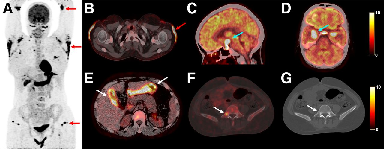
She underwent whole-body 18F-FDG PET/CT (Fig. 1) for assessment of disease extent, and multiple cutaneous lesions were detected. PET/CT also detected metabolically active lesions in the sella and suprasellar region consistent with the clinical picture of panhypopituitarism. Additionally, PET/CT detected lesions bilaterally in the mesial temporal lobes, diffuse mural thickening in the gallbladder and stomach, and marrow-based lesions that were not clinically suspected.

At baseline, 18F-FDG PET/CT maximum-intensity projection (A) and axial PET/CT (B) revealed multiple sites of cutaneous involvement (red arrows) bilaterally in eyelid or infraorbital region, cubital fossae, axillary and breast fold regions, back, and pelvic and groin regions. PET/CT also detected tracer-avid lesions in sella, in suprasellar region, and bilaterally in mesial temporal lobes (C and D, blue arrows), diffuse mural thickening in gallbladder and stomach (E, white arrows), and marrow-based lesions (F and G, white arrows show representative lesion in right pedicle of L5 vertebra).
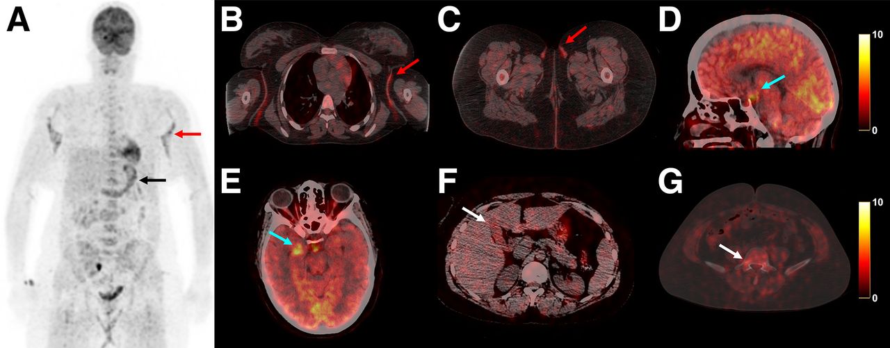
The patient received treatment with cladribine (0.14 mg/kg/d for 5 d of a week, repeated every month). She was also on low-dose oral steroids, desmopressin, levothyroxine, and estrogen hormonal supplementation. Subsequent 18F-FDG PET/CT (Fig. 2) done for response assessment after 4 cycles of cladribine therapy showed a significant decrease in the extent and metabolic activity of most previously noted lesions, suggestive of a favorable response to therapy.

(A) Posttreatment 18F-FDG PET/CT maximum-intensity projection showed significant decrease in extent and tracer avidity of most previously noted lesions. Residual disease with mild metabolic activity was noted as cutaneous involvement bilaterally in axillae (SUVmax of 5.6 vs. 25.4 previously; ∼78% decrease), breast folds, and hip and groin regions (A–C, red arrows); stomach involvement (A, black arrow); sellar lesions (SUVmax of 7.6 vs. 45.6 previously; ∼83.3% decrease); and right mesial temporal lesions (D and E, blue arrows). Resolution of previously tracer-avid gallbladder mural thickening (F, white arrow) and marrow-based lesions (G, white arrow) was also seen.
DISCUSSION
Histiocytosis is a heterogeneous group of disorders characterized by pathologic infiltration and accumulation of cells of the monocytic lineage in different tissues (1). Xanthoma disseminatum is a rare, normolipidemic, mucocutaneous form of non-Langerhans cell histiocytosis (2). Existing reports have established potential central nervous system (most commonly pituitary involvement with diabetes inspidus), ocular, hepatic, gastrointestinal, renal, and osseous involvement (3,4). In the index case, 18F-FDG PET/CT demonstrated metabolically active cutaneous and central nervous system involvement. Additionally, stomach and gallbladder mural thickening and marrow-based lesions were also detected, which were not clinically suspected. Cladribine (2-chlorodeoxyadenosine) is a synthetic purine nucleoside analog that inhibits DNA synthesis and has shown promising results in xanthoma disseminatum (5). This case report highlights the clinical utility of 18F-FDG PET/CT in the evaluation of disease extent and response assessment of histiocytic disorders, which can be challenging, often requiring a multimodality approach (6).
CONCLUSION
18F-FDG PET/CT can provide a comprehensive structural and functional whole-body evaluation in rare histiocytic disorders and may be used for accurate assessment of disease extent and treatment response.
DISCLOSURE
No potential conflict of interest relevant to this article was reported.
Footnotes
Published online Sep. 12, 2023.
REFERENCES
- 1.
- 2.
- 3.
- 4.
- 5.
- 6.
- Received for publication June 9, 2023.
- Revision received July 27, 2023.