


Abstract
A patient who had sickle cell disease and had spleen uptake on bone scans is described, and additional causes for that finding are discussed.
It is uncommon for bone-seeking agents to be taken up by extraosseous tissues such as the spleen, liver, breast, and lungs. To make a diagnosis, it is helpful to study the patient’s medical history, laboratory results, and other imaging tests.
CASE REPORT
A 46-y-old man with a history of sickle cell disease had a total left-knee replacement 5 y previously. He was referred to the hospital with pain in the left knee. A bone scan was required because of a suspicion of a prosthetic joint infection with osteomyelitis.
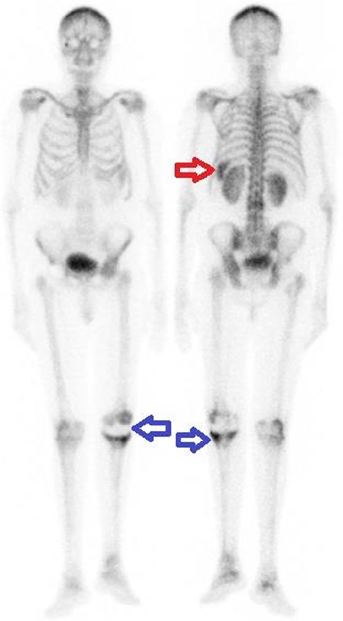
After intravenous injection of 750 MBq of 99mTc-methylene diphosphonate (99mTc-MDP), flow and blood-pool images of the knee, as well as delayed whole-body views and SPECT images, were acquired. There were signs of hyperemia, increased perfusion, and osteoblastic activity supporting a prosthetic joint infection with osteomyelitis. On a whole-body scan, the left upper quadrant of the abdomen, directly superolateral to the left kidney, showed diffusely enhanced radiotracer uptake, which suggested splenic uptake (Fig. 1). In addition, SPECT images showed diffusely increased uptake localized to the spleen (Fig. 2).

Anterior (left) and posterior (right) bone scans demonstrate splenic uptake in left upper quadrant (red arrow). There is also photopenic area in left knee prosthesis and increased radiotracer uptake in periprosthetic bone structures (blue arrows).

Transaxial (left) and coronal (right) SPECT images show activity uptake of spleen (arrows).
The hemogram revealed normocytic–normochromic anemia, which supported sickle cell anemia. On hemoglobin electrophoresis, hemoglobin S levels were high (69.4%). The patient was also receiving chelation therapy and had a history of iron excess from several transfusions.
The peripheral blood smear demonstrated normocytic–normochromic anemia with sickle cells, Howell–Jolly bodies, and Pappenheimer bodies.
The study protocol was approved by the Ethics Committee of the Mersin Provincial Health Directorate-Mersin City Training and Research Hospital, and the subject gave written informed consent.
DISCUSSION
Hemosiderosis, hemochromatosis, and sickle cell disease are the most frequent causes of uptake in the spleen on a bone scan. Neoplastic deposits in the spleen (such as those from breast cancer or Hodgkin disease), splenic hemangiomas, splenic artery calcification, frequent platelet and red blood cell transfusions, and thalassemia major are among some of the unusual causes of splenic uptake (1). Deficiency in glucose-6-phosphate dehydrogenase is one uncommon cause of splenic uptake.
Additionally, when 99mTc-MDP is administered shortly after the injection of gadolinium-diethylenetriamine pentaacetic acid MRI contrast agents, or even when 99mTc-MDP is given before the injection, hepatic and splenic uptake of 99mTc-MDP has been observed (2,3). Recent previous nuclear medicine studies (4,5), such as with 99mTc-sulfur colloid or 111In-labeled white blood cells, might also be a cause of splenic absorption. Additionally, both will show liver uptake.
It has been assumed that calcium deposition is the mechanism through which bone-seeking chemicals are taken up by the spleen in sickle cell anemia. It is possible that tiny calcium deposits that cannot be seen radiographically may potentially cause 99mTc-MDP uptake. In necrotic muscle cells, calcium ions are bound by the mitochondria in crystalline structures such as those of hydroxyapatite, according to several ultrastructural investigations (4).
Recurrent transfusions and increased iron deposition from the sequestration of aberrant red blood cells inside the spleen cause hemosiderosis. Numerous searches, including one by Jones et al. (5), suggested that iron deposits could be the reason for the absorption of radionuclides that seek bone.
CONCLUSION
In extraosseous tissues such as the spleen, liver, breast, and lungs, unexpected intake of bone-seeking agents is rare. The patient’s medical history, laboratory results, and additional imaging workup are all reviewed to help make the diagnosis. Many disorders, including sickle cell disease, which is covered in our case study, can result in spleen uptake on a bone scan. It is assumed that previous infarcts or microscopic or macroscopic splenic calcifications are the reason. Another possibility is iron overload in individuals getting blood transfusions.
DISCLOSURE
No potential conflict of interest relevant to this article was reported.
Footnotes
Published online Feb. 13, 2024.
- Received for publication October 26, 2023.
- Revision received January 11, 2024.
In this issue


Jump to section
Related Articles
Cited By...
- No citing articles found.