
Main menu

  • Home
  • Content
    • Current
      • JNMT Supplement
    • Ahead of print
    • Past Issues
    • Continuing Education
    • JNMT Podcast
    • SNMMI Annual Meeting Abstracts
  • Subscriptions
    • Subscribers
    • Rates
    • Journal Claims
    • Institutional and Non-member
  • Authors
    • Submit to JNMT
    • Information for Authors
    • Assignment of Copyright
    • AQARA Requirements
  • Info
    • Reviewers
    • Permissions
    • Advertisers
    • Corporate & Special Sales
  • About
    • About Us
    • Editorial Board
    • Contact Information
  • More
    • Alerts
    • Feedback
    • Help
    • SNMMI Journals
  • SNMMI
    • JNMT
    • JNM
    • SNMMI Journals
    • SNMMI

User menu

  • Subscribe
  • My alerts
  • Log in
  • My Cart

Search

  • Advanced search
Journal of Nuclear Medicine Technology
  • SNMMI
    • JNMT
    • JNM
    • SNMMI Journals
    • SNMMI
  • Subscribe
  • My alerts
  • Log in
  • My Cart
Journal of Nuclear Medicine Technology

Advanced Search

  • Home
  • Content
    • Current
    • Ahead of print
    • Past Issues
    • Continuing Education
    • JNMT Podcast
    • SNMMI Annual Meeting Abstracts
  • Subscriptions
    • Subscribers
    • Rates
    • Journal Claims
    • Institutional and Non-member
  • Authors
    • Submit to JNMT
    • Information for Authors
    • Assignment of Copyright
    • AQARA Requirements
  • Info
    • Reviewers
    • Permissions
    • Advertisers
    • Corporate & Special Sales
  • About
    • About Us
    • Editorial Board
    • Contact Information
  • More
    • Alerts
    • Feedback
    • Help
    • SNMMI Journals
  • Watch or Listen to JNMT Podcast
  • Visit SNMMI on Facebook
  • Join SNMMI on LinkedIn
  • Follow SNMMI on Twitter
  • Subscribe to JNMT RSS feeds
Review ArticleTeaching Case Studies

Detection of 18F-FDG Dose Leakage Using a Topical Device

Stephanie Sanchez and Geoffrey M. Currie
Journal of Nuclear Medicine Technology September 2020, 48 (3) 283-284; DOI: https://doi.org/10.2967/jnmt.119.240283
Stephanie Sanchez
Faculty of Science, Charles Sturt University, Wagga Wagga, Australia
  • Find this author on Google Scholar
  • Find this author on PubMed
  • Search for this author on this site
Geoffrey M. Currie
Faculty of Science, Charles Sturt University, Wagga Wagga, Australia
  • Find this author on Google Scholar
  • Find this author on PubMed
  • Search for this author on this site
  • Article
  • Figures & Data
  • Info & Metrics
  • PDF
Loading

Abstract

PET and SUV depend on reliable pharmacokinetic modeling, part of which is predictable dose delivery. Partial extravasation of the intravenous dose administered undermines the predictability of dose delivery and, potentially, the accuracy of the SUV calculation. Use of the Lara device with topical sensors is a simple, noninvasive way to determine partial dose extravasation. As part of routine monitoring of 18F-FDG PET administrations, an interesting case was identified that mimicked extravasation but represented dose leakage during infusion via an automatic injector. The Lara device provided a useful tool for more timely critical evaluation and problem solving, extending advantages to the patient and practice.

  • nuclear medicine
  • PET
  • 18F-FDG
  • extravasation
  • Lara
  • auto-injector

PET and quantitative values such as the SUV depend on reliable pharmacokinetic modeling, part of which is predictable dose delivery. Partial extravasation of the intravenous dose administration undermines the predictability of dose delivery and, potentially, the accuracy of SUV calculations (1). Visually reviewing images after a PET procedure is perhaps the most common way to identify extravasation; however, because the injection site may be outside the field of view for whole-body PET, which is typically performed with the patient’s arms hyperextended above the head (Fig. 1), this method is not always accurate (2). A simple tool for detecting extravasation is the Lara (Lucerno Dynamics) device, which uses topically applied scintillation sensors to monitor activity migration inward from the injection site during the uptake phase. Data from the United States using this device suggests that as many as 23% of doses are partially extravasated and go undetected (3).

FIGURE 1.
  • Download figure
  • Open in new tab
  • Download powerpoint
FIGURE 1.

Partial extravasation (arrow) of 18F-FDG in patient in whom reconstructed study excluded injection site from field of view.

CASE REPORT

A 58-y-old man with a history of follicular lymphoma presented for whole-body 18F-FDG PET for follow-up evaluation 2 mo after chemotherapy. The patient was administered 331 MBq of 18F-FDG via a 20-gauge cannula in the right cubital fossa. As part of standard care, Lara sensors were applied to the patient using adhesive pads 7 cm proximal to the injection site and the same site on the contralateral arm (control arm). The automatic injector (KARL100; Tema Sinergie) was connected to the cannula to begin the injection, and Lara monitoring continued through the uptake phase as standard protocol. The automatic injector administers the correct dose via a Rad-Inject pump (Imaxeon). The information gathered from the Lara device was entered into the software platform, and a time–activity curve was produced for the injection arm and the control arm (Fig. 2A). Under normal circumstances (Fig. 2B), the injection-arm curve is expected to peak early and then drop rapidly to approximately the level of the control arm as the bolus moves away from the point of injection. In this patient (Fig. 2A), the injection arm activity peaked and then reduced rapidly but remained significantly higher than, and did not converge with, the control arm. This finding is consistent with retention of the 18F-FDG at the injection site. The patient was prepared for scanning, and whole-body imaging was performed (Fig. 3A). On examination of the time–activity curve (Fig. 2A), closer attention was paid to the whole-body PET scan, which was noted to be of low count and poor quality (Fig. 3A) and to show evidence of skin contamination by 18F-FDG (Fig. 3B). Further investigation revealed that a large portion of the intended 18F-FDG dose was resident in the pillow on which the patient’s arm had rested during the injection and uptake phase. In this case, the dose had not extravasated; rather, a leaking connector had caused part of the dose to reach the patient’s systemic circulation and the remainder to be shunted outside the injection apparatus and absorbed into the pillow (with some skin contamination on the arm and flank). The patient was rescheduled, and the subsequent 18F-FDG PET scan was performed without incident (Fig. 4).

FIGURE 2.
  • Download figure
  • Open in new tab
  • Download powerpoint
FIGURE 2.

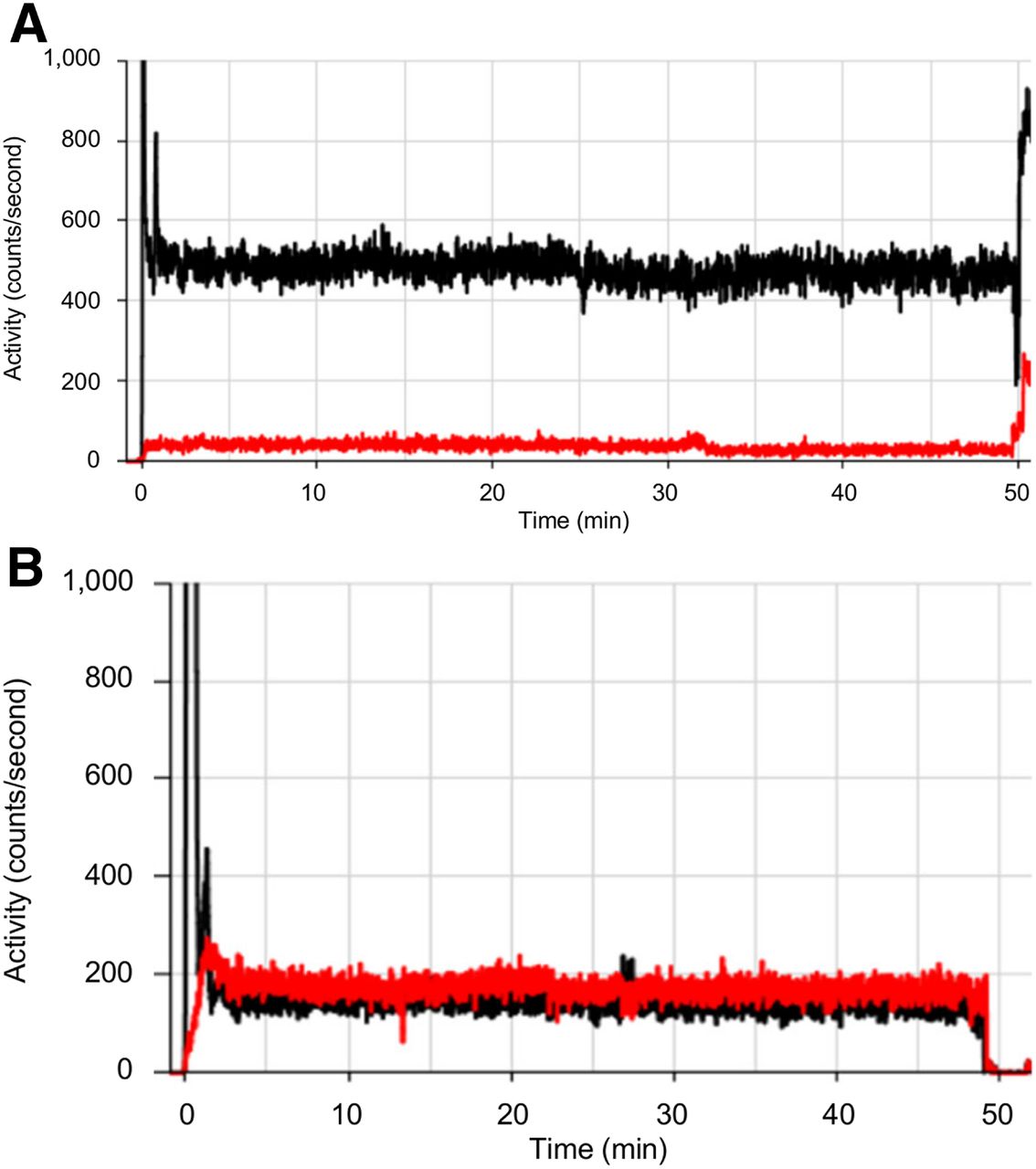
Time–activity curves for injection site (black) and control arm (red) showing rapid peak of injection bolus followed by static residual activity in injection arm, suggestive of extravasation (A), and normalization to control arm, suggestive of no extravasation (B).

FIGURE 3.
  • Download figure
  • Open in new tab
  • Download powerpoint
FIGURE 3.

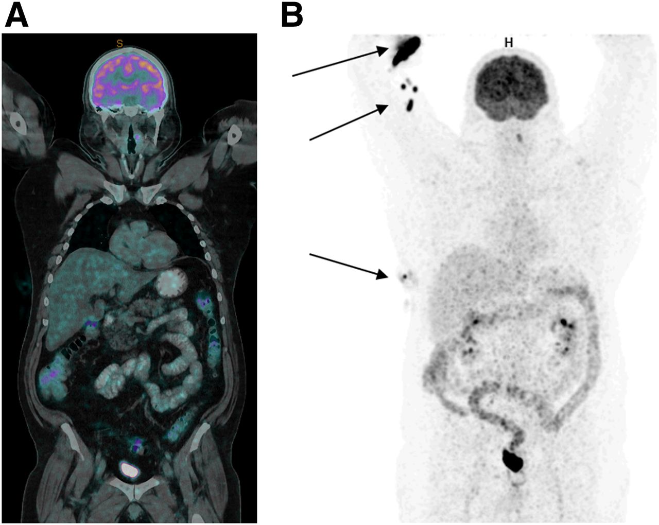
Whole-body (truncated at hips) imaging at 60 min after intravenous administration of 18F-FDG. (A) PET/CT image of low count and poor quality. (B) PET image showing skin contamination of radiopharmaceutical (arrows).

FIGURE 4.
  • Download figure
  • Open in new tab
  • Download powerpoint
FIGURE 4.

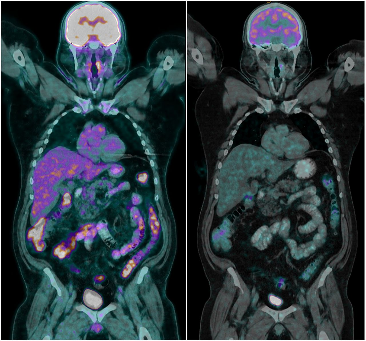
Repeated 18F-FDG PET/CT scan (left) demonstrating superior counts and quality compared with original scan (right), which had count loss associated with leaked activity.

DISCUSSION

Although the automatic injector set-up with a large-bore cannula and high-volume flush is thought to minimize partially extravasated doses compared with hand injection, hand injection in this case would have provided the opportunity to observe the leak, take corrective measures, circumvent most of the leakage, and prevent the need to repeat the study. Automatic injectors also reduce the radiation dose to the staff; therefore, the added value of using them needs to be offset by measures to ensure that unsupervised injection lines remain patent.

CONCLUSION

The Lara device provided a useful tool for more timely critical evaluation and problem solving, extending advantages to the patient and practice.

DISCLOSURE

This project was funded by an ANSTO/ANZSNM research grant. No other potential conflict of interest relevant to this article was reported.

Footnotes

  • Published online Apr. 20, 2020.

REFERENCES

  1. 1.↵
    1. Williams JM,
    2. Arlinghaus LR,
    3. Rani SD,
    4. et al
    . Towards real-time topical detection and characterization of FDG dose infiltration prior to PET imaging. Eur J Nucl Med Mol Imaging. 2016;43:2374–2380.
    OpenUrl
  2. 2.↵
    1. Lattanze RK,
    2. Osman MM,
    3. Ryan KA,
    4. Frye S,
    5. Townsend D
    . Usefulness of topically applied sensors to assess the quality of 18F-FDG injections and validation against dynamic positron emission tomography (PET) images. Front Med (Lausanne). 2018;5:303.
    OpenUrl
  3. 3.↵
    1. Wong TZ,
    2. Benefield T,
    3. Masters S,
    4. et al
    . Quality improvement initiatives to assess and improve PET/C injection infiltration rates at multiple centers. J Nucl Med Technol. 2019;47:326–331.
    OpenUrlAbstract/FREE Full Text
  • Received for publication December 6, 2019.
  • Accepted for publication March 5, 2020.
PreviousNext
Back to top

In this issue

Journal of Nuclear Medicine Technology: 48 (3)
Journal of Nuclear Medicine Technology
Vol. 48, Issue 3
September 1, 2020
  • Table of Contents
  • About the Cover
  • Index by author
Print
Download PDF
Article Alerts
Sign In to Email Alerts with your Email Address
Email Article

Thank you for your interest in spreading the word on Journal of Nuclear Medicine Technology.

NOTE: We only request your email address so that the person you are recommending the page to knows that you wanted them to see it, and that it is not junk mail. We do not capture any email address.

Enter multiple addresses on separate lines or separate them with commas.
Detection of 18F-FDG Dose Leakage Using a Topical Device
(Your Name) has sent you a message from Journal of Nuclear Medicine Technology
(Your Name) thought you would like to see the Journal of Nuclear Medicine Technology web site.
Citation Tools
Detection of 18F-FDG Dose Leakage Using a Topical Device
Stephanie Sanchez, Geoffrey M. Currie
Journal of Nuclear Medicine Technology Sep 2020, 48 (3) 283-284; DOI: 10.2967/jnmt.119.240283

Citation Manager Formats

  • BibTeX
  • Bookends
  • EasyBib
  • EndNote (tagged)
  • EndNote 8 (xml)
  • Medlars
  • Mendeley
  • Papers
  • RefWorks Tagged
  • Ref Manager
  • RIS
  • Zotero
Share
Detection of 18F-FDG Dose Leakage Using a Topical Device
Stephanie Sanchez, Geoffrey M. Currie
Journal of Nuclear Medicine Technology Sep 2020, 48 (3) 283-284; DOI: 10.2967/jnmt.119.240283
Twitter logo Facebook logo LinkedIn logo Mendeley logo
  • Tweet Widget
  • Facebook Like
  • Google Plus One
Bookmark this article

Jump to section

  • Article
    • Abstract
    • CASE REPORT
    • DISCUSSION
    • CONCLUSION
    • DISCLOSURE
    • Footnotes
    • REFERENCES
  • Figures & Data
  • Info & Metrics
  • PDF

Related Articles

  • No related articles found.
  • PubMed
  • Google Scholar

Cited By...

  • No citing articles found.
  • Google Scholar

More in this TOC Section

  • High-Sensitivity Troponin Elevation in a Young Woman with Typical Chest Pain: The Heart of the Matter
  • Pulmonary Adenocarcinoma Revealed by Parathyroid Scintigraphy: An Incidental Case to Remember
  • Prominent Right Ventricular Tracer Uptake: A Harbinger of Multivessel Coronary Artery Disease
Show more Teaching Case Studies

Similar Articles

Keywords

  • nuclear medicine
  • PET
  • 18F-FDG
  • extravasation
  • LARA
  • auto-injector
SNMMI

© 2025 SNMMI

Powered by HighWire