


Abstract
We anecdotally observed an increased accumulation of 99mTc-tetrofosmin in the stomach of myocardial perfusion patients when their uptake phase coincided with preparation of hamburgers in an adjacent room for gastric emptying studies on other patients. The potential for a scent-stimulated alteration of gastric biodistribution required further investigation. Methods: An experimental group and a control group were enrolled (20 patients per group). The experimental group could smell food being prepared during the uptake phase. Stomach, heart, and background regions were drawn in multiple projections, and the resulting data were evaluated. Results: The experimental and control groups did not significantly differ in stomach counts per pixel, background-corrected counts per pixel, or heart-to-stomach ratio. Further analysis of the data revealed that women had a significantly higher increase in stomach counts (P = 0.022) and background-corrected stomach counts (P = 0.018) than men. Conclusion: Women had a greater increase in gastric 99mTc-tetrofosmin activity than men during the radiopharmaceutical uptake phase, but there was no causal relationship between an increase in activity and olfactory stimulation from the cooking of food.
We anecdotally observed several instances of an increased accumulation of 99mTc-tetrofosmin in the stomach (Fig. 1) of myocardial perfusion patients when their uptake phase coincided with preparation of hamburgers in an adjacent room for gastric emptying studies on other patients. The potential for a scent-stimulated alteration of gastric biodistribution prompted further investigation.
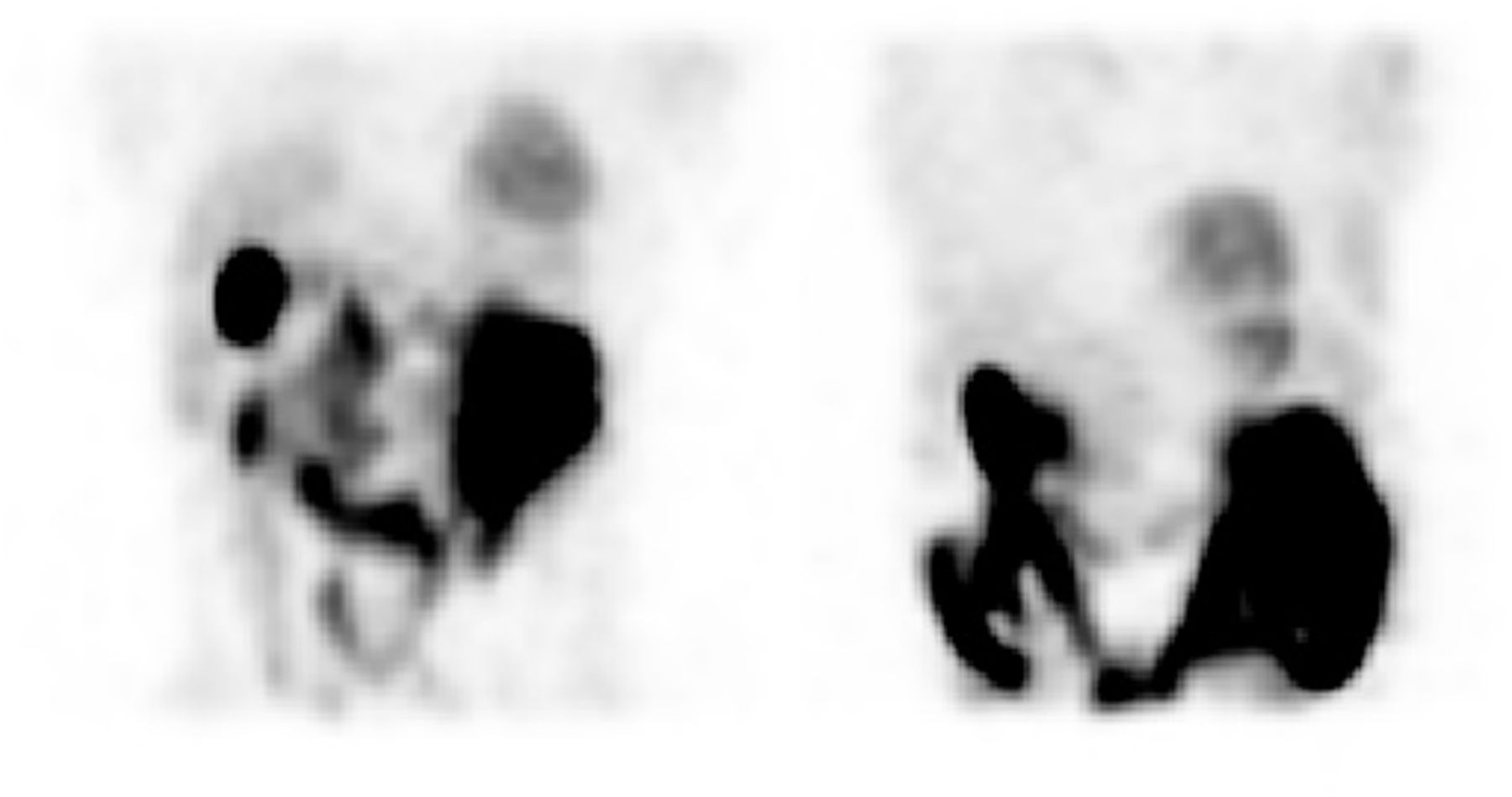
Typical myocardial perfusion rest study showing prominent cardiac activity and minimal gastric activity (left), compared with marked gastric activity when uptake phase coincided with hamburger preparation (right).
Despite the benefits of myocardial perfusion imaging, image quality continues to suffer from several artifacts (1,2). Radiopharmaceutical artifacts are more difficult to manage than technical errors. 99mTc-tetrofosmin localizes not only within the heart but also in various pathologic tissues (e.g., soft-tissue tumors) and organs (e.g., liver, gallbladder, stomach, and bowel). This can become problematic, especially when aberrant activity overlies the myocardium.
The impact of the scent of food on 99mTc-tetrofosmin biodistribution has not, to our knowledge, been previously reported in the literature, although olfactory stimulation during the uptake phase of the radiopharmaceutical in a fasting patient may indeed have a significant impact on biodistribution. During the cephalic phase of digestion (occurring when the scent, sight, taste, or thought of food is present), a significant increase of gastric acid secretions is induced by the vagus nerve. These secretions include pepsinogen, gastrin, hydrochloric acid, and other digestive enzymes (3). The final step of hydrochloric acid production involves the H+/K+/adenosine triphosphatase proton pump (4). This pump utilizes the energy of adenosine triphosphate to countertransport H+ out of the cell and K+ ions across the cell membrane (4). The uptake mechanism of 99mTc-tetrofosmin in gastric tissue is unknown. Some have suggested that 99mTc-sestamibi localizes in the mitochondria of parietal cells and is excreted via the H+/K+/adenosine triphosphatase pump into the gastric lumen (5,6), and it has therefore been hypothesized that 99mTc-tetrofosmin may localize in a similar manner (6).
99mTc-tetrofosmin activity from the stomach can also be attributed to disease states such as dyspepsia, gastritis, and enterogastric bile reflux (7,8). Gholamrezanezhad et al. (7) conducted a study involving 1,056 myocardial perfusion patients, and although the authors found a low incidence of gastric wall uptake, they concluded that in most patients these uptake patterns were associated with dyspeptic symptoms. These findings are consistent with a previous study, by Côté and Dumont (8), who identified gastric wall hyperactivity due to known dyspepsia in 13 of 819 patients. Several case studies (9–11) have also demonstrated significant activity in the gastric wall or cavity during myocardial perfusion imaging due to gastrointestinal pathologic states.
It has been proposed that medications such as type 2 histamine receptor antagonists and proton pump inhibitors may also increase activity in the stomach during myocardial perfusion imaging (12). Mouden et al. (6) investigated gastric wall uptake in myocardial perfusion patients who had taken proton pump inhibitors or type 2 histamine receptor antagonists over a prolonged time; the authors found that 17 of 127 patients had clinically relevant gastric uptake of 99mTc-tetrofosmin.
The purpose of this study was to retrospectively quantitate gastric activity in patients undergoing myocardial perfusion stress testing who could smell hamburgers being cooked during the uptake phase, versus a control group who could not smell the food.
MATERIALS AND METHODS
Twenty consecutive patients were retrospectively enrolled for each of the two groups: experimental and control. Twenty-two were male (55%) and 18 female (45%). The mean age of the population was 61 y, with a 95% CI of 56.7–65.7 y and a range 27–87 y. Hospital and university institutional ethics approvals were obtained.
Before presenting to the department for the stress test, the patients were required to have stopped taking any medications that would interfere with the study (when possible), abstain from caffeine-containing products for 24 h, and fast from midnight or for at least 4 h before the study (with the exception of diabetic patients). These requirements also represented the inclusion and exclusion criteria for the study.
All patients underwent a 1-d rest (350 MBq)/stress (1,000 MBq) protocol with a 30- to 50-min delay between radiopharmaceutical (99mTc-tetrofosmin) administration and the start of imaging. The patients were positioned supine with the camera in cardiac configuration mode, and both SPECT and low-dose CT images were acquired. The SPECT images were obtained on a 64 × 64 matrix at 22 s per step. The low-dose CT image was used for the purpose of attenuation correction and was acquired using a kilovoltage of 140, a current of 1 mA, and a pitch of 1.9.
During the delay between radiopharmaceutical administration and imaging, the patients in the experimental group were seated in a waiting room that was adjacent to an area in which meals were being prepared for gastric emptying procedures on other patients. The meal being prepared was a hamburger comprising 100 g of beef; a bun with lettuce, tomato, and egg; and dressings such as mayonnaise, salt, pepper, or tomato sauce. Only the egg was cooked within the department; however, the bun was toasted and the meat heated. Anecdotal observation suggests that the reheating of the meat was the primarily olfactory stimulus in the waiting room.
The raw data acquired during rest imaging were analyzed using Xeleris software (GE Healthcare). Planar projections at 0°, 22.5°, and 45° angles obtained from the maximum-intensity projection were used, and regions of interest were drawn around the stomach, heart, and soft-tissue background to determine counting rates. The regions of interest were drawn at 3 angles to correct for motion, and each region of interest was also redrawn 3 times. Raw mean and background-corrected mean counts per pixel were determined, and the heart-to-stomach ratio was calculated.
Statistical significance was calculated using χ2 analysis for nominal data and the Student t test for continuous data. Grouped data were evaluated using F test ANOVA. A P value of less than 0.05 was considered significant. The differences between independent means were calculated with a 95% confidence interval (CI). Intervalue correlation was evaluated with χ2 analysis, and intervalue reliability was measured using the Cohen κ-coefficient. A matched-pairs t test evaluated for statistically significant correlations between paired data.
RESULTS
Across the two cohorts, the mean counts per pixel were 28.6 for background (95% CI, 25.5–31.7; range, 16.0–57.9), 105.3 for heart (95% CI, 88.2–122.3; range, 44.5–287.0), and 105.3 for stomach (95% CI, 80.3–105.3; range, 10.5–429.9). The overlap in 95% CIs indicates no statistically significant difference between heart and stomach counts per pixel in the group as a whole. This remained true for the mean background-corrected counts per pixel: 76.6 for heart (95% CI, 62.0–91.2; range, 23.7–240.2) and 76.6 for stomach (95% CI, 53.1–100.2; a range, −28.0 to 383.0). The mean background-corrected heart-to-stomach ratio was −0.15 (95% CI, −2.82 to 2.51; range, −46.9 to 11.2).
A comparison of the experimental group with the control group revealed no statistically significant differences between them (Table 1). The fact that there were far more women in the control group than in the experimental group, although not statistically significant (P = 0.06), may have confounded the data. Importantly, there was no statistically significant difference between the experimental and control groups in stomach count per pixel, background-corrected count per pixel, or heart-to-stomach ratio. Indeed, the mean count per pixel was lower in the experimental group than in the control group, but this difference was likely a reflection of the sex distribution.
Key Parameters Comparing Control and Experimental Groups
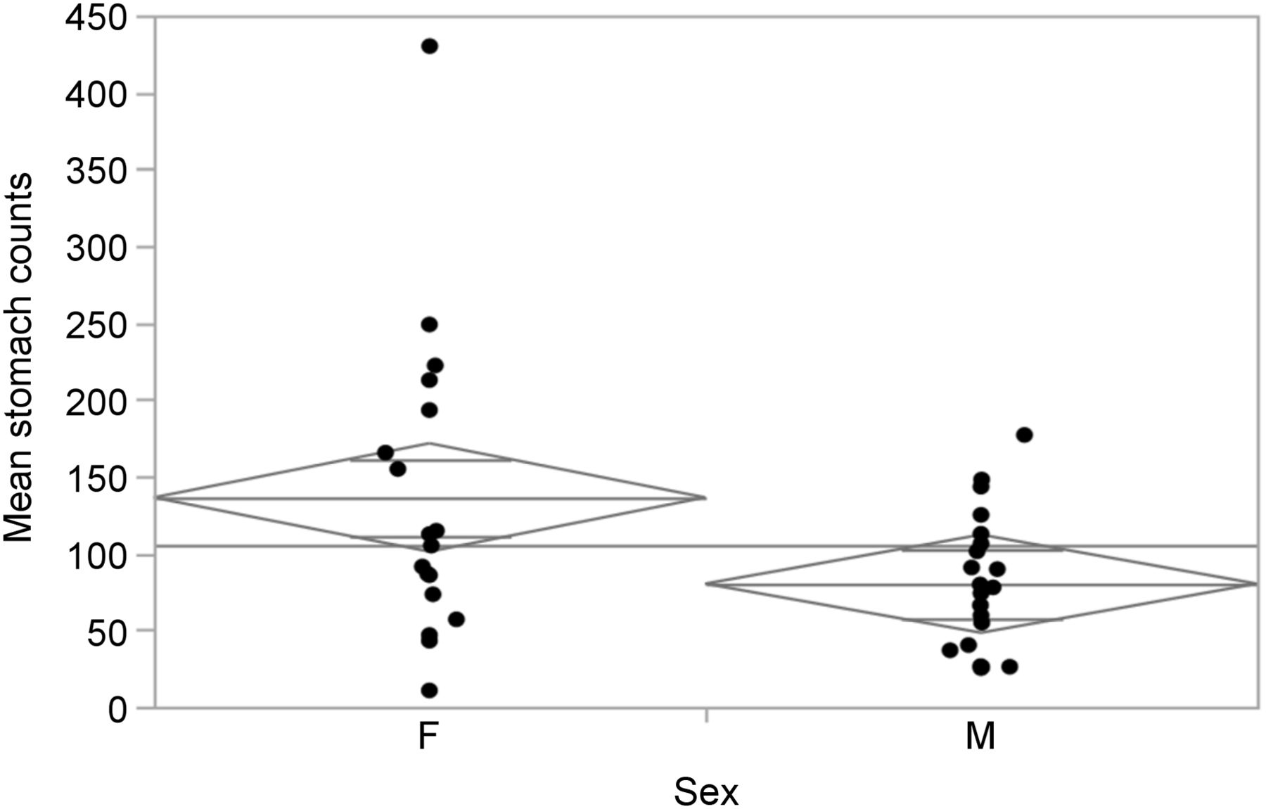
A closer examination of the parameters with respect to sex (Table 2) revealed no statistically significant difference in age, background counts, or heart counts between the sexes. It did, however, reveal a statistically significant increase in stomach count (P = 0.022) (Fig. 2) and background-corrected stomach count (P = 0.018) (Fig. 3) in women. The disproportionate representation of women in the control group likely explains the skewed data outlined in Table 1. Elimination of a potential outlier (Figs. 2 and 3) did not alter this finding, with statistically significant increases found for both stomach count (P = 0.038) and background-corrected stomach count (P = 0.029).
Key Parameters Comparing Women and Men

One-way analysis of mean stomach counts per pixel against sex.
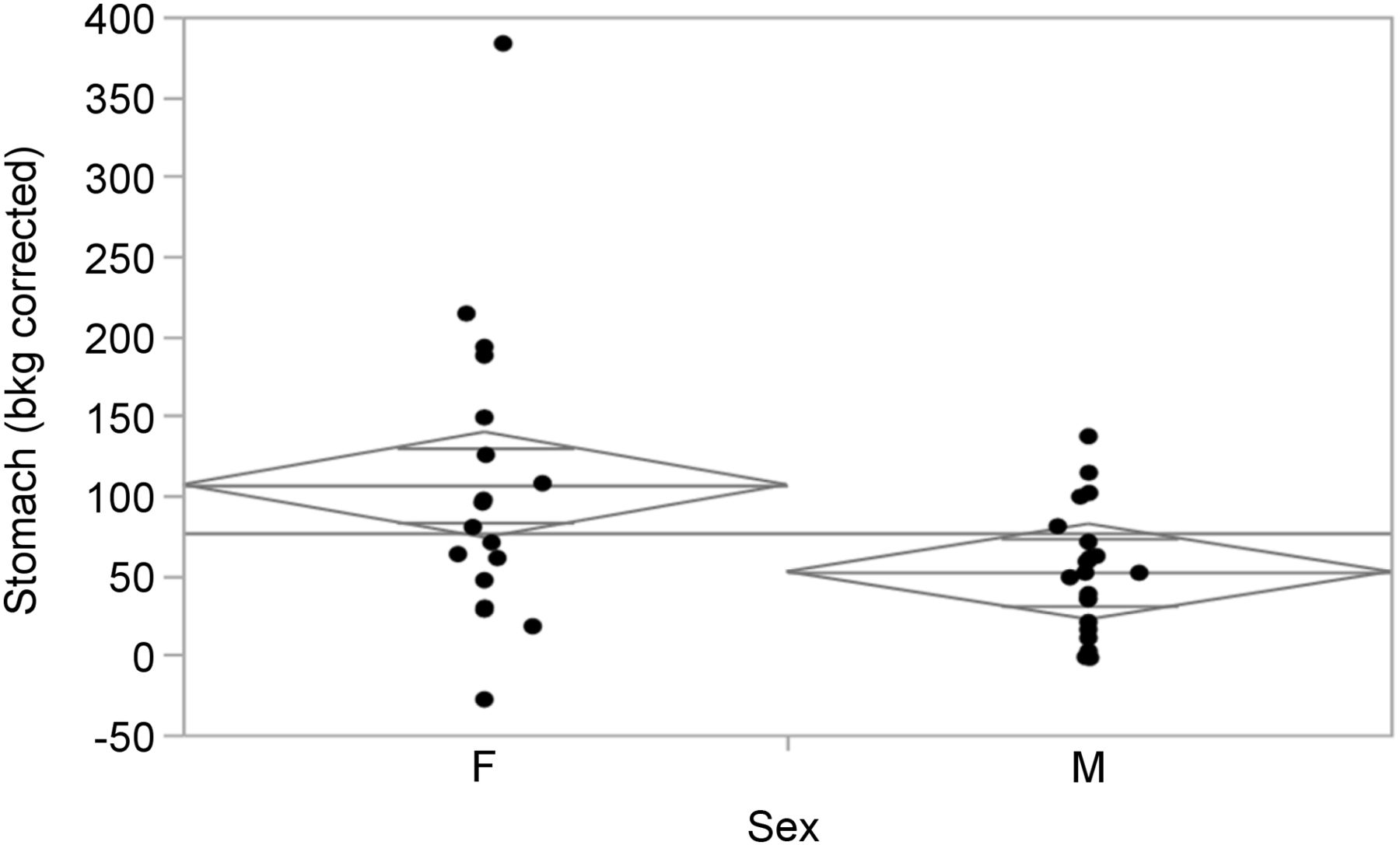
One-way analysis of mean background-corrected stomach counts per pixel against sex.
No statistically significant relationship was demonstrated between age and background count, heart count, stomach count, background-corrected heart count, background-corrected stomach count, or heart-to-stomach ratio (all P > 0.17 and R2 < 0.05). No statistically significant differences were noted between regions of interest (intraoperator variability, P > 0.157 and R2 > 0.99) or between count distributions based on angular projection (P > 0.748); however, as expected, the counts for both 22.5° and 45° were statistically higher than the count for 0°, and the count for 45° was statistically higher than the count for 22.5° (P < 0.04).
DISCUSSION
This study yielded several important findings and observations. First, it disproved the hypothesis that olfactory stimulation caused the anecdotally observed 99mTc-tetrofosmin increase in the stomach during the uptake phase. Second, it revealed a statistically significant relationship between the female sex and increased stomach counts (including after background correction), supporting our observation that the first few patients in whom we suspected a scent-related effect were all women. The mechanism of this phenomenon is unclear but likely relates to hormone-driven changes in gastrointestinal function. Previous studies showed slower gastric transit in women than men, and this finding was independent of pregnancy status or phase of the menstrual cycle (13). The implication is that it is not the concentration of sex hormones that is important but simply the presence of them. The sex hormones are thought to affect several hormones, receptors, and enzymes that have an impact on digestion and can increase uptake of 99mTc-tetrofosmin in the stomach. It is also possible that the observation relates simply to an increased prevalence of pathologic states that can increase 99mTc-tetrofosmin uptake in the stomach; however, data on comorbidity were not available. Interestingly, there was a weak positive correlation between age and stomach counts in women that extended beyond the menopause window (R2 = 0.16, or 16% of the increase in stomach counts can be attributed to age). This correlation between age and stomach counts was not seen in men (R2 = 0.003).
A third key outcome of this study is that it justified the need for research on the part of medical radiation technologists. Because policies can be formulated and decisions made on the basis of even anecdotal observations, what we observe should be rigorously evaluated with appropriately structured research. Medical radiation technologists indeed have the responsibility of engaging in and undertaking such research, as it falls within both their scope of practice and their capabilities. This study provided insight into why this role of the medical radiation technologist is essential. Rather than being based on only observations or assumptions, problem solving is most effective when all the details are known. Variations and problems ranging from the simple to the complex are encountered frequently in daily practice, and these provide suitable fodder for technologist-driven research. The outcomes of such research are able to better inform and improve the outcomes for patients.
Another key insight gleaned by the study is the need to account for research confounders. Too often, small research projects overlook potential confounders—in this case, sex. A simpler study might still have found that there was no substance to the anecdotal observation but might have failed to link sex as a confounder. Although it is difficult to account for all confounders, and a large, randomized, controlled trial is beyond the scope of many clinical practitioners, our study showed how the inclusion of a simple control group can provide deeper insight.
Study Limitations
The major limitation of this study was the small population. The imperfect matching of the experimental and control groups, although providing deeper insight, represents a limitation. The retrospective nature of the study prohibited the gleaning of important information on medications and comorbidity to better explain the sex-based differences observed.
Recommendations
Additional studies with a more tightly controlled design are recommended to further our understanding of the effects of sex hormones on the biodistribution of 99mTc-tetrofosmin and other myocardial perfusion agents.
CONCLUSION
Women had a greater increase in gastric 99mTc-tetrofosmin activity than men during the radiopharmaceutical uptake phase, but there was no causal relationship between an increase in activity and olfactory stimulation from the cooking of food.
DISCLOSURE
No potential conflict of interest relevant to this article was reported.
Footnotes
Published online Aug. 4, 2016.
REFERENCES
- Received for publication April 18, 2016.
- Accepted for publication July 19, 2016.


