
Main menu

  • Home
  • Content
    • Current
      • JNMT Supplement
    • Ahead of print
    • Past Issues
    • Continuing Education
    • JNMT Podcast
    • SNMMI Annual Meeting Abstracts
  • Subscriptions
    • Subscribers
    • Rates
    • Journal Claims
    • Institutional and Non-member
  • Authors
    • Submit to JNMT
    • Information for Authors
    • Assignment of Copyright
    • AQARA Requirements
  • Info
    • Reviewers
    • Permissions
    • Advertisers
    • Corporate & Special Sales
  • About
    • About Us
    • Editorial Board
    • Contact Information
  • More
    • Alerts
    • Feedback
    • Help
    • SNMMI Journals
  • SNMMI
    • JNMT
    • JNM
    • SNMMI Journals
    • SNMMI

User menu

  • Subscribe
  • My alerts
  • Log in
  • My Cart

Search

  • Advanced search
Journal of Nuclear Medicine Technology
  • SNMMI
    • JNMT
    • JNM
    • SNMMI Journals
    • SNMMI
  • Subscribe
  • My alerts
  • Log in
  • My Cart
Journal of Nuclear Medicine Technology

Advanced Search

  • Home
  • Content
    • Current
    • Ahead of print
    • Past Issues
    • Continuing Education
    • JNMT Podcast
    • SNMMI Annual Meeting Abstracts
  • Subscriptions
    • Subscribers
    • Rates
    • Journal Claims
    • Institutional and Non-member
  • Authors
    • Submit to JNMT
    • Information for Authors
    • Assignment of Copyright
    • AQARA Requirements
  • Info
    • Reviewers
    • Permissions
    • Advertisers
    • Corporate & Special Sales
  • About
    • About Us
    • Editorial Board
    • Contact Information
  • More
    • Alerts
    • Feedback
    • Help
    • SNMMI Journals
  • Watch or Listen to JNMT Podcast
  • Visit SNMMI on Facebook
  • Join SNMMI on LinkedIn
  • Follow SNMMI on Twitter
  • Subscribe to JNMT RSS feeds
Review ArticleTeaching Case Studies

Appearance of Meningioma on 99mTc-HMPAO SPECT: Correlation with MRI

Zaid Al-Faham, Mohammad A. Kassir, Daniel Wood and Helena R. Balon
Journal of Nuclear Medicine Technology June 2016, 44 (2) 90-91; DOI: https://doi.org/10.2967/jnmt.115.163287
Zaid Al-Faham
Department of Diagnostic Radiology and Molecular Imaging, William Beaumont School of Medicine and Beaumont Health System, Oakland University, Royal Oak, Michigan
  • Find this author on Google Scholar
  • Find this author on PubMed
  • Search for this author on this site
Mohammad A. Kassir
Department of Diagnostic Radiology and Molecular Imaging, William Beaumont School of Medicine and Beaumont Health System, Oakland University, Royal Oak, Michigan
  • Find this author on Google Scholar
  • Find this author on PubMed
  • Search for this author on this site
Daniel Wood
Department of Diagnostic Radiology and Molecular Imaging, William Beaumont School of Medicine and Beaumont Health System, Oakland University, Royal Oak, Michigan
  • Find this author on Google Scholar
  • Find this author on PubMed
  • Search for this author on this site
Helena R. Balon
Department of Diagnostic Radiology and Molecular Imaging, William Beaumont School of Medicine and Beaumont Health System, Oakland University, Royal Oak, Michigan
  • Find this author on Google Scholar
  • Find this author on PubMed
  • Search for this author on this site
  • Article
  • Figures & Data
  • Info & Metrics
  • PDF
Loading

Abstract

On brain perfusion SPECT, a primary brain lesion presents as a localized defect that corresponds to the mass lesion. 99mTc-HMPAO images generally show a focal defect in the region of abnormality, whether containing necrotic tissue, recurrent tumor, or both. Further characterization with MR imaging is needed to confirm the diagnosis, as demonstrated in this case report.

  • meningioma
  • SPECT
  • brain
  • HMPAO

We report a case of meningioma suspected on 99mTc-hexamethylpropyleneamine oxime (HMPAO) brain SPECT. Few cases of meningioma appear on SPECT scans. Brain SPECT may provide complementary information for the diagnosis of brain tumors.

CASE REPORT

A 34-y-old woman diagnosed with Lyme disease 15 y previously presented with a 2-month history of chronic headaches, tremor, numbness, and adenopathy. Blood biochemistry was within normal limits. A 99mTc-HMPAO SPECT brain scan was performed because of suspected Lyme neuroborreliosis. The patient was injected with 1,073 MBq (29 mCi) of 99mTc-HMPAO, and SPECT images of the brain were subsequently obtained.

DISCUSSION

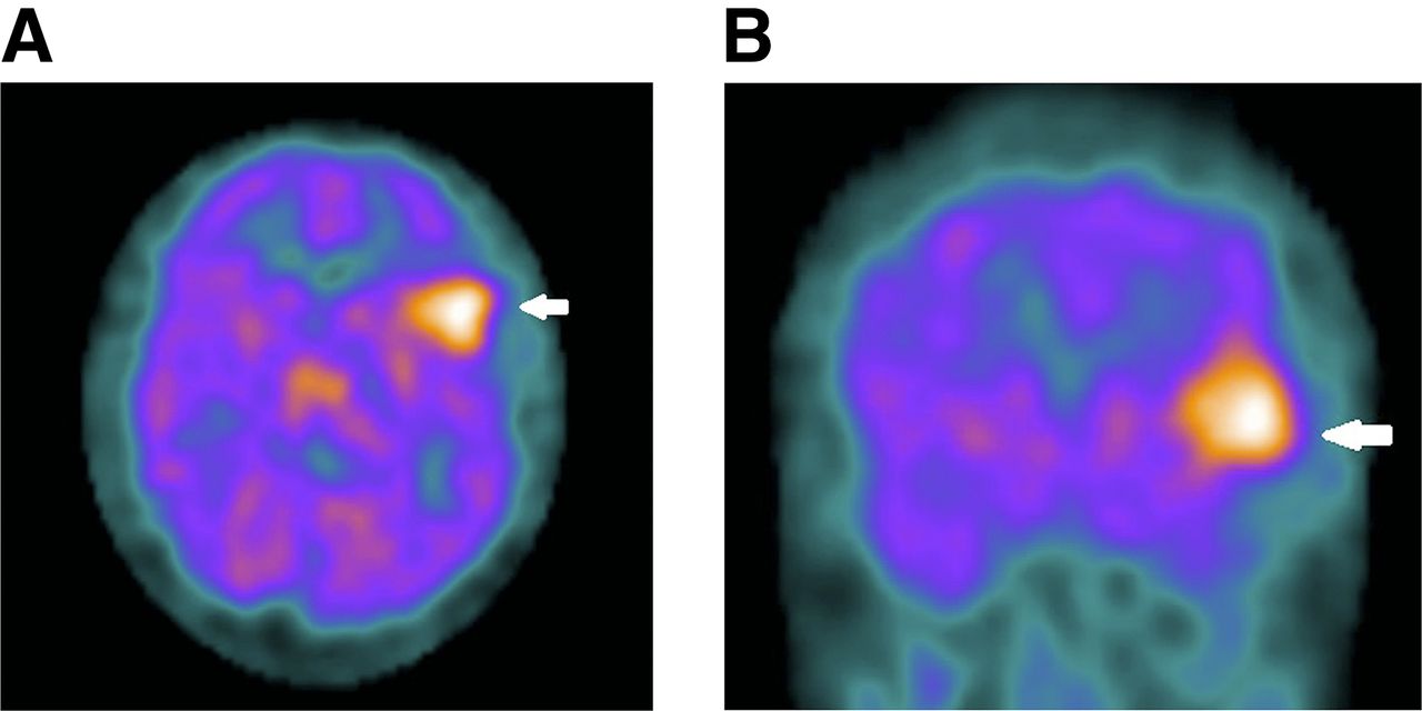
The SPECT images showed a large focus of radiotracer uptake in the left frontotemporoparietal region (Fig. 1). A differential diagnosis of infection, seizure focus, and neoplasm was given, with high suspicion of meningioma. MR imaging of the brain was subsequently performed and demonstrated a solid, enhancing extraaxial mass in the same area, with an appearance consistent with meningioma (Fig. 2).

FIGURE 1.
  • Download figure
  • Open in new tab
  • Download powerpoint
FIGURE 1.

SPECT images of brain obtained with 99mTc-HMPAO show intense focal radiotracer uptake in left frontal area (arrows) on representative axial (A) and coronal (B) slices.

FIGURE 2.
  • Download figure
  • Open in new tab
  • Download powerpoint
FIGURE 2.

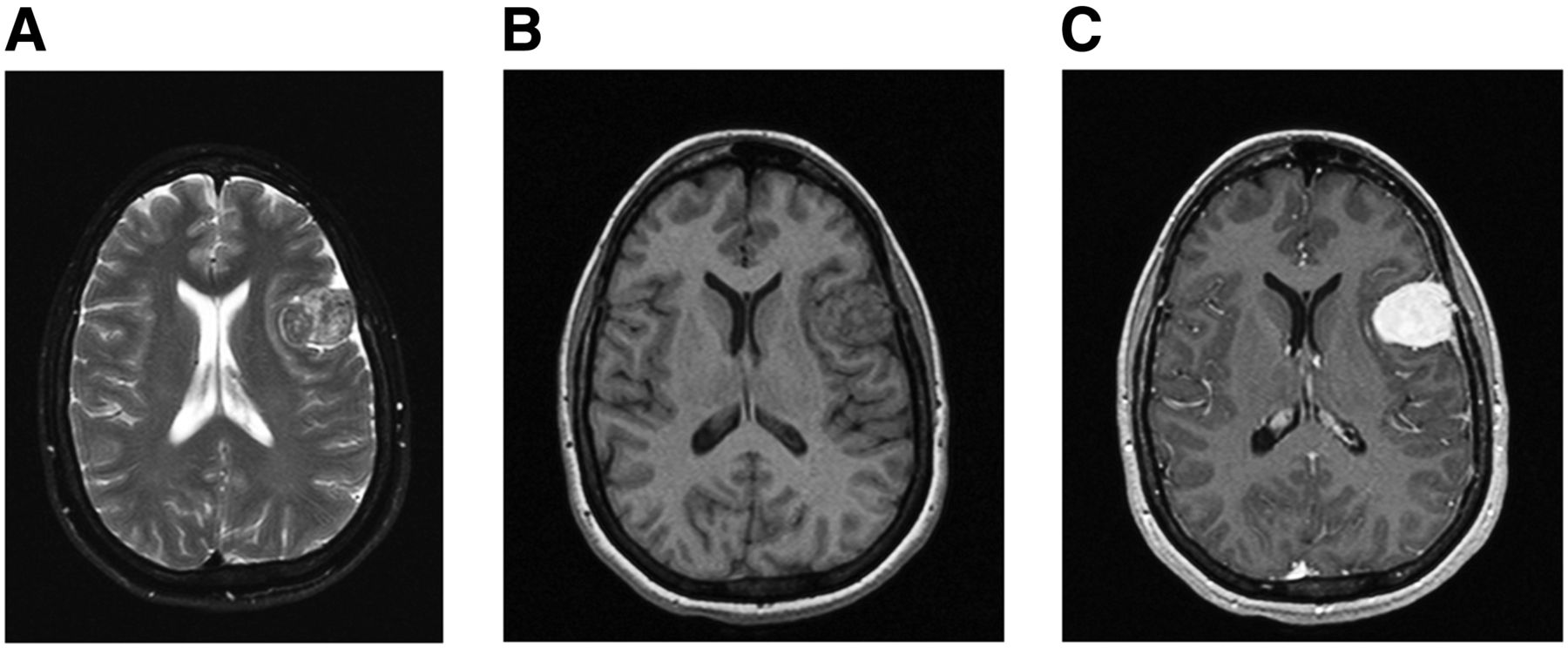
Axial T2-weighted (A) and axial unenhanced (B) and enhanced (C) T1-weighted MR images demonstrate solid 3.2 × 3.5 cm enhancing extraaxial mass with enhancing dural tail overlying left frontal lobe.

99mTc-HMPAO is a lipophilic compound that passes the intact blood–brain barrier freely (1,2). It can be labeled with 99mTc to assess high-flow lesions (1,2). Meningiomas typically present with high radiotracer uptake on 99mTc-HMPAO SPECT (1,2). Conversely, in patients with calcified meningioma there will be decreased radiotracer uptake (2). Patients with stroke also can have decreased to absent 99mTc-HMPAO uptake. Findings are time-dependent and have the highest prognostic value during the first 48 h after a stroke; strokes due to occlusion of a major cerebral artery produce focal absence of perfusion, and a normal brain perfusion result on SPECT during the first 6 h after a stroke implies minor or lacunar stroke (3). There are some reports of brain tumors with increased 99mTc-HMPAO accumulation other than meningioma, such as glioblastoma multiform, pituitary adenoma, high-grade astrocytoma, and multiple myeloma (1). Shih et al. showed cases of meningiomas without 99mTc-HMPAO uptake, possibly explained by a skull defect due to previous craniotomy, replacement of normal brain tissue by tumor, or functioning arteriovenous shunting due to tumor vascular blush, as demonstrated on cerebral angiograms (4). Further characterization with MR imaging was necessary to confirm the diagnosis.

MR images demonstrated a solid, 3.2 × 3.5 cm enhancing extraaxial mass with an enhancing dural tail overlying the left frontal lobe. Meningioma typically appears isointense to hypointense to gray matter on T1-weighted images and isointense to hyperintense on T2-weighted images (5). A dural tail is a nonspecific finding that often suggests meningioma but can be seen with other tumors. Meningiomas can also be associated with mass effect and cerebral edema (6). Our findings were most consistent with the diagnosis of meningioma.

CONCLUSION

On SPECT brain perfusion imaging, a primary brain lesion presents as a localized defect that corresponds to the mass lesion. 99mTc-HMPAO images generally show a focal defect in the region of abnormality, whether containing necrotic tissue, recurrent tumor, or both. Further characterization with MR imaging is needed to confirm the diagnosis.

DISCLOSURE

No potential conflict of interest relevant to this article was reported.

Footnotes

  • Published online Oct. 15, 2015.

REFERENCES

  1. 1.↵
    1. Takeshita T,
    2. Kaminaga T,
    3. Kikuchi Y,
    4. Furui S
    . Multiple myeloma showing increased accumulation of Tc-99m hexamethylpropylene amine oxime on brain SPECT. Clin Nucl Med. 2000;25:495–496.
    OpenUrlCrossRefPubMed
  2. 2.↵
    1. Suess E,
    2. Malessa S,
    3. Ungersböck K,
    4. et al
    . Technetium-99m-d, 1-hexamethylpropyleneamine oxime (HMPAO) uptake and glutathione content in brain tumors. J Nucl Med. 1991;32:1675–1681.
    OpenUrlAbstract/FREE Full Text
  3. 3.↵
    1. Alexandrov AV,
    2. Black SE,
    3. Ehrlich LE,
    4. et al
    . Simple visual analysis of brain perfusion on HMPAO SPECT predicts early outcome in acute stroke. Stroke. 1996;27:1537–1542.
    OpenUrlAbstract/FREE Full Text
  4. 4.↵
    1. Shih WJ,
    2. Lee JK,
    3. Milan P
    . Discordant technetium-99m-MIBI and technetium-99m-HMPAO uptake of recurrent occipital meningioma on brain SPECT images. J Nucl Med. 1996;37:1183–1185.
    OpenUrlAbstract/FREE Full Text
  5. 5.↵
    1. Keogh BP,
    2. Henson JW
    . Clinical manifestations and diagnostic imaging of brain tumors. Hematol Oncol Clin N Am 2012;26:733-755.
    OpenUrlCrossRefPubMed
  6. 6.↵
    1. Wallace EW
    . The dural tail sign. Radiology. 2004;233:56–57.
    OpenUrlCrossRefPubMed
  • Received for publication July 2, 2015.
  • Accepted for publication September 10, 2015.
PreviousNext
Back to top

In this issue

Journal of Nuclear Medicine Technology: 44 (2)
Journal of Nuclear Medicine Technology
Vol. 44, Issue 2
June 1, 2016
  • Table of Contents
  • About the Cover
  • Index by author
Print
Download PDF
Article Alerts
Sign In to Email Alerts with your Email Address
Email Article

Thank you for your interest in spreading the word on Journal of Nuclear Medicine Technology.

NOTE: We only request your email address so that the person you are recommending the page to knows that you wanted them to see it, and that it is not junk mail. We do not capture any email address.

Enter multiple addresses on separate lines or separate them with commas.
Appearance of Meningioma on 99mTc-HMPAO SPECT: Correlation with MRI
(Your Name) has sent you a message from Journal of Nuclear Medicine Technology
(Your Name) thought you would like to see the Journal of Nuclear Medicine Technology web site.
Citation Tools
Appearance of Meningioma on 99mTc-HMPAO SPECT: Correlation with MRI
Zaid Al-Faham, Mohammad A. Kassir, Daniel Wood, Helena R. Balon
Journal of Nuclear Medicine Technology Jun 2016, 44 (2) 90-91; DOI: 10.2967/jnmt.115.163287

Citation Manager Formats

  • BibTeX
  • Bookends
  • EasyBib
  • EndNote (tagged)
  • EndNote 8 (xml)
  • Medlars
  • Mendeley
  • Papers
  • RefWorks Tagged
  • Ref Manager
  • RIS
  • Zotero
Share
Appearance of Meningioma on 99mTc-HMPAO SPECT: Correlation with MRI
Zaid Al-Faham, Mohammad A. Kassir, Daniel Wood, Helena R. Balon
Journal of Nuclear Medicine Technology Jun 2016, 44 (2) 90-91; DOI: 10.2967/jnmt.115.163287
Twitter logo Facebook logo LinkedIn logo Mendeley logo
  • Tweet Widget
  • Facebook Like
  • Google Plus One
Bookmark this article

Jump to section

  • Article
    • Abstract
    • CASE REPORT
    • DISCUSSION
    • CONCLUSION
    • DISCLOSURE
    • Footnotes
    • REFERENCES
  • Figures & Data
  • Info & Metrics
  • PDF

Related Articles

  • No related articles found.
  • PubMed
  • Google Scholar

Cited By...

  • No citing articles found.
  • Google Scholar

More in this TOC Section

  • High-Sensitivity Troponin Elevation in a Young Woman with Typical Chest Pain: The Heart of the Matter
  • Prominent Right Ventricular Tracer Uptake: A Harbinger of Multivessel Coronary Artery Disease
  • SPECT/CT for Discrimination Between Active and Inactive Os Trigonum in Posterior Ankle Syndrome
Show more Teaching Case Studies

Similar Articles

Keywords

  • meningioma
  • SPECT
  • brain
  • HMPAO
SNMMI

© 2025 SNMMI

Powered by HighWire