JNMT
HOME HELP FEEDBACK SUBSCRIPTIONS ARCHIVE SEARCH TABLE OF CONTENTS
 QUICK SEARCH:   [advanced]


This Article
Right arrow Abstract Freely available
Right arrow Figures Only
Right arrow Full Text (PDF)
Right arrow Alert me when this article is cited
Right arrow Alert me if a correction is posted
Services
Right arrow Email this article to a friend
Right arrow Similar articles in this journal
Right arrow Similar articles in PubMed
Right arrow Alert me to new issues of the journal
Right arrow Download to citation manager
Right arrow reprints & permissions
Citing Articles
Right arrow Citing Articles via Google Scholar
Google Scholar
Right arrow Articles by Kuo, P. H.
Right arrow Articles by Cheng, D. W.
Right arrow Search for Related Content
PubMed
Right arrow PubMed Citation
Right arrow Articles by Kuo, P. H.
Right arrow Articles by Cheng, D. W.
Journal of Nuclear Medicine Technology Volume 33, Number 4, 2005 230-231
© 2005 by Society of Nuclear Medicine

Artifactual Spinal Metastases Imaged by PET/CT: A Case Report

Phillip H. Kuo, MD, PhD and David W. Cheng, MD, PhD

Department of Diagnostic Radiology, Yale University School of Medicine, New Haven, Connecticut


    ABSTRACT
 TOP
 ABSTRACT
 INTRODUCTION
 CASE REPORT
 DISCUSSION
 REFERENCES
 
A 55-y-old patient with multiple myeloma presented for restaging after chemotherapy and radiation. The patient had undergone vertebroplasty of multiple thoracic vertebrae because of painful compression fractures. The 18F-FDG PET images showed increased activity at the T8 and T10–T12 vertebral bodies. Comparison of the attenuation-corrected and non–attenuation-corrected images demonstrated that the activity was due to an artifact of attenuation correction. The CT scan correlated the sites of vertebroplasty to the 4 foci of increased uptake of 18F-FDG. The increasing use of vertebroplasty for malignant spinal fractures warrants vigilance for this artifact.

Key Words: PET; CT; myeloma; vertebroplasty; artifact


    INTRODUCTION
 TOP
 ABSTRACT
 INTRODUCTION
 CASE REPORT
 DISCUSSION
 REFERENCES
 
Improper attenuation correction of high-density materials within patients can result in artifacts on CT images. Vigilance for these artifacts is essential to prevent misdiagnosis of disease recurrence.


    CASE REPORT
 TOP
 ABSTRACT
 INTRODUCTION
 CASE REPORT
 DISCUSSION
 REFERENCES
 
A 55-y-old patient with multiple myeloma received chemotherapy and radiation and then presented for restaging by 18F-FDG PET/CT. Studies have demonstrated the utility of 18F-FDG PET in the evaluation of multiple myeloma (14). Before the therapy, the multiple myeloma had caused extensive lytic lesions in the patient’s spine, resulting in compression fractures at multiple levels. Subsequently, the patient underwent vertebroplasty of multiple thoracic vertebrae to relieve the debilitating pain of these compression fractures. In vertebroplasty, a cement mixture consisting mostly of polymethylmethacrylate is injected through a needle into porous bone, with the porosity commonly caused by osteoporosis but also by malignancy as in this case (58). This cement reinforces the bone to reduce further fracture.

The PET/CT scan was performed on a Discovery ST 16-slice PET/CT camera (GE Healthcare), with the protocol for attenuation correction adjusted for use of oral contrast material. The 18F-FDG PET images showed increased activity at the T8 and T10–T12 vertebral bodies (Fig. 1). Comparison of the attenuation-corrected and non–attenuation-corrected images demonstrated that the activity was due to an artifact of attenuation correction (Fig. 1). The CT scan correlated the sites of vertebroplasty to the 4 foci of increased uptake of 18F-FDG. The cement for the vertebroplasty was determined to have a density of 2,000–3,000 HU (much denser than normal bone). PET/CT uses the transmission scan from the CT as a density map for attenuation correction of the measured activity (9). In this case, the attenuation correction algorithm added back too many counts to compensate for attenuation by the extremely dense cement.


View larger version (70K):
[in this window]
[in a new window]
 
FIGURE 1. 
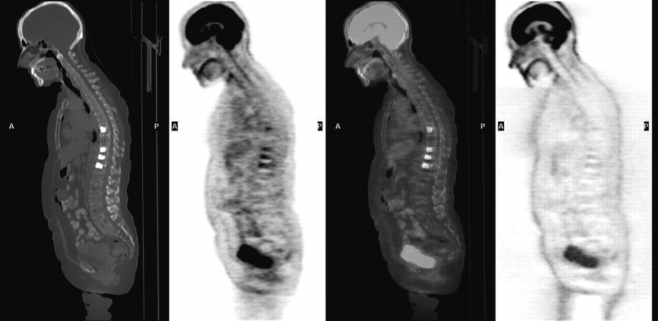
Images of CT, attenuation-corrected PET, fused PET/CT, and non–attenuation-corrected PET in sagittal plane are shown, respectively, from left to right. Increased activity at T8 and T10–T12 was not present on non–attenuation-corrected images and therefore was an artifact of attenuation correction from vertebroplasty material.


    DISCUSSION
 TOP
 ABSTRACT
 INTRODUCTION
 CASE REPORT
 DISCUSSION
 REFERENCES
 
The increasing use of vertebroplasty for benign and malignant compression fractures warrants vigilance for this artifact. If the artifact had not been caught in this case, the patient would have been misdiagnosed with recurrence of disease and received more chemotherapy. Finally, the lesson learned from this case can also be applied to other causes of artifactual metastases from improper attenuation correction of high-density materials within patients (1013).


    FOOTNOTES
 
For correspondence or reprints contact: Phillip H. Kuo, MD, PhD, Department of Diagnostic Radiology, Yale University School of Medicine, 333 Cedar St., P.O. Box 208042, New Haven, CT 06520-8042.

E-mail: phillip.kuo{at}yale.edu


    REFERENCES
 TOP
 ABSTRACT
 INTRODUCTION
 CASE REPORT
 DISCUSSION
 REFERENCES
 

  1. Jadvar H, Conti PS. Diagnostic utility of FDG PET in multiple myeloma. Skeletal Radiol. 2002;31:690–694.[Medline]
  2. Durie BG, Waxman AD, D’Agnolo A, et al. Whole-body 18F-FDG PET identifies high-risk myeloma. J Nucl Med. 2002;43:1457–1463.[Abstract/Free Full Text]
  3. Schirrmeister H, Bommer M, Buck AK, et al. Initial results in the assessment of multiple myeloma using 18F-FDG PET. Eur J Nucl Med Mol Imaging. 2002;29:361–366.[Medline]
  4. Orchard K, Barrington S, Buscombe J, et al. Fluoro-deoxyglucose positron emission tomography imaging for the detection of occult disease in multiple myeloma. Br J Haematol. 2002;117:133–135.[Medline]
  5. Diamond TH, Hartwell T, Clarke W, et al. Percutaneous vertebroplasty for acute vertebral body fracture and deformity in multiple myeloma: a short report. Br J Haematol. 2004;124:485–487.[Medline]
  6. Shimony JS, Gilula LA, Zeller AJ, et al. Percutaneous vertebroplasty for malignant compression fractures with epidural involvement. Radiology. 2004;232:846–853.[Abstract/Free Full Text]
  7. Jensen ME, Kallmes DE. Percutaneous vertebroplasty in the treatment of malignant spine disease. Cancer J. 2002;8:194–206.[Medline]
  8. Fourney DR, Schomer DF, Nader R, et al. Percutaneous vertebroplasty and kyphoplasty for painful vertebral body fractures in cancer patients. J Neurosurg. 2003;98:21–30.
  9. Kinahan PE, Hasegawa BH, Beyer T. X-ray-based attenuation correction for positron emission tomography/computed tomography scanners. Semin Nucl Med. 2003;33:166–179.[Medline]
  10. Bockisch A, Beyer T, Antoch G, et al. Positron emission tomography/computed tomography: imaging protocols, artifacts, and pitfalls. Mol Imaging Biol. 2004;6:188–199.[Medline]
  11. Goerres GW, Ziegler SI, Burger C, et al. Artifacts at PET and PET/CT caused by metallic hip prosthetic material. Radiology. 2003;226:577–584.[Abstract/Free Full Text]
  12. Goerres GW, Hany TF, Kamel E, et al. Head and neck imaging with PET and PET/CT: artefacts from dental metallic implants. Eur J Nucl Med Mol Imaging. 2002;29:367–370.[Medline]
  13. Antoch G, Freudenberg LS, Egelhof T, et al. Focal tracer uptake: a potential artifact in contrast-enhanced dual-modality PET/CT scans. J Nucl Med. 2002;43:1339–1342.[Abstract/Free Full Text]


This Article
Right arrow Abstract Freely available
Right arrow Figures Only
Right arrow Full Text (PDF)
Right arrow Alert me when this article is cited
Right arrow Alert me if a correction is posted
Services
Right arrow Email this article to a friend
Right arrow Similar articles in this journal
Right arrow Similar articles in PubMed
Right arrow Alert me to new issues of the journal
Right arrow Download to citation manager
Right arrow reprints & permissions
Citing Articles
Right arrow Citing Articles via Google Scholar
Google Scholar
Right arrow Articles by Kuo, P. H.
Right arrow Articles by Cheng, D. W.
Right arrow Search for Related Content
PubMed
Right arrow PubMed Citation
Right arrow Articles by Kuo, P. H.
Right arrow Articles by Cheng, D. W.


HOME HELP FEEDBACK SUBSCRIPTIONS ARCHIVE SEARCH TABLE OF CONTENTS
THE JOURNAL OF NUCLEAR MEDICINE JOURNAL OF NUCLEAR MEDICINE TECHNOLOGY