
Main menu

  • Home
  • Content
    • Current
      • JNMT Supplement
    • Ahead of print
    • Past Issues
    • SNMMI Annual Meeting Abstracts
  • Subscriptions
    • Subscribers
    • Rates
    • Journal Claims
    • Institutional and Non-member
  • Authors
    • Submit to JNMT
    • Information for Authors
    • Assignment of Copyright
    • AQARA Requirements
  • Info
    • Permissions
    • Continuing Education
    • Advertisers
    • Corporate & Special Sales
  • About
    • About Us
    • Editorial Board
    • Editorial Contact
  • More
    • Alerts
    • Feedback
    • Help
    • SNMMI Journals
  • SNMMI
    • JNMT
    • JNM
    • SNMMI Journals
    • SNMMI

User menu

  • Subscribe
  • My alerts
  • Log in
  • Log out
  • My Cart

Search

  • Advanced search
Journal of Nuclear Medicine Technology
  • SNMMI
    • JNMT
    • JNM
    • SNMMI Journals
    • SNMMI
  • Subscribe
  • My alerts
  • Log in
  • Log out
  • My Cart
Journal of Nuclear Medicine Technology

Advanced Search

  • Home
  • Content
    • Current
    • Ahead of print
    • Past Issues
    • SNMMI Annual Meeting Abstracts
  • Subscriptions
    • Subscribers
    • Rates
    • Journal Claims
    • Institutional and Non-member
  • Authors
    • Submit to JNMT
    • Information for Authors
    • Assignment of Copyright
    • AQARA Requirements
  • Info
    • Permissions
    • Continuing Education
    • Advertisers
    • Corporate & Special Sales
  • About
    • About Us
    • Editorial Board
    • Editorial Contact
  • More
    • Alerts
    • Feedback
    • Help
    • SNMMI Journals
  • Follow SNMMI on Twitter
  • Visit SNMMI on Facebook
  • Join SNMMI on LinkedIn
  • Subscribe to JNMT RSS feeds
Review ArticleContinuing Education

Gastrointestinal Motility, Part 1: Esophageal Transit and Gastric Emptying

Alan H. Maurer
Journal of Nuclear Medicine Technology March 2016, 44 (1) 1-11; DOI: https://doi.org/10.2967/jnumed.112.114314
Alan H. Maurer
Nuclear Medicine and Molecular Imaging, Temple University Hospital and School of Medicine, Philadelphia, Pennsylvania
  • Find this author on Google Scholar
  • Find this author on PubMed
  • Search for this author on this site
  • Article
  • Figures & Data
  • Info & Metrics
  • PDF
Loading

Article Figures & Data

Figures

  • Tables
  • FIGURE 1.
    • Download figure
    • Open in new tab
    • Download powerpoint
    FIGURE 1.

    Normal esophageal transit (single swallow). Sequential dynamic images (left, 0–25 s) demonstrate normal bolus transit through esophagus. Composite image (center) is produced by summing all images from the initial 30 s. Regions of interest (dotted lines) that define upper, middle, and lower thirds of esophagus are shown. Time–activity curves (right) show counts recorded in each region as bolus progresses down esophagus. Esophageal transit time (11 s) is measured from time–activity curves of leading to trailing edges of upper and lower thirds of esophagus.

  • FIGURE 2.
    • Download figure
    • Open in new tab
    • Download powerpoint
    FIGURE 2.

    Normal global esophageal emptying (multiple swallows). Sequential images at 15 s per image are shown (left). Region of interest (rectangular box) is drawn over entire esophagus. From this region, time–activity curve (right) is generated showing percentage of activity retained in esophagus at each time. Amount of activity retained after multiple swallows can be used to help characterize primary esophageal motor disorders (Fig. 3; Table 1) or to follow therapeutic interventions as in achalasia.
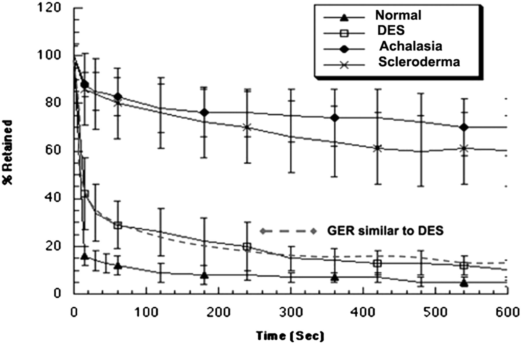
  • FIGURE 3.
    • Download figure
    • Open in new tab
    • Download powerpoint
    FIGURE 3.

    Esophageal emptying for primary esophageal motility disorders. Mean data for healthy subjects are shown compared with diffuse esophageal spasm (DES), achalasia, and scleroderma. Emptying curve for patients with esophagitis from gastroesophageal reflux is similar to DES. (Adapted from (75).)
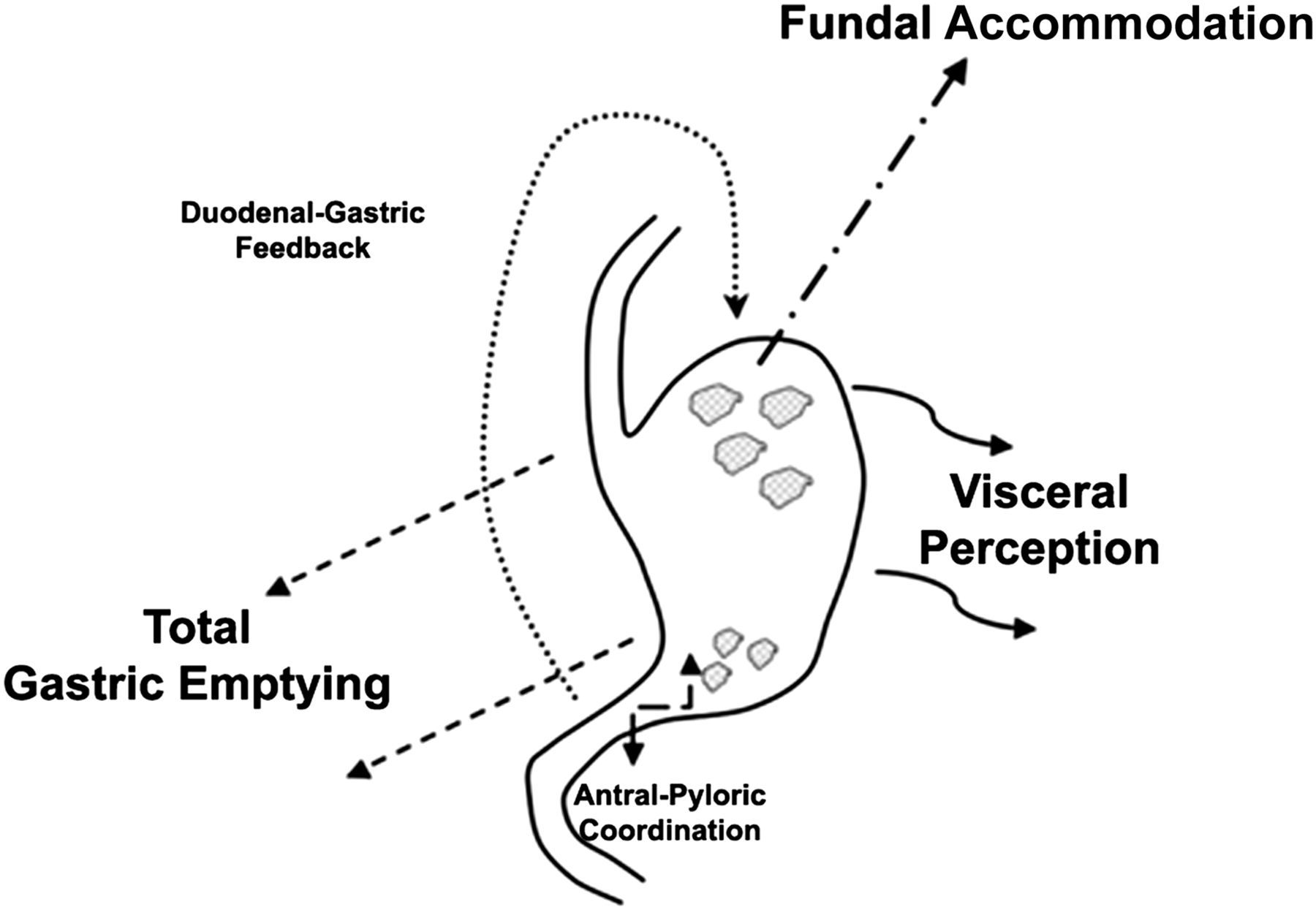
  • FIGURE 4.
    • Download figure
    • Open in new tab
    • Download powerpoint
    FIGURE 4.

    Multiple factors associated with GE are considered important to explain dyspeptic patient symptoms. Total GE, impaired fundal accommodation, and visceral hypersensitivity are 3 major factors currently under study. Antral–duodenal coordination and duodenal–gastric feedback mechanisms are also considered important but are not as well characterized. (Modified from (76).)
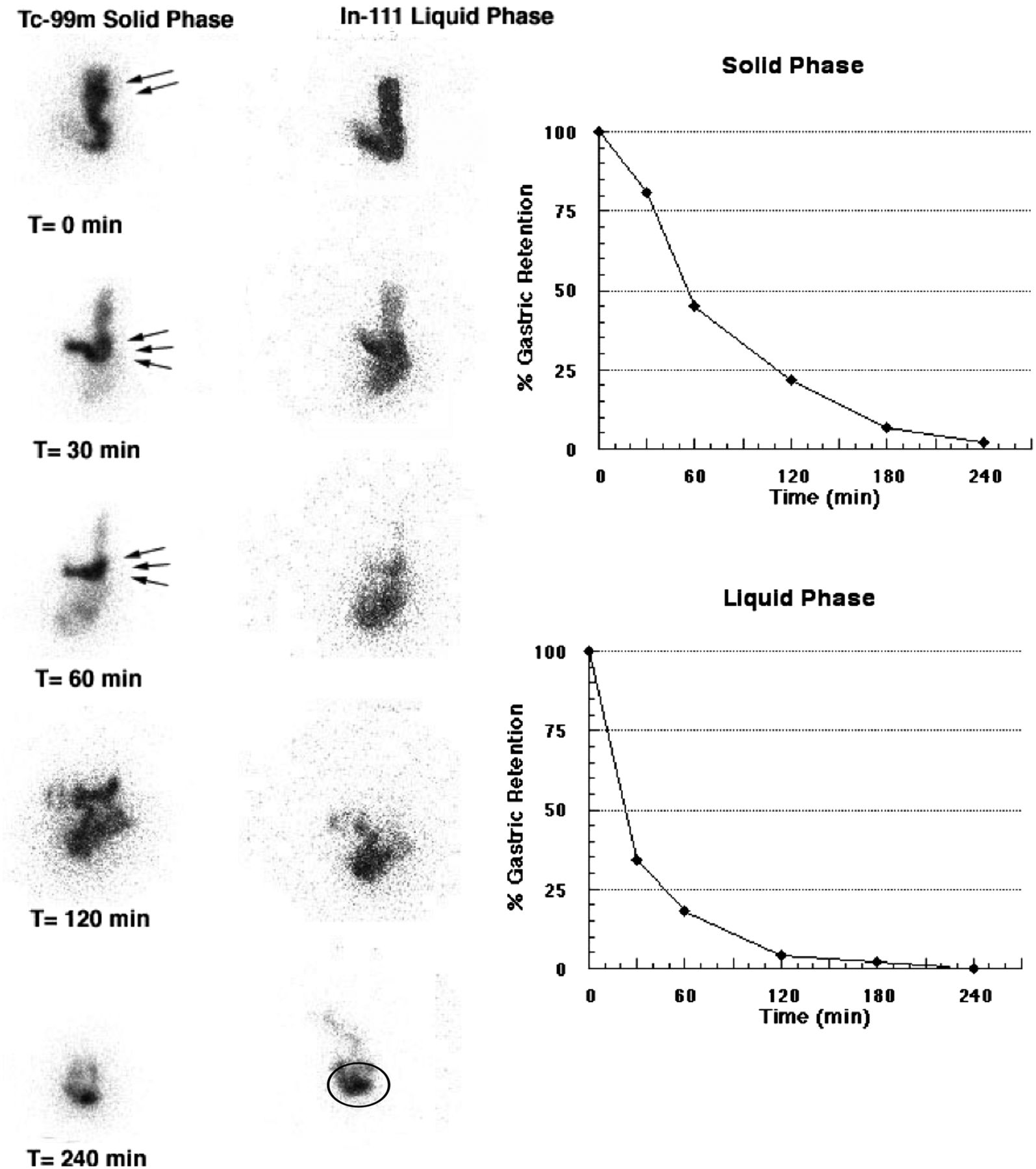
  • FIGURE 5.
    • Download figure
    • Open in new tab
    • Download powerpoint
    FIGURE 5.

    Normal dual-isotope, solid–liquid GE study (anterior views only). These images demonstrate early rapid distribution of liquids throughout stomach at 0 min. Liquid emptying curve is monoexponential. In contrast, solids show preferential early fundal localization (accommodation) (double arrows). Over time, solids progress down into antrum (triple arrows). Solid emptying curve is sigmoidal because of early lag phase for solids. Over time, one can observe small-bowel transit of solids and liquids, with buildup of activity in the terminal ileum (oval region of interest).
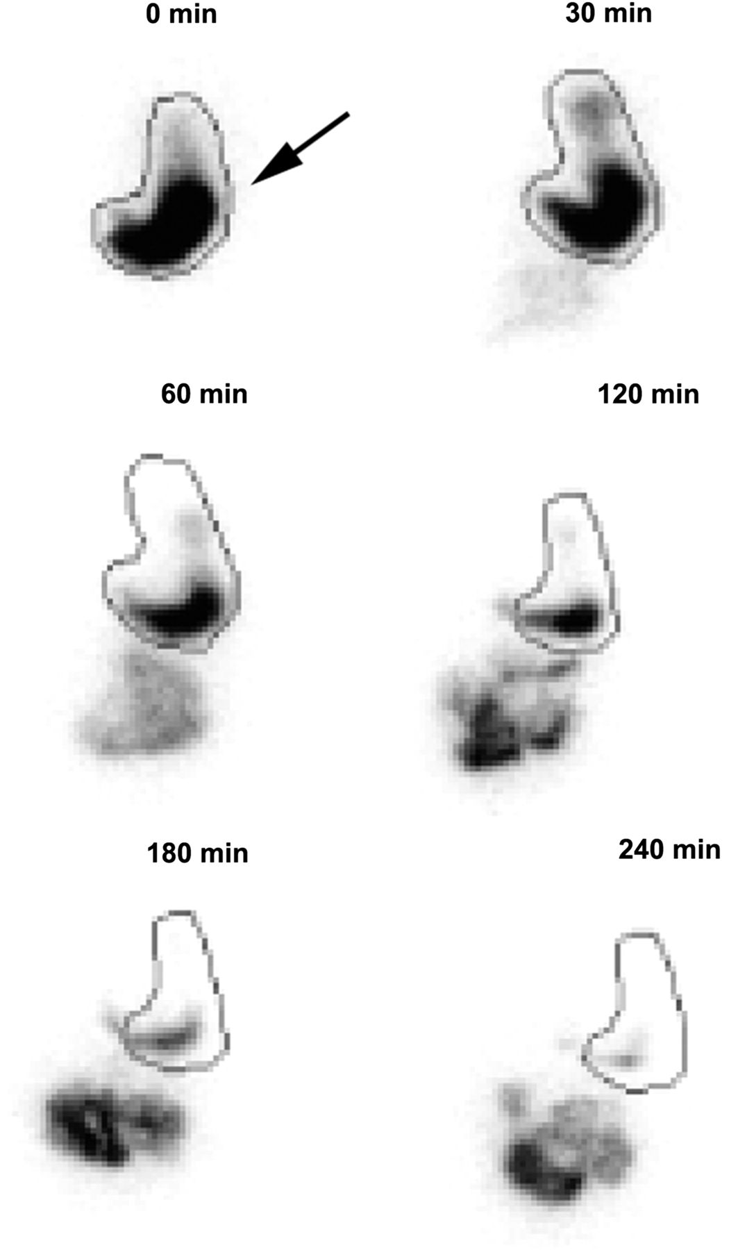
  • FIGURE 6.
    • Download figure
    • Open in new tab
    • Download powerpoint
    FIGURE 6.

    Impaired fundal accommodation (anterior views only). First postmeal ingestion image (0 min) shows lack of normal fundal accommodation, with most of meal being seen in distal stomach rather than in fundus (arrow). Overall GE was normal, with 42% of meal retained at 120 min and 8% at 240 min.

Tables

  • Figures
    • View popup
    TABLE 1

    Rome III Classification of Functional Gastrointestinal Disorders

    AdultInfantChild/adolescent
    A: EsophagealG1: Infant regurgitationH1: Vomiting and aerophagia
     A1: HeartburnG2: Infant rumination H1a: Rumination
     A2: Chest painG3: Cyclic vomiting H1b: Cyclic vomiting
     A3: DysphagiaG4: Infant colic H1c: Aerophagia
     A4: GlobusG5: Functional diarrheaH2: Abdominal pain
    B: GastroduodenalG6: Infant dyschezia H2a: Dyspepsia
     B1: DyspepsiaG7: Functional constipation H2b: Irritable bowel
     B2: Belching H2c: Abdominal migraine
     B3: Nausea/vomiting H2d: Childhood abdominal pain
     B4: RuminationH3: Constipation and incontinence
    C: Bowel H3a: Functional constipation
     C1: Irritable bowel H3b: Nonretentive fecal incontinence
     C2: Bloating
     C3: Constipation
     C4: Diarrhea
     C5: Unspecified
    D: Functional abdominal pain
    E: Biliary
    F: Anorectal
    • View popup
    TABLE 2

    Diagnostic Criteria for Esophageal Transit Scintigraphy (11)

    ConditionVisual bolus transit analysis from dynamic displayEsophageal transit timeEsophageal retention at 10 min
    NormalNormal aboral bolus transit through upper, middle, and lower thirds of esophagus with normal relaxation of lower esophageal sphincter<14 s<18%
    Nonspecific esophageal motility disorderAny localized abnormal retrograde–antegrade bolus movement (normal movement is mild, transient, and retrograde in distal esophagus before relaxation of lower esophageal sphincter, which clears rapidly)>14 s>18%
    Isolated lower esophageal sphincter dysfunctionNormal bolus transit in upper and middle esophagus with delayed transit localized at gastroesophageal junction>14 sUsually <18%; may see mild retention of <30%
    SclerodermaMarked delay in bolus transit, typically localized to distal esophagus>30 s>30%, with marked improvement when upright
    Diffuse esophageal spasmRepetitive retrograde–antegrade contractions throughout esophagus>14 sNormal or mild retention, <30%
    AchalasiaMarked delay in bolus transit throughout esophagus (may progress normally in upper esophagus from oropharyngeal propulsion)>30 s>50%, with no improvement when upright
PreviousNext
Back to top

In this issue

Journal of Nuclear Medicine Technology: 44 (1)
Journal of Nuclear Medicine Technology
Vol. 44, Issue 1
March 1, 2016
  • Table of Contents
  • About the Cover
  • Index by author
Print
Download PDF
Article Alerts
Sign In to Email Alerts with your Email Address
Email Article

Thank you for your interest in spreading the word on Journal of Nuclear Medicine Technology.

NOTE: We only request your email address so that the person you are recommending the page to knows that you wanted them to see it, and that it is not junk mail. We do not capture any email address.

Enter multiple addresses on separate lines or separate them with commas.
Gastrointestinal Motility, Part 1: Esophageal Transit and Gastric Emptying
(Your Name) has sent you a message from Journal of Nuclear Medicine Technology
(Your Name) thought you would like to see the Journal of Nuclear Medicine Technology web site.
Citation Tools
Gastrointestinal Motility, Part 1: Esophageal Transit and Gastric Emptying
Alan H. Maurer
Journal of Nuclear Medicine Technology Mar 2016, 44 (1) 1-11; DOI: 10.2967/jnumed.112.114314

Citation Manager Formats

  • BibTeX
  • Bookends
  • EasyBib
  • EndNote (tagged)
  • EndNote 8 (xml)
  • Medlars
  • Mendeley
  • Papers
  • RefWorks Tagged
  • Ref Manager
  • RIS
  • Zotero
Share
Gastrointestinal Motility, Part 1: Esophageal Transit and Gastric Emptying
Alan H. Maurer
Journal of Nuclear Medicine Technology Mar 2016, 44 (1) 1-11; DOI: 10.2967/jnumed.112.114314
del.icio.us logo Digg logo Reddit logo Twitter logo CiteULike logo Facebook logo Google logo Mendeley logo
  • Tweet Widget
  • Facebook Like
  • Google Plus One
Bookmark this article

Jump to section

  • Article
    • Abstract
    • CLINICAL INDICATIONS
    • GENERAL METHODOLOGY
    • ESOPHAGEAL TRANSIT
    • GASTRIC EMPTYING
    • ANCILLARY TESTS OF GASTRIC FUNCTION
    • CONCLUSION
    • Footnotes
    • REFERENCES
  • Figures & Data
  • Info & Metrics
  • PDF

Related Articles

  • No related articles found.
  • PubMed
  • Google Scholar

Cited By...

  • No citing articles found.
  • Google Scholar

More in this TOC Section

  • SNMMI Clinical Trials Network Research Series for Technologists: Clinical Research Primer—Use of Imaging Agents in Therapeutic Drug Development and Approval
  • Diuretic Renal Scintigraphy Protocol Considerations
  • Root Cause Analysis in Nuclear Medicine for Sentinel Events
Show more Continuing Education

Similar Articles

Keywords

  • gastrointestinal scintigraphy
  • esophageal transit scintigraphy
  • Gastric Emptying Scintigraphy
SNMMI

© 2023 Journal of Nuclear Medicine Technology

Powered by HighWire