
Main menu

  • Home
  • Content
    • Current
      • JNMT Supplement
    • Ahead of print
    • Past Issues
    • Continuing Education
    • JNMT Podcast
    • SNMMI Annual Meeting Abstracts
  • Subscriptions
    • Subscribers
    • Rates
    • Journal Claims
    • Institutional and Non-member
  • Authors
    • Submit to JNMT
    • Information for Authors
    • Assignment of Copyright
    • AQARA Requirements
  • Info
    • Reviewers
    • Permissions
    • Advertisers
    • Corporate & Special Sales
  • About
    • About Us
    • Editorial Board
    • Contact Information
  • More
    • Alerts
    • Feedback
    • Help
    • SNMMI Journals
  • SNMMI
    • JNMT
    • JNM
    • SNMMI Journals
    • SNMMI

User menu

  • Subscribe
  • My alerts
  • Log in
  • Log out
  • My Cart

Search

  • Advanced search
Journal of Nuclear Medicine Technology
  • SNMMI
    • JNMT
    • JNM
    • SNMMI Journals
    • SNMMI
  • Subscribe
  • My alerts
  • Log in
  • Log out
  • My Cart
Journal of Nuclear Medicine Technology

Advanced Search

  • Home
  • Content
    • Current
    • Ahead of print
    • Past Issues
    • Continuing Education
    • JNMT Podcast
    • SNMMI Annual Meeting Abstracts
  • Subscriptions
    • Subscribers
    • Rates
    • Journal Claims
    • Institutional and Non-member
  • Authors
    • Submit to JNMT
    • Information for Authors
    • Assignment of Copyright
    • AQARA Requirements
  • Info
    • Reviewers
    • Permissions
    • Advertisers
    • Corporate & Special Sales
  • About
    • About Us
    • Editorial Board
    • Contact Information
  • More
    • Alerts
    • Feedback
    • Help
    • SNMMI Journals
  • Watch or Listen to JNMT Podcast
  • Visit SNMMI on Facebook
  • Join SNMMI on LinkedIn
  • Follow SNMMI on Twitter
  • Subscribe to JNMT RSS feeds
Review ArticleImaging

Cholescintigraphy for Differentiation of True Duodenal Obstruction from Pseudo Obstruction

Paul Rodrigue, Pankaj Nepal and Richard Assaker
Journal of Nuclear Medicine Technology December 2020, 48 (4) 344-345; DOI: https://doi.org/10.2967/jnmt.120.246132
Paul Rodrigue
Department of Radiology, St. Vincent’s Medical Center, Bridgeport, Connecticut
  • Find this author on Google Scholar
  • Find this author on PubMed
  • Search for this author on this site
Pankaj Nepal
Department of Radiology, St. Vincent’s Medical Center, Bridgeport, Connecticut
  • Find this author on Google Scholar
  • Find this author on PubMed
  • Search for this author on this site
Richard Assaker
Department of Radiology, St. Vincent’s Medical Center, Bridgeport, Connecticut
  • Find this author on Google Scholar
  • Find this author on PubMed
  • Search for this author on this site
  • Article
  • Figures & Data
  • Info & Metrics
  • PDF
Loading

Abstract

Small-bowel obstruction is a fairly common cause of abdominal pain. Differentiating between true duodenal obstruction and pseudo obstruction is important given differing treatment strategies. Here, we present augmentation of a hepatobiliary scan with a small amount of oral liquids and delayed imaging to differentiate between these two entities.

  • gastrointestinal
  • cholecystitis
  • cholescintigraphy
  • common bile duct obstruction
  • duodenal obstruction
  • malrotation

We present the case of a 51-y-old woman with bilious vomiting and right-upper-quadrant abdominal pain. Hepatobiliary scanning showed lack of radiotracer transit to the small bowel, with mild reflux into the stomach. Giving the patient a small amount of water before obtaining delayed imaging was successful in excluding true duodenal obstruction secondary to variant anatomy such as malrotation.

CASE REPORT

A 51-y-old woman presented with upper abdominal pain, bilious vomiting, and possible gallbladder wall thickening on recent ultrasound. Abdominal CT (Fig. 1) performed during the same emergency room visit demonstrated a large calculus in the neck of the gallbladder, compressing the adjacent duodenum. Immediately after intravenous administration of 214.6 MBq (5.8 mCi) of 99mTc-mebrofenin (2-[[2-(3-bromo-2,4,6-trimethylanilino)-2-oxoethyl]-(carboxymethyl)amino]acetic acid), the patient was imaged over 60 min (Fig. 2).

FIGURE 1.
  • Download figure
  • Open in new tab
  • Download powerpoint
FIGURE 1.

Abdominal CT performed earlier during same emergency room visit demonstrated large calculus in neck of gallbladder, compressing adjacent duodenum.

FIGURE 2.
  • Download figure
  • Open in new tab
  • Download powerpoint
FIGURE 2.

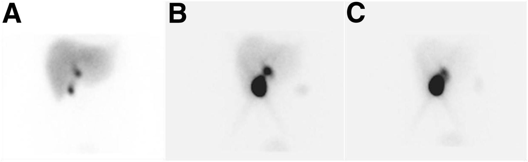
Hepatobiliary scintigraphy images after intravenous administration of 214.6 MBq (5.8 mCi) of 99mTc mebrofenin. (A) Normal uptake and excretion of radiotracer by liver, along with gallbladder and common bile duct, was visualized by 10 min after radiotracer injection. (B) At approximately 25 min, incidental reflux into stomach was visualized. (C) No bowel activity distal to common bile duct was visualized at 60 min after radiotracer injection.

The images demonstrated normal uptake and excretion of radiotracer by the liver. Radiotracer was visualized within the gallbladder and common bile duct by 10 min after injection. At approximately 25 min, incidental reflux into the stomach was visualized. No bowel activity distal to the common bile duct was visualized within the first 60 min of imaging. The patient consumed approximately 118 mL (4 oz) of water orally at the conclusion of the initial imaging and was sent back to her hospital room. She returned 120 min later for delayed images 3 h after radiotracer injection. Delayed images in the anterior projection demonstrated radiotracer activity throughout the small bowel and into the ascending colon, excluding true small-bowel obstruction and showing that the gallstone in the neck of the gallbladder was responsible for this pseudo obstruction.

DISCUSSION

99mTc-mebrofenin, an intravenously administered radiotracer, is commonly used to trace the production and flow of bile through the biliary system into the small intestine. For timely visualization of the gallbladder in adults, a minimum of 2 h and preferably 6 h of fasting is necessary before administration of the radiotracer. However, fasting for longer than 24 h, such as is seen in morbidly ill hospitalized patients, including those on total parenteral nutrition, can cause diagnostic pitfalls from nonvisualization of the gallbladder. In such cases, the patient may be pretreated with sincalide. Opioid medications also interfere with interpretive accuracy; such interference can be minimized by delaying the study for up to 4 half-lives of the medication. If needed, the opioid effect can be reversed with naloxone hydrochloride (1).

Nuclear studies using oral radiopharmaceuticals have previously been shown useful for imaging the transit of a food bolus through the gastrointestinal tract. In this instance, we offer cholescintigraphy as an alternative method for excluding true small-bowel obstruction. Numerous gastrointestinal pathologic conditions have long been described using cholescintigraphy (2,3). For patients with a history of bilious vomiting, hepatobiliary scanning may differentiate between acute cholecystitis, acute proximal gastrointestinal tract obstruction, and other undiagnosed conditions, such as malrotation (4,5). After consultation with the radiology department, the decision was made to order a hepatobiliary 99mTc-mebrofenin scan, which would offer a high sensitivity and specificity for evaluation of both these conditions (3,6). Giving the patient a small amount of water orally may help to differentiate between true upper gastrointestinal obstruction or pseudo obstruction, as was true in this case. Delayed imaging at 3 h (Fig.3) after initial injection of the radiotracer demonstrated transit of radiotracer throughout the small bowel and into the proximal colon. Subsequently, the patient was taken to the operating room for laparoscopic cholecystectomy.

FIGURE 3.
  • Download figure
  • Open in new tab
  • Download powerpoint
FIGURE 3.

Delayed hepatobiliary scintigraphy image at 3 h after initial injection of radiotracer demonstrated transit of radiotracer throughout small bowel and into proximal colon.

CONCLUSION

Augmentation of a nuclear hepatobiliary scan with a small amount of oral liquids and delayed imaging may be helpful in differentiating between true duodenal obstruction and pseudo obstruction.

DISCLOSURE

No potential conflict of interest relevant to this article was reported.

Acknowledgments

We acknowledge our patients, the great source of learning.

Footnotes

  • Published online Jul. 24, 2020.

REFERENCES

  1. 1.↵
    1. Tulchinsky M,
    2. Ciak BW,
    3. Delbeke D,
    4. et al
    . SNM practice guideline for hepatobiliary scintigraphy 4.0. J Nucl Med Technol. 2010;38:210‐218.
    OpenUrlAbstract/FREE Full Text
  2. 2.↵
    1. Fisher RS,
    2. Malmud LS
    . Scintigraphic techniques for the study of gastrointestinal motor function. Adv Intern Med. 1986;31:395–418.
    OpenUrlPubMed
  3. 3.↵
    1. Chandramouly BS,
    2. Burgess CK,
    3. Vaccaro RM
    . Diagnostic significance of unusual small intestinal pattern on technetium-99m DISIDA hepatobiliary imaging studies. Clin Nucl Med. 1987;12:852–856.
    OpenUrlPubMed
  4. 4.↵
    1. Weissmann HS,
    2. Sugarman LA,
    3. Frank MS,
    4. Freeman LM
    . Serendipity in technetium-99m dimethyl iminodiacetic acid cholescintigraphy: diagnosis of nonbiliary disorders in suspected acute cholecystitis. Radiology. 1980;135:449–454.
    OpenUrlPubMed
  5. 5.↵
    1. Fakhri AA,
    2. Hussain A,
    3. Taiyebi A,
    4. Fakhri AF
    . Duodenal obstruction on 99mTc-DISIDA cholescintigraphy: a noninvasive approach to bowel obstruction diagnosis. J Nucl Med Technol. 2016;44:265–266.
    OpenUrlAbstract/FREE Full Text
  6. 6.↵
    1. Ziessman HA
    . Hepatobiliary scintigraphy in 2014. J Nucl Med. 2014;55:967–975.
    OpenUrlAbstract/FREE Full Text
  • Received for publication May 1, 2020.
  • Accepted for publication May 27, 2020.
PreviousNext
Back to top

In this issue

Journal of Nuclear Medicine Technology: 48 (4)
Journal of Nuclear Medicine Technology
Vol. 48, Issue 4
December 1, 2020
  • Table of Contents
  • About the Cover
  • Index by author
Print
Download PDF
Article Alerts
Sign In to Email Alerts with your Email Address
Email Article

Thank you for your interest in spreading the word on Journal of Nuclear Medicine Technology.

NOTE: We only request your email address so that the person you are recommending the page to knows that you wanted them to see it, and that it is not junk mail. We do not capture any email address.

Enter multiple addresses on separate lines or separate them with commas.
Cholescintigraphy for Differentiation of True Duodenal Obstruction from Pseudo Obstruction
(Your Name) has sent you a message from Journal of Nuclear Medicine Technology
(Your Name) thought you would like to see the Journal of Nuclear Medicine Technology web site.
Citation Tools
Cholescintigraphy for Differentiation of True Duodenal Obstruction from Pseudo Obstruction
Paul Rodrigue, Pankaj Nepal, Richard Assaker
Journal of Nuclear Medicine Technology Dec 2020, 48 (4) 344-345; DOI: 10.2967/jnmt.120.246132

Citation Manager Formats

  • BibTeX
  • Bookends
  • EasyBib
  • EndNote (tagged)
  • EndNote 8 (xml)
  • Medlars
  • Mendeley
  • Papers
  • RefWorks Tagged
  • Ref Manager
  • RIS
  • Zotero
Share
Cholescintigraphy for Differentiation of True Duodenal Obstruction from Pseudo Obstruction
Paul Rodrigue, Pankaj Nepal, Richard Assaker
Journal of Nuclear Medicine Technology Dec 2020, 48 (4) 344-345; DOI: 10.2967/jnmt.120.246132
Twitter logo Facebook logo LinkedIn logo Mendeley logo
  • Tweet Widget
  • Facebook Like
  • Google Plus One
Bookmark this article

Jump to section

  • Article
    • Abstract
    • CASE REPORT
    • DISCUSSION
    • CONCLUSION
    • DISCLOSURE
    • Acknowledgments
    • Footnotes
    • REFERENCES
  • Figures & Data
  • Info & Metrics
  • PDF

Related Articles

  • No related articles found.
  • PubMed
  • Google Scholar

Cited By...

  • 2020 - A Year Like No Other
  • Google Scholar

More in this TOC Section

  • Early 10-Minute Postinjection [18F]F-FAPI-42 uEXPLORER Total-Body PET/CT Scanning Protocol for Staging Lung Cancer Using HYPER Iterative Reconstruction
  • Single- Versus Dual-Time-Point Imaging for Transthyretin Cardiac Amyloid Using 99mTc-Pyrophosphate
  • Does Arthrography Improve Accuracy of SPECT/CT for Diagnosis of Aseptic Loosening in Patients with Painful Knee Arthroplasty: A Systematic Review and Metaanalysis
Show more Imaging

Similar Articles

Keywords

  • gastrointestinal
  • cholecystitis
  • cholescintigraphy
  • common bile duct obstruction
  • duodenal obstruction
  • malrotation
SNMMI

© 2025 SNMMI

Powered by HighWire