
Main menu

  • Home
  • Content
    • Current
      • JNMT Supplement
    • Ahead of print
    • Past Issues
    • Continuing Education
    • JNMT Podcast
    • SNMMI Annual Meeting Abstracts
  • Subscriptions
    • Subscribers
    • Rates
    • Journal Claims
    • Institutional and Non-member
  • Authors
    • Submit to JNMT
    • Information for Authors
    • Assignment of Copyright
    • AQARA Requirements
  • Info
    • Reviewers
    • Permissions
    • Advertisers
    • Corporate & Special Sales
  • About
    • About Us
    • Editorial Board
    • Contact Information
  • More
    • Alerts
    • Feedback
    • Help
    • SNMMI Journals
  • SNMMI
    • JNMT
    • JNM
    • SNMMI Journals
    • SNMMI

User menu

  • Subscribe
  • My alerts
  • Log in
  • My Cart

Search

  • Advanced search
Journal of Nuclear Medicine Technology
  • SNMMI
    • JNMT
    • JNM
    • SNMMI Journals
    • SNMMI
  • Subscribe
  • My alerts
  • Log in
  • My Cart
Journal of Nuclear Medicine Technology

Advanced Search

  • Home
  • Content
    • Current
    • Ahead of print
    • Past Issues
    • Continuing Education
    • JNMT Podcast
    • SNMMI Annual Meeting Abstracts
  • Subscriptions
    • Subscribers
    • Rates
    • Journal Claims
    • Institutional and Non-member
  • Authors
    • Submit to JNMT
    • Information for Authors
    • Assignment of Copyright
    • AQARA Requirements
  • Info
    • Reviewers
    • Permissions
    • Advertisers
    • Corporate & Special Sales
  • About
    • About Us
    • Editorial Board
    • Contact Information
  • More
    • Alerts
    • Feedback
    • Help
    • SNMMI Journals
  • Watch or Listen to JNMT Podcast
  • Visit SNMMI on Facebook
  • Join SNMMI on LinkedIn
  • Follow SNMMI on Twitter
  • Subscribe to JNMT RSS feeds
Review ArticleTeaching Case Studies

18F-FDG and Amyloid PET in Dementia

Lance D Burrell and Dawn Holley
Journal of Nuclear Medicine Technology June 2018, 46 (2) 151-152; DOI: https://doi.org/10.2967/jnmt.117.205187
Lance D Burrell
1Center for Quantitative Cancer Imaging, Huntsman Cancer Institute, University of Utah, Salt Lake City, Utah; and
CNMT, PET, RT(CT)
  • Find this author on Google Scholar
  • Find this author on PubMed
  • Search for this author on this site
Dawn Holley
2Stanford University, Stanford, California
CNMT, RT(N)(CT)
  • Find this author on Google Scholar
  • Find this author on PubMed
  • Search for this author on this site
  • Article
  • Figures & Data
  • Info & Metrics
  • PDF
Loading

Abstract

We present a case of memory loss and aphasia in a 72-y-old man and discuss how PET imaging supported a diagnosis of Alzheimer dementia despite conflicting clinical findings.

  • correlative imaging
  • neurology
  • PET/CT
  • amyloid
  • dementia
  • FDG

PET imaging of the brain is useful for the clinical evaluation of dementia (1). Amyloid PET imaging adds information to the current imaging standard: 18F-FDG PET metabolic imaging (2).

CASE REPORT

A 72-y-old man presented with complaints of memory loss, aphasia, and involuntary movements. Neuropsychologic testing found multiple areas of cognitive deficit and the indication of functional decline. These findings supported a diagnosis of early dementia with an unclear etiology. Some indicators suggested Alzheimer disease as the pathologic condition, whereas others suggested frontotemporal dementia. Relatively early onset of the cognitive symptoms is more suggestive of frontotemporal dementia than of Alzheimer disease.

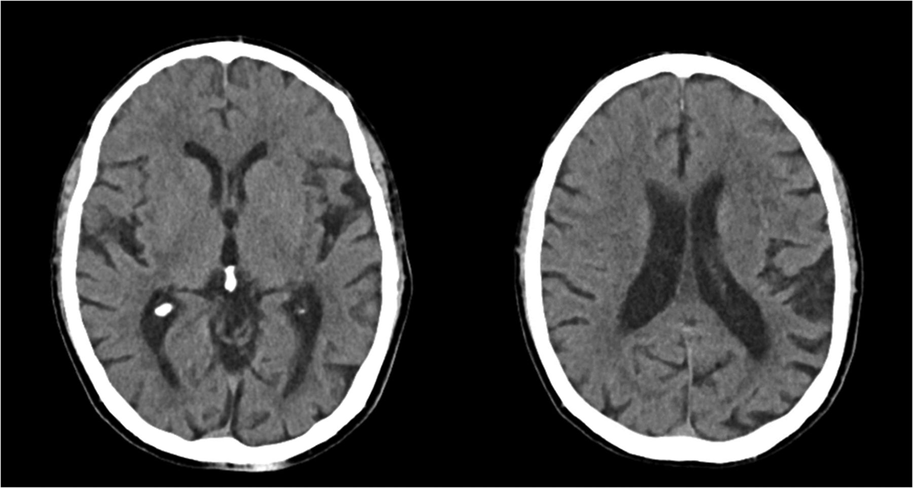
CT imaging revealed global atrophy of the cerebrum and cerebellum, without evidence of lobar-pattern dementia (Fig. 1). The report from the second clinical visit stated that this patient had an atypical dementia syndrome with prominent language deficits. Included in the differential diagnosis were language-predominant Alzheimer disease and progressive aphasia due to frontotemporal dementia. Cognitive decline had been present for at least 6 mo. A dementia syndrome was present, and the clinical criteria were met for both Alzheimer disease and frontotemporal dementia.

FIGURE 1.
  • Download figure
  • Open in new tab
  • Download powerpoint
FIGURE 1.

Midline axial CT images of brain showing cerebral atrophy.

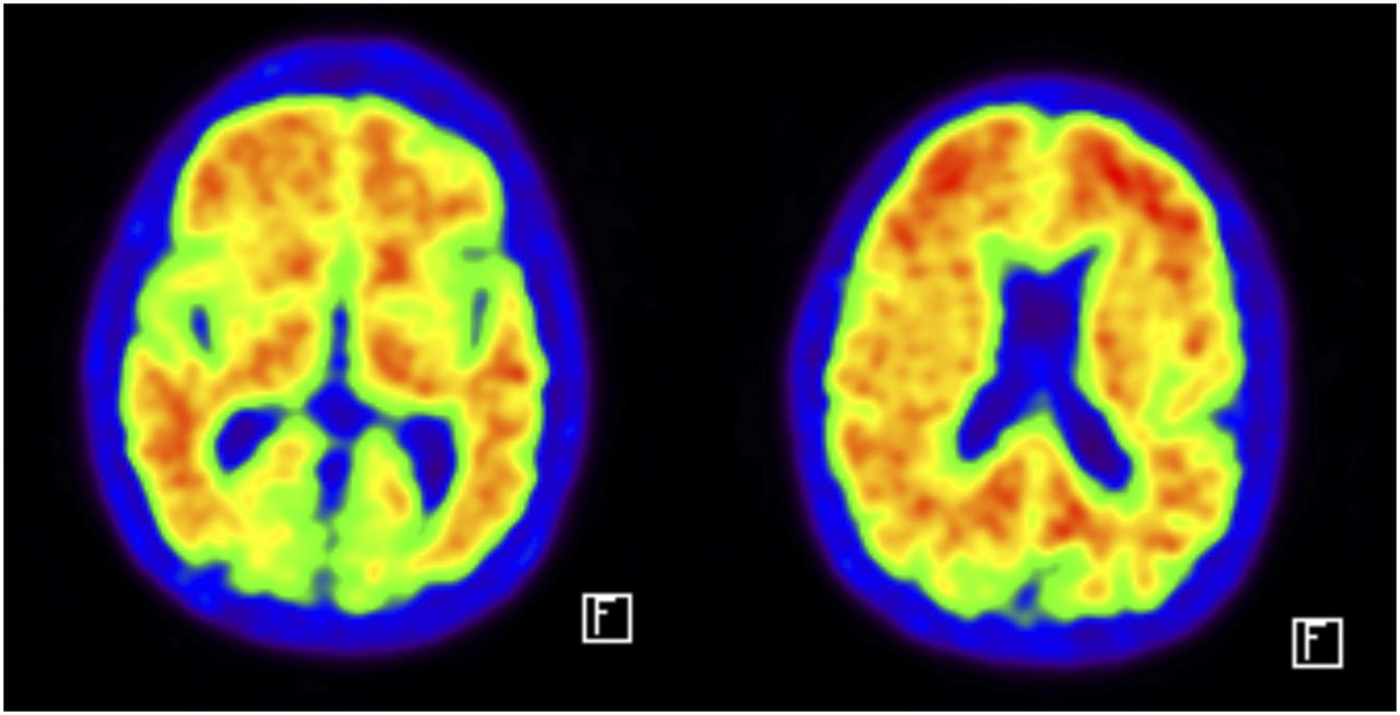
An 18F-FDG PET scan was ordered. The patient fasted for 6 h, and the resulting blood glucose level was 84 mg/dL. 18F-FDG (354.46 MBq [9.58 mCi]) was injected intravenously. Thirty minutes later, a 30-min PET/CT scan of the brain was obtained. The 18F-FDG PET report indicated that the observed metabolic pattern was not classic for a specific neurodegenerative process. Early frontotemporal dementia could not be totally excluded but was unlikely. The lack of a metabolic reduction in the frontal and anterior temporal cortex made the diagnosis of primary progressive aphasia, a subtype of frontotemporal dementia, unlikely. Early Alzheimer disease was a possible diagnosis because of the metabolic reductions noted in the left lateral temporal, parietal, and precuneus cortex (Fig. 2).

FIGURE 2.
  • Download figure
  • Open in new tab
  • Download powerpoint
FIGURE 2.

Midline axial 18F-FDG PET images of brain showing cortical metabolic reductions.

To further delineate the characteristics of this patient’s dementia, a β-amyloid PET scan was ordered. 18F-flutemetamol (Vizamyl; GE Healthcare) (177.23 MBq [4.79 mCi]) was injected intravenously. Ninety minutes later, a 20-min PET/CT scan of the brain was acquired. The scan was positive for cortical β-amyloid, with diffuse prominent uptake throughout the cerebral cortex and basal ganglia (Fig. 3).

FIGURE 3.
  • Download figure
  • Open in new tab
  • Download powerpoint
FIGURE 3.

Midline axial 18F-flutemetamol PET images of brain showing β-amyloid plaques.

DISCUSSION

PET imaging is useful in atypical cases when clinical evaluation, neuropsychologic testing, and anatomic imaging point to more than one potential diagnosis. PET imaging should be leveraged in the appropriate clinical scenario. In this case study, the patient clinically had symptoms of both Alzheimer disease and frontotemporal dementia. The information about metabolic changes that was added by 18F-FDG PET imaging aligned better with Alzheimer disease, and the β-amyloid PET imaging further supported this diagnosis. This high degree of clinical diagnostic certainty based on PET imaging, despite an atypical presentation, allowed clinicians to initiate proactive care planning.

CONCLUSION

Clinical symptoms of cognitive impairment can point to a variety of diagnoses. Appropriate use of advanced PET imaging can support clinical decision making and lead to the most accurate diagnosis for the patient.

DISCLOSURE

No potential conflict of interest relevant to this article was reported.

Acknowledgments

This case study was prepared as part of the Brain Tech TIME initiative of the SNMMI-TS.

Footnotes

  • Published online Dec. 22, 2017.

REFERENCES

  1. 1.↵
    1. Bohnen NI,
    2. Djang D,
    3. Herholz K,
    4. Anzai Y,
    5. Minoshima S
    . Effectiveness and safety of 18F-FDG PET in the evaluation of dementia: a review of the recent literature. J Nucl Med. 2012;53:59–71.
    OpenUrlAbstract/FREE Full Text
  2. 2.↵
    1. Johnson KA,
    2. Minoshima S,
    3. Bohnen N,
    4. et al
    . Appropriate use criteria for amyloid PET: a report of the Amyloid Imaging Task Force, the Society of Nuclear Medicine and Molecular Imaging, and the Alzheimer’s Association. J Nucl Med. 2013;54:476–490.
    OpenUrlAbstract/FREE Full Text
  • Received for publication November 10, 2017.
  • Accepted for publication November 27, 2017.
PreviousNext
Back to top

In this issue

Journal of Nuclear Medicine Technology: 46 (2)
Journal of Nuclear Medicine Technology
Vol. 46, Issue 2
June 1, 2018
  • Table of Contents
  • About the Cover
  • Index by author
Print
Download PDF
Article Alerts
Sign In to Email Alerts with your Email Address
Email Article

Thank you for your interest in spreading the word on Journal of Nuclear Medicine Technology.

NOTE: We only request your email address so that the person you are recommending the page to knows that you wanted them to see it, and that it is not junk mail. We do not capture any email address.

Enter multiple addresses on separate lines or separate them with commas.
18F-FDG and Amyloid PET in Dementia
(Your Name) has sent you a message from Journal of Nuclear Medicine Technology
(Your Name) thought you would like to see the Journal of Nuclear Medicine Technology web site.
Citation Tools
18F-FDG and Amyloid PET in Dementia
Lance D Burrell, Dawn Holley
Journal of Nuclear Medicine Technology Jun 2018, 46 (2) 151-152; DOI: 10.2967/jnmt.117.205187

Citation Manager Formats

  • BibTeX
  • Bookends
  • EasyBib
  • EndNote (tagged)
  • EndNote 8 (xml)
  • Medlars
  • Mendeley
  • Papers
  • RefWorks Tagged
  • Ref Manager
  • RIS
  • Zotero
Share
18F-FDG and Amyloid PET in Dementia
Lance D Burrell, Dawn Holley
Journal of Nuclear Medicine Technology Jun 2018, 46 (2) 151-152; DOI: 10.2967/jnmt.117.205187
Twitter logo Facebook logo LinkedIn logo Mendeley logo
  • Tweet Widget
  • Facebook Like
  • Google Plus One
Bookmark this article

Jump to section

  • Article
    • Abstract
    • CASE REPORT
    • DISCUSSION
    • CONCLUSION
    • DISCLOSURE
    • Acknowledgments
    • Footnotes
    • REFERENCES
  • Figures & Data
  • Info & Metrics
  • PDF

Related Articles

  • No related articles found.
  • PubMed
  • Google Scholar

Cited By...

  • No citing articles found.
  • Google Scholar

More in this TOC Section

  • High-Sensitivity Troponin Elevation in a Young Woman with Typical Chest Pain: The Heart of the Matter
  • Prominent Right Ventricular Tracer Uptake: A Harbinger of Multivessel Coronary Artery Disease
  • SPECT/CT for Discrimination Between Active and Inactive Os Trigonum in Posterior Ankle Syndrome
Show more Teaching Case Studies

Similar Articles

Keywords

  • correlative imaging
  • Neurology
  • PET/CT
  • amyloid
  • dementia
  • FDG
SNMMI

© 2025 SNMMI

Powered by HighWire