  • Main menu
  • User menu
  • Search
  • English ▼
    • English
    • Afrikaans
    • Albanian
    • Amharic
    • Arabic
    • Armenian
    • Azerbaijani
    • Basque
    • Belarusian
    • Bengali
    • Bosnian
    • Bulgarian
    • Catalan
    • Cebuano
    • Chichewa
    • Chinese (Simplified)
    • Chinese (Traditional)
    • Corsican
    • Croatian
    • Czech
    • Danish
    • Dutch
    • Esperanto
    • Estonian
    • Filipino
    • Finnish
    • French
    • Frisian
    • Galician
    • Georgian
    • German
    • Greek
    • Gujarati
    • Haitian Creole
    • Hausa
    • Hawaiian
    • Hebrew
    • Hindi
    • Hmong
    • Hungarian
    • Icelandic
    • Igbo
    • Indonesian
    • Irish
    • Italian
    • Japanese
    • Javanese
    • Kannada
    • Kazakh
    • Khmer
    • Korean
    • Kurdish (Kurmanji)
    • Kyrgyz
    • Lao
    • Latin
    • Latvian
    • Lithuanian
    • Luxembourgish
    • Macedonian
    • Malagasy
    • Malay
    • Malayalam
    • Maltese
    • Maori
    • Marathi
    • Mongolian
    • Myanmar (Burmese)
    • Nepali
    • Norwegian
    • Pashto
    • Persian
    • Polish
    • Portuguese
    • Punjabi
    • Romanian
    • Russian
    • Samoan
    • Scottish Gaelic
    • Serbian
    • Sesotho
    • Shona
    • Sindhi
    • Sinhala
    • Slovak
    • Slovenian
    • Somali
    • Spanish
    • Sudanese
    • Swahili
    • Swedish
    • Tajik
    • Tamil
    • Telugu
    • Thai
    • Turkish
    • Ukrainian
    • Urdu
    • Uzbek
    • Vietnamese
    • Welsh
    • Xhosa
    • Yiddish
    • Yoruba
    • Zulu

Main menu

  • Home
  • Content
    • Current
      • JNMT Supplement
    • Ahead of print
    • Past Issues
    • Continuing Education
    • JNMT Podcast
    • SNMMI Annual Meeting Abstracts
  • Subscriptions
    • Subscribers
    • Rates
    • Journal Claims
    • Institutional and Non-member
  • Authors
    • Submit to JNMT
    • Information for Authors
    • Assignment of Copyright
    • AQARA Requirements
  • Info
    • Reviewers
    • Permissions
    • Advertisers
    • Corporate & Special Sales
  • About
    • About Us
    • Editorial Board
    • Contact Information
  • More
    • Alerts
    • Feedback
    • Help
    • SNMMI Journals
  • SNMMI
    • JNMT
    • JNM
    • SNMMI Journals
    • SNMMI

User menu

  • Subscribe
  • My alerts
  • Log in
  • Log out
  • My Cart

Search

  • Advanced search
Journal of Nuclear Medicine Technology
  • SNMMI
    • JNMT
    • JNM
    • SNMMI Journals
    • SNMMI
  • Subscribe
  • My alerts
  • Log in
  • Log out
  • My Cart
Journal of Nuclear Medicine Technology

Advanced Search

English ▼
  • English
  • Afrikaans
  • Albanian
  • Amharic
  • Arabic
  • Armenian
  • Azerbaijani
  • Basque
  • Belarusian
  • Bengali
  • Bosnian
  • Bulgarian
  • Catalan
  • Cebuano
  • Chichewa
  • Chinese (Simplified)
  • Chinese (Traditional)
  • Corsican
  • Croatian
  • Czech
  • Danish
  • Dutch
  • Esperanto
  • Estonian
  • Filipino
  • Finnish
  • French
  • Frisian
  • Galician
  • Georgian
  • German
  • Greek
  • Gujarati
  • Haitian Creole
  • Hausa
  • Hawaiian
  • Hebrew
  • Hindi
  • Hmong
  • Hungarian
  • Icelandic
  • Igbo
  • Indonesian
  • Irish
  • Italian
  • Japanese
  • Javanese
  • Kannada
  • Kazakh
  • Khmer
  • Korean
  • Kurdish (Kurmanji)
  • Kyrgyz
  • Lao
  • Latin
  • Latvian
  • Lithuanian
  • Luxembourgish
  • Macedonian
  • Malagasy
  • Malay
  • Malayalam
  • Maltese
  • Maori
  • Marathi
  • Mongolian
  • Myanmar (Burmese)
  • Nepali
  • Norwegian
  • Pashto
  • Persian
  • Polish
  • Portuguese
  • Punjabi
  • Romanian
  • Russian
  • Samoan
  • Scottish Gaelic
  • Serbian
  • Sesotho
  • Shona
  • Sindhi
  • Sinhala
  • Slovak
  • Slovenian
  • Somali
  • Spanish
  • Sudanese
  • Swahili
  • Swedish
  • Tajik
  • Tamil
  • Telugu
  • Thai
  • Turkish
  • Ukrainian
  • Urdu
  • Uzbek
  • Vietnamese
  • Welsh
  • Xhosa
  • Yiddish
  • Yoruba
  • Zulu
  • Home
  • Content
    • Current
    • Ahead of print
    • Past Issues
    • Continuing Education
    • JNMT Podcast
    • SNMMI Annual Meeting Abstracts
  • Subscriptions
    • Subscribers
    • Rates
    • Journal Claims
    • Institutional and Non-member
  • Authors
    • Submit to JNMT
    • Information for Authors
    • Assignment of Copyright
    • AQARA Requirements
  • Info
    • Reviewers
    • Permissions
    • Advertisers
    • Corporate & Special Sales
  • About
    • About Us
    • Editorial Board
    • Contact Information
  • More
    • Alerts
    • Feedback
    • Help
    • SNMMI Journals
  • Watch or Listen to JNMT Podcast
  • Visit SNMMI on Facebook
  • Join SNMMI on LinkedIn
  • Follow SNMMI on Twitter
  • Subscribe to JNMT RSS feeds
Research ArticleRadiopharmacy & Adjunctive Medications

Can 68Ga-DOTA Peptides Be Potential Radiotracers for PET Imaging of the Spleen?

Ismet Sarikaya, Abdelhamid H. Elgazzar, Ali Sarikaya, Naheel Alnafisi and Saud Alenezi
Journal of Nuclear Medicine Technology June 2018, 46 (2) 133-135; DOI: https://doi.org/10.2967/jnmt.117.203448
Ismet Sarikaya
1Department of Nuclear Medicine, Kuwait University Faculty of Medicine, Safat, Kuwait
  • Find this author on Google Scholar
  • Find this author on PubMed
  • Search for this author on this site
Abdelhamid H. Elgazzar
1Department of Nuclear Medicine, Kuwait University Faculty of Medicine, Safat, Kuwait
  • Find this author on Google Scholar
  • Find this author on PubMed
  • Search for this author on this site
Ali Sarikaya
2Department of Nuclear Medicine, Trakya University Faculty of Medicine, Edirne, Turkey and
  • Find this author on Google Scholar
  • Find this author on PubMed
  • Search for this author on this site
Naheel Alnafisi
3Department of Nuclear Medicine, Mubarak Al-Kabeer Hospital, Jabriya, Kuwait
  • Find this author on Google Scholar
  • Find this author on PubMed
  • Search for this author on this site
Saud Alenezi
1Department of Nuclear Medicine, Kuwait University Faculty of Medicine, Safat, Kuwait
  • Find this author on Google Scholar
  • Find this author on PubMed
  • Search for this author on this site
  • Article
  • Figures & Data
  • Info & Metrics
  • PDF
Loading

Abstract

On radionuclide somatostatin receptor imaging studies, the spleen shows high physiologic uptake. Reducing the intensity of the image settings helps to better assess the distribution of radiotracer in the spleen. In our routine studies, we incidentally recognized that 68Ga-DOTANOC PET provides higher-resolution splenic images than 111In-octreotide SPECT. Autoradiography and immunohistochemistry studies have shown that somatostatin receptors are located mainly in the red pulp of the spleen. The distribution of 68Ga-DOTANOC in the spleen appears to correlate with the distribution of red pulp. In this article, we present 68Ga-DOTANOC PET/CT spleen images of our patients.

  • spleen
  • somatostatin receptor
  • red pulp
  • 68Ga-DOTANOC
  • PET/CT

Somatostatin receptor (SSTR) imaging with radiolabeled somatostatin analogs has been used since the late 1980s. It is used mainly for the detection, localization, staging, and follow-up of neuroendocrine tumors. 123I-Tyr-3-octreotide was used in early studies but later was replaced by 111In-pentetreotide (octreotide scintigraphy [OctreoScan; Mallinckrodt]). Currently, there are new 68Ga-labeled somatostatin analogs for PET imaging (1). Conventional SSTR imaging with 111In-pentetreotide is still widely used in the detection of neuroendocrine tumors, but new 68Ga-labeled PET radiotracers (68Ga-DOTATATE, 68Ga-DOTATOC, and 68Ga-DOTANOC) have been increasingly used and replacing conventional SSTR imaging in many centers because PET cameras have better properties than γ-cameras with SPECT imaging and the radiotracers have better properties than 111In-pentetreotide. Compared with 111In-pentetreotide imaging, PET with 68Ga-DOTA peptides detects more lesions, shows higher uptake in the lesions, and provides a shorter acquisition time and lower radiation exposure (2,3).

High splenic uptake on radionuclide SSTR imaging studies is a physiologic finding. Because we incidentally recognized that splenic images are of higher resolution with 68Ga-DOTANOC PET/CT than with 111In-octreotide, we decided to present spleen images of our patients in this article.

MATERIALS AND METHODS

68Ga-DOTANOC PET/CT images were obtained for 7 patients with suspected or biopsy-proven neuroendocrine tumors. Radiolabeling was performed at another institute (Kuwait Cancer Control Center). Images were obtained on a Philips time-of-flight PET/CT camera. PET images were obtained 60 min after intravenous injection of 111–185 MBq (3–5 mCi) of 68Ga-DOTANOC. Before PET image acquisition, a low-dose CT scan was obtained for attenuation correction and anatomic localization purposes. The PET acquisition was 3 min per bed position from skull base to mid thigh. The images were corrected for attenuation on the basis of the CT data, reconstructed using a standard iterative algorithm, and reformatted into transaxial, coronal, and sagittal views. Maximum-intensity-projection images were also generated. Both attenuation-corrected and uncorrected PET images, as well as PET/CT images, were visually evaluated. Because of intense uptake in the spleen, the intensity of the image settings was reduced to better assess the distribution of activity in the splenic parenchyma.

RESULTS

Five patients were male (aged 16, 28, 41, 55, and 62 y), and 2 were female (aged 48 and 73 y). Figure 1 shows a normal distribution of 68Ga-DOTANOC, Figure 2 shows basic splenic anatomy, Figure 3 shows 68Ga-DOTANOC PET/CT images of the spleen in 3 of our patients, and Figure 4 shows 111In-pentetreotide images for comparison with the 68Ga-DOTANOC images. On 68Ga-DOTANOC PET/CT images, the spleen shows high uptake with a slightly heterogeneous distribution in the parenchyma. Areas showing more prominent uptake in the parenchyma and subcapsular region are probably due to red pulp, because they correlate with the distribution of the red pulp. Small areas of lower uptake located more centrally and in the hilar region likely represent vascular structures and white pulp.

FIGURE 1.
  • Download figure
  • Open in new tab
  • Download powerpoint
FIGURE 1.

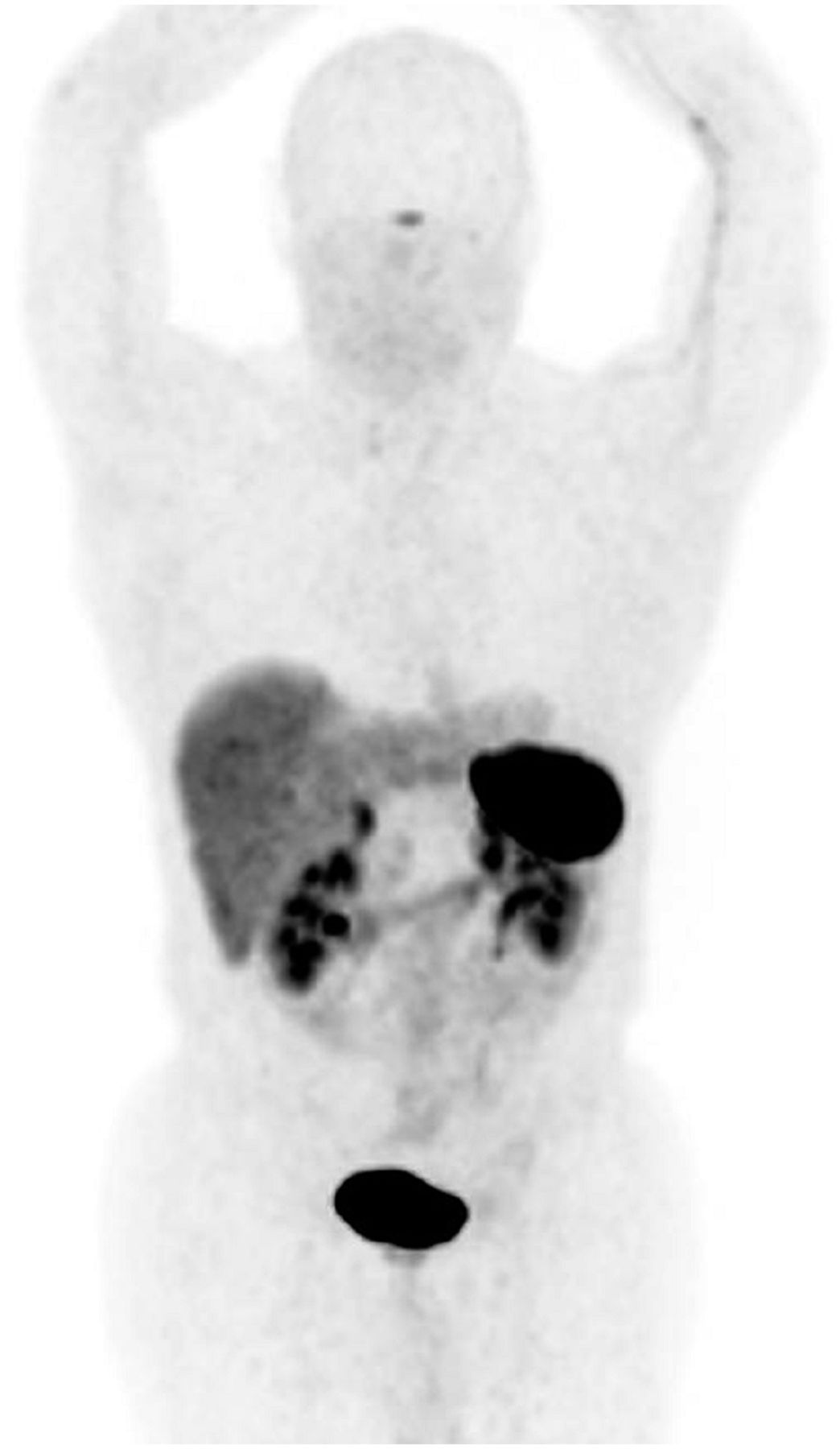
68Ga-DOTANOC whole-body PET maximum-intensity projection demonstrates normal distribution of activity in spleen, liver, pituitary gland, adrenal glands, pancreatic head, and bowel, with excreted activity in kidneys and bladder.

FIGURE 2.
  • Download figure
  • Open in new tab
  • Download powerpoint
FIGURE 2.

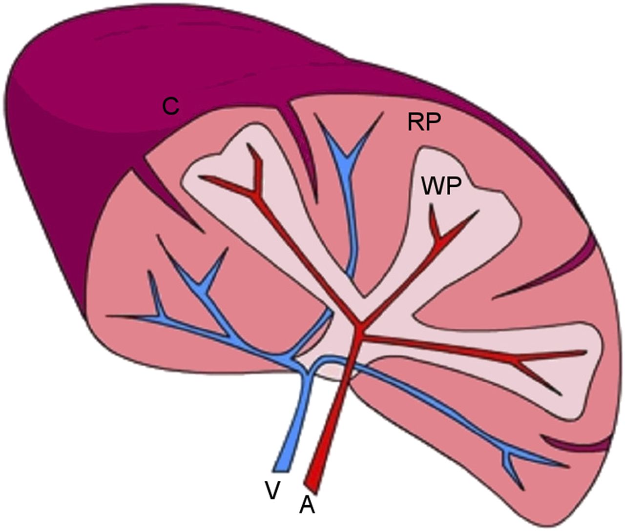
Basic anatomy of spleen: red pulp (RP), white pulp (WP), vein (V), artery (A), and splenic capsule (C).

FIGURE 3.
  • Download figure
  • Open in new tab
  • Download powerpoint
FIGURE 3.

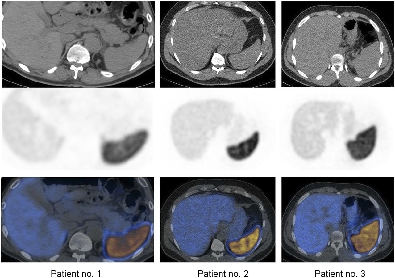
Selected transaxial CT (top), 68Ga-DOTANOC PET (middle), and 68Ga-DOTANOC PET/CT (bottom) images of 3 patients. PET demonstrates physiologic high uptake in spleen, with slight heterogeneous distribution of activity. Slightly more prominent uptake in periphery of spleen and focally throughout splenic parenchyma is likely due to subcapsular and parenchymal red pulp. Central and hilar areas showing less uptake are likely due to splenic vascular structures or white pulp.

FIGURE 4.
  • Download figure
  • Open in new tab
  • Download powerpoint
FIGURE 4.

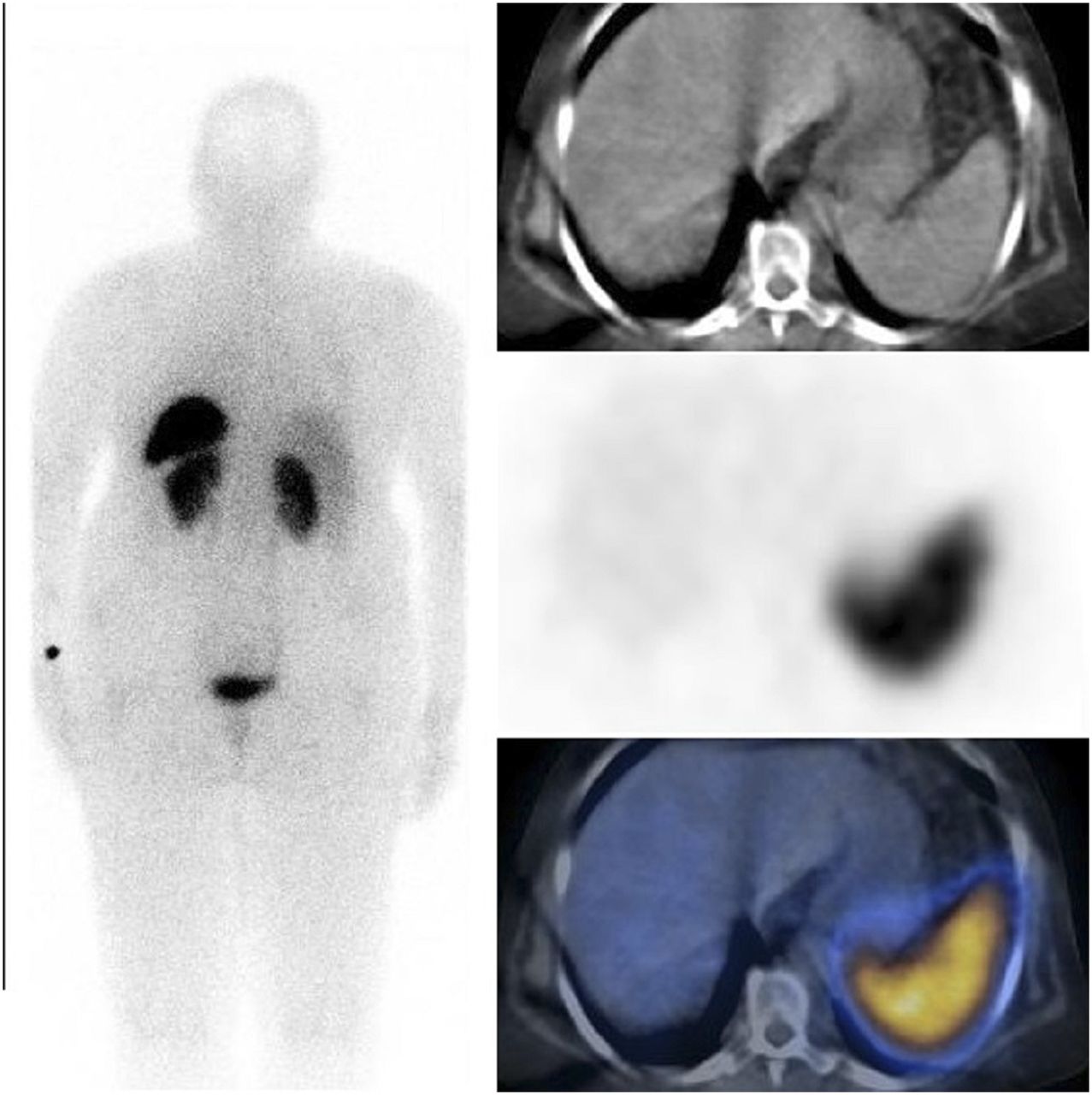
111In-pentetreotide posterior whole-body and selected transaxial CT, SPECT, and SPECT/CT images. Physiologic uptake in spleen is high and image resolution poor, compared with higher-resolution 68Ga-DOTANOC images.

In our patients, the SUVmax of the spleen and the waiting period between the 68Ga-DOTANOC injection and the acquisition start were, respectively, 41.3 and 60 min, 41.4 and 95 min, 58.8 and 76 min, 62.1 and 80 min, 67.7 and 93 min, 69 and 129 min, and 70.4 and 109 min.

DISCUSSION

The spleen has 2 main compartments, the red pulp and the white pulp (Fig. 1). The red pulp is a blood filter that removes foreign materials and damaged and aged red blood cells (RBCs) from the circulation. It is also a storage site for iron, RBCs, and platelets. White pulp surrounds the central arterioles and comprises a periarteriolar lymphoid sheath, the follicles, and the marginal zone. Immune responses to blood-borne antigens occur in the white pulp (4).

Autoradiography and immunohistochemistry studies have demonstrated that SSTRs are located mainly in the red pulp of the spleen (5–7). Reubi et al. reported that red pulp contains diffusely distributed SSTRs (7). The most abundant SSTR subtype in the spleen was SSTR2 (79.7%), followed by SSTR1 (19.6%), SSTR4 (0.6%), SSTR3 (0.1%), and SSTR5 (0.0%) (8). Quantitative reverse transcription polymerase chain reaction also has shown a significantly higher expression of SST2A messenger RNA in the spleen (9). Fluorescence immunocytochemistry has revealed somatostatin-positive cells in clusters within the white pulp and in a more dispersed pattern within the red pulp in both rats and chickens (10). In one study, there was a marked constriction of the splenic vascular bed after somatostatin administration, with a 50% decrease in blood flow; the investigators suggested that this effect of somatostatin was due to direct action on vascular receptor sites (11). Given the high amount of SSTRs in the spleen, somatostatin is expected to locate SSTRs on other sites in red pulp in addition to vascular structures.

68Ga-DOTANOC PET/CT provides high-quality images of the spleen. The distribution of 68Ga-DOTANOC appears to correlate with the distribution of red pulp in the spleen. Given the excellent splenic images provided by SSTR PET/CT with 68Ga-labeled DOTANOC or other DOTA peptides, this modality may be an alternative to standard radionuclide splenic imaging studies with 99mTc-labeled heat-damaged RBCs (selective spleen scintigraphy) or 99mTc-sulfur colloid. Although standard radionuclide splenic imaging studies are the procedure of choice to image the spleen, they have certain limitations. 99mTc-labeled heat-damaged RBC studies are laborious and time-consuming and require strict sterile technique (12). Insufficient or excessive damage to RBCs is not uncommon and can cause a suboptimal study. Colloid scans with 99mTc-sulfur colloid are less sensitive than selective spleen imaging in the identification of small splenic tissues (13). High hepatic uptake in colloid scans can mask the visualization of adjacent small splenic tissues. PET imaging of the spleen with 68Ga-DOTA peptides not only provides higher-quality images than selective spleen SPECT imaging but also is quicker and easier to perform. The radiation dose to the spleen also appears to be lower with 68Ga-DOTANOC PET imaging (without including CT) than with 99mTc-labeled heat-damaged RBC scintigraphy, at 0.0725 mGy/MBq (0.269 rad/mCi) versus 0.56 mGy/MBq (2.1 rad/mCi), respectively (14,15). However, the main limitations of PET/CT imaging of the spleen with 68Ga-DOTA peptides are the high cost of this study and the limited availability. Currently, PET/CT cameras are widely available, but preparation of these radiotracers requires a costly 68Ga generator and a radiolabeling synthesis unit.

CONCLUSION

SSTR imaging with PET radiotracers provides high-resolution splenic images, and we expect that it can be an alternative to standard radionuclide splenic imaging studies to assess morphologic abnormalities of the spleen and detect splenosis and accessory spleen. However, the main limitations of PET/CT imaging of the spleen with 68Ga-DOTA peptides are the high cost and limited availability.

DISCLOSURE

No potential conflict of interest relevant to this article was reported.

Footnotes

  • Published online Mar. 29, 2018.

REFERENCES

  1. 1.↵
    1. Becker W,
    2. Marienhagen J,
    3. Scheubel R,
    4. et al
    . Octreotide scintigraphy localizes somatostatin receptor-positive islet cell carcinomas. Eur J Nucl Med. 1991;18:924–927.
    OpenUrlPubMedGoogle Scholar
  2. 2.↵
    1. Kowalski J,
    2. Henze M,
    3. Schuhmacher J,
    4. Maecke HR,
    5. Hofmann M,
    6. Haberkorn U
    . Evaluation of positron emission tomography imaging using [68Ga]-DOTA-D-Phe1-Tyr3-octreotide in comparison to [111In]-DTPAOC SPECT: first results in patients with neuroendocrine tumors. Mol Imaging Biol. 2003;5:42–48.
    OpenUrlCrossRefPubMedGoogle Scholar
  3. 3.↵
    1. Buchmann I,
    2. Henze M,
    3. Engelbrecht S,
    4. et al
    . Comparison of 68Ga-DOTATOC PET and 111In-DTPAOC (OctreoScan) SPECT in patients with neuroendocrine tumours. Eur J Nucl Med Mol Imaging. 2007;34:1617–1626.
    OpenUrlCrossRefPubMedGoogle Scholar
  4. 4.↵
    1. Cesta MF
    . Normal structure, function, and histology of the spleen. Toxicol Pathol. 2006;34:455–465.
    OpenUrlCrossRefPubMedGoogle Scholar
  5. 5.↵
    1. Reubi JC,
    2. Waser B,
    3. Horisberger U,
    4. et al
    . In vitro autoradiographic and in vivo scintigraphic localization of somatostatin receptors in human lymphatic tissue. Blood. 1993;82:2143–2151.
    OpenUrlAbstract/FREE Full TextGoogle Scholar
  6. 6.
    1. Melis M,
    2. Kaemmerer D,
    3. de Swart J,
    4. et al
    . Localization of radiolabeled somatostatin analogs in the spleen. Clin Nucl Med. 2016;41:e111–e114.
    OpenUrlGoogle Scholar
  7. 7.↵
    1. Reubi JC,
    2. Horisberger U,
    3. Kappeler A,
    4. Laissue JA
    . Localization of receptors for vasoactive intestinal peptide, somatostatin, and substance P in distinct compartments of human lymphoid organs. Blood. 1998;92:191–197.
    OpenUrlAbstract/FREE Full TextGoogle Scholar
  8. 8.↵
    1. Boy C,
    2. Heusner TA,
    3. Poeppel TD,
    4. et al
    . 68Ga-DOTATOC PET/CT and somatostatin receptor (sst1-sst5) expression in normal human tissue: correlation of sst2 mRNA and SUVmax. Eur J Nucl Med Mol Imaging. 2011;38:1224–1236.
    OpenUrlCrossRefPubMedGoogle Scholar
  9. 9.↵
    1. Ferone D,
    2. Pivonello R,
    3. Kwekkeboom DJ,
    4. et al
    . Immunohistochemical localization and quantitative expression of somatostatin receptors in normal human spleen and thymus: implications for the in vivo visualization during somatostatin receptor scintigraphy. J Endocrinol Invest. 2012;35:528–534.
    OpenUrlPubMedGoogle Scholar
  10. 10.↵
    1. Aguila MC,
    2. Dees WL,
    3. Haensly WE,
    4. McCann SM
    . Evidence that somatostatin is localized and synthesized in lymphoid organs. Proc Natl Acad Sci USA. 1991;88:11485–11489.
    OpenUrlAbstract/FREE Full TextGoogle Scholar
  11. 11.↵
    1. Samnegård H,
    2. Tydén G,
    3. Thulin L,
    4. Friman L,
    5. Udén R
    . Effect of somatostatin on regional splanchnic blood flows in man: angiographic studies. Acta Chir Scand Suppl. 1980;500:71–73.
    OpenUrlPubMedGoogle Scholar
  12. 12.↵
    1. MacDonald A,
    2. Burrell S
    . Infrequently performed studies in nuclear medicine: part 1. J Nucl Med Technol. 2008;36:132–143.
    OpenUrlAbstract/FREE Full TextGoogle Scholar
  13. 13.↵
    1. Massey MD,
    2. Stevens JS
    . Residual spleen found on denatured red blood cell scan following negative colloid scans. J Nucl Med. 1991;32:2286–2287.
    OpenUrlAbstract/FREE Full TextGoogle Scholar
  14. 14.↵
    1. Balon HR,
    2. Brown TL,
    3. Goldsmith SJ,
    4. et al
    . The SNM practice guideline for somatostatin receptor scintigraphy 2.0. J Nucl Med Technol. 2011;39:317–324.
    OpenUrlAbstract/FREE Full TextGoogle Scholar
  15. 15.↵
    1. Pettinato C,
    2. Sarnelli A,
    3. Di Donna M,
    4. et al
    . 68Ga-DOTANOC: biodistribution and dosimetry in patients affected by neuroendocrine tumors. Eur J Nucl Med Mol Imaging. 2008;35:72–79.
    OpenUrlCrossRefPubMedGoogle Scholar
  • Received for publication October 10, 2017.
  • Accepted for publication January 6, 2018.
PreviousNext
Back to top

In this issue

Journal of Nuclear Medicine Technology: 46 (2)
Journal of Nuclear Medicine Technology
Vol. 46, Issue 2
June 1, 2018
  • Table of Contents
  • About the Cover
  • Index by author
Print
Download PDF
Article Alerts
Email Article
Citation Tools
Share
Can 68Ga-DOTA Peptides Be Potential Radiotracers for PET Imaging of the Spleen?
Ismet Sarikaya, Abdelhamid H. Elgazzar, Ali Sarikaya, Naheel Alnafisi, Saud Alenezi
Journal of Nuclear Medicine Technology Jun 2018, 46 (2) 133-135; DOI: 10.2967/jnmt.117.203448
Twitter logo Facebook logo LinkedIn logo Mendeley logo
  • Tweet Widget
Bookmark this article

Jump to section

  • Article
    • Abstract
    • MATERIALS AND METHODS
    • RESULTS
    • DISCUSSION
    • CONCLUSION
    • DISCLOSURE
    • Footnotes
    • REFERENCES
  • Figures & Data
  • Info & Metrics
  • PDF

Related Articles

  • No related articles found.
  • PubMed
  • Google Scholar

Cited By...

  • No citing articles found.
  • Google Scholar

More in this TOC Section

  • Assessment of Residual [99mTc]Tc-Tetrofosmin Activity in Routine Nuclear Medicine Practice at Hospital Kuala Lumpur
  • Investigation of the Importance of Applying Various Methods of Calculation in Determining the Blood-Absorbed Dose for Patients with Differentiated Thyroid Carcinoma
  • Influential Factors in the Preparation of 68Ga-DOTATATE
Show more Radiopharmacy & Adjunctive Medications

Similar Articles

  • TECHNOLOGIST NEWS: Technologist Awards
  • Technologist News
  • Technologist News
  • Technologist News
  • Technologist News
See more

Keywords

  • spleen
  • somatostatin receptor
  • red pulp
  • 68Ga-DOTANOC
  • PET/CT
SNMMI

© 2025 SNMMI

Powered by HighWire
Alerts for this Article
Sign In to Email Alerts with your Email Address
Email this Article

Thank you for your interest in spreading the word on Journal of Nuclear Medicine Technology.

NOTE: We only request your email address so that the person you are recommending the page to knows that you wanted them to see it, and that it is not junk mail. We do not capture any email address.

Enter multiple addresses on separate lines or separate them with commas.
Can 68Ga-DOTA Peptides Be Potential Radiotracers for PET Imaging of the Spleen?
(Your Name) has sent you a message from Journal of Nuclear Medicine Technology
(Your Name) thought you would like to see the Journal of Nuclear Medicine Technology web site.
Citation Tools
Can 68Ga-DOTA Peptides Be Potential Radiotracers for PET Imaging of the Spleen?
Ismet Sarikaya, Abdelhamid H. Elgazzar, Ali Sarikaya, Naheel Alnafisi, Saud Alenezi
Journal of Nuclear Medicine Technology Jun 2018, 46 (2) 133-135; DOI: 10.2967/jnmt.117.203448

Citation Manager Formats

  • BibTeX
  • Bookends
  • EasyBib
  • EndNote (tagged)
  • EndNote 8 (xml)
  • Medlars
  • Mendeley
  • Papers
  • RefWorks Tagged
  • Ref Manager
  • RIS
  • Zotero

We use cookies on this site to enhance your user experience

By clicking any link on this page you are giving your consent for us to set cookies.