  • Main menu
  • User menu
  • Search
  • English ▼
    • English
    • Afrikaans
    • Albanian
    • Amharic
    • Arabic
    • Armenian
    • Azerbaijani
    • Basque
    • Belarusian
    • Bengali
    • Bosnian
    • Bulgarian
    • Catalan
    • Cebuano
    • Chichewa
    • Chinese (Simplified)
    • Chinese (Traditional)
    • Corsican
    • Croatian
    • Czech
    • Danish
    • Dutch
    • Esperanto
    • Estonian
    • Filipino
    • Finnish
    • French
    • Frisian
    • Galician
    • Georgian
    • German
    • Greek
    • Gujarati
    • Haitian Creole
    • Hausa
    • Hawaiian
    • Hebrew
    • Hindi
    • Hmong
    • Hungarian
    • Icelandic
    • Igbo
    • Indonesian
    • Irish
    • Italian
    • Japanese
    • Javanese
    • Kannada
    • Kazakh
    • Khmer
    • Korean
    • Kurdish (Kurmanji)
    • Kyrgyz
    • Lao
    • Latin
    • Latvian
    • Lithuanian
    • Luxembourgish
    • Macedonian
    • Malagasy
    • Malay
    • Malayalam
    • Maltese
    • Maori
    • Marathi
    • Mongolian
    • Myanmar (Burmese)
    • Nepali
    • Norwegian
    • Pashto
    • Persian
    • Polish
    • Portuguese
    • Punjabi
    • Romanian
    • Russian
    • Samoan
    • Scottish Gaelic
    • Serbian
    • Sesotho
    • Shona
    • Sindhi
    • Sinhala
    • Slovak
    • Slovenian
    • Somali
    • Spanish
    • Sudanese
    • Swahili
    • Swedish
    • Tajik
    • Tamil
    • Telugu
    • Thai
    • Turkish
    • Ukrainian
    • Urdu
    • Uzbek
    • Vietnamese
    • Welsh
    • Xhosa
    • Yiddish
    • Yoruba
    • Zulu

Main menu

  • Home
  • Content
    • Current
      • JNMT Supplement
    • Ahead of print
    • Past Issues
    • Continuing Education
    • JNMT Podcast
    • SNMMI Annual Meeting Abstracts
  • Subscriptions
    • Subscribers
    • Rates
    • Journal Claims
    • Institutional and Non-member
  • Authors
    • Submit to JNMT
    • Information for Authors
    • Assignment of Copyright
    • AQARA Requirements
  • Info
    • Reviewers
    • Permissions
    • Advertisers
    • Corporate & Special Sales
  • About
    • About Us
    • Editorial Board
    • Contact Information
  • More
    • Alerts
    • Feedback
    • Help
    • SNMMI Journals
  • SNMMI
    • JNMT
    • JNM
    • SNMMI Journals
    • SNMMI

User menu

  • Subscribe
  • My alerts
  • Log in
  • My Cart

Search

  • Advanced search
Journal of Nuclear Medicine Technology
  • SNMMI
    • JNMT
    • JNM
    • SNMMI Journals
    • SNMMI
  • Subscribe
  • My alerts
  • Log in
  • My Cart
Journal of Nuclear Medicine Technology

Advanced Search

English ▼
  • English
  • Afrikaans
  • Albanian
  • Amharic
  • Arabic
  • Armenian
  • Azerbaijani
  • Basque
  • Belarusian
  • Bengali
  • Bosnian
  • Bulgarian
  • Catalan
  • Cebuano
  • Chichewa
  • Chinese (Simplified)
  • Chinese (Traditional)
  • Corsican
  • Croatian
  • Czech
  • Danish
  • Dutch
  • Esperanto
  • Estonian
  • Filipino
  • Finnish
  • French
  • Frisian
  • Galician
  • Georgian
  • German
  • Greek
  • Gujarati
  • Haitian Creole
  • Hausa
  • Hawaiian
  • Hebrew
  • Hindi
  • Hmong
  • Hungarian
  • Icelandic
  • Igbo
  • Indonesian
  • Irish
  • Italian
  • Japanese
  • Javanese
  • Kannada
  • Kazakh
  • Khmer
  • Korean
  • Kurdish (Kurmanji)
  • Kyrgyz
  • Lao
  • Latin
  • Latvian
  • Lithuanian
  • Luxembourgish
  • Macedonian
  • Malagasy
  • Malay
  • Malayalam
  • Maltese
  • Maori
  • Marathi
  • Mongolian
  • Myanmar (Burmese)
  • Nepali
  • Norwegian
  • Pashto
  • Persian
  • Polish
  • Portuguese
  • Punjabi
  • Romanian
  • Russian
  • Samoan
  • Scottish Gaelic
  • Serbian
  • Sesotho
  • Shona
  • Sindhi
  • Sinhala
  • Slovak
  • Slovenian
  • Somali
  • Spanish
  • Sudanese
  • Swahili
  • Swedish
  • Tajik
  • Tamil
  • Telugu
  • Thai
  • Turkish
  • Ukrainian
  • Urdu
  • Uzbek
  • Vietnamese
  • Welsh
  • Xhosa
  • Yiddish
  • Yoruba
  • Zulu
  • Home
  • Content
    • Current
    • Ahead of print
    • Past Issues
    • Continuing Education
    • JNMT Podcast
    • SNMMI Annual Meeting Abstracts
  • Subscriptions
    • Subscribers
    • Rates
    • Journal Claims
    • Institutional and Non-member
  • Authors
    • Submit to JNMT
    • Information for Authors
    • Assignment of Copyright
    • AQARA Requirements
  • Info
    • Reviewers
    • Permissions
    • Advertisers
    • Corporate & Special Sales
  • About
    • About Us
    • Editorial Board
    • Contact Information
  • More
    • Alerts
    • Feedback
    • Help
    • SNMMI Journals
  • Watch or Listen to JNMT Podcast
  • Visit SNMMI on Facebook
  • Join SNMMI on LinkedIn
  • Follow SNMMI on Twitter
  • Subscribe to JNMT RSS feeds
Research ArticleImaging

Metabolic Signature on 18F-FDG PET/CT, HER2 Status, and Survival in Gastric Adenocarcinomas

Romulo Celli, Monica Colunga, Natalie Patel, Mehdi Djekidel and Dhanpat Jain
Journal of Nuclear Medicine Technology December 2016, 44 (4) 234-238; DOI: https://doi.org/10.2967/jnmt.116.181479
Romulo Celli
1Department of Pathology, Yale School of Medicine, New Haven, Connecticut
  • Find this author on Google Scholar
  • Find this author on PubMed
  • Search for this author on this site
Monica Colunga
2Sciences Po, Paris, France; and
  • Find this author on Google Scholar
  • Find this author on PubMed
  • Search for this author on this site
Natalie Patel
1Department of Pathology, Yale School of Medicine, New Haven, Connecticut
  • Find this author on Google Scholar
  • Find this author on PubMed
  • Search for this author on this site
Mehdi Djekidel
3QMC Quality Medical Consulting, Hamden, Connecticut
  • Find this author on Google Scholar
  • Find this author on PubMed
  • Search for this author on this site
Dhanpat Jain
1Department of Pathology, Yale School of Medicine, New Haven, Connecticut
  • Find this author on Google Scholar
  • Find this author on PubMed
  • Search for this author on this site
  • Article
  • Figures & Data
  • Info & Metrics
  • PDF
Loading

Abstract

The human epidermal growth factor 2 (HER2)–overexpressing (HER2-positive [HER2+]) gastric (GC) and gastroesophageal junction adenocarcinomas (GEJC) are felt to represent a more aggressive form of disease, which may correlate to increased metabolic activity. Whether tumor SUVmax measured by 18F-FDG PET/CT could be a preoperative parameter used to predict HER2 status of GC/GEJC is unknown. Methods: Pathology reports of HER2+ GC/GEJC biopsies and resections from 31 patients were reviewed and compared with HER2-negative (HER2−) cases distributed evenly over the same time period. We analyzed their SUVmax intensity and then compared the HER2 status and SUVmax parameters and their association with survival. Results: After matching for age and sex, there was no difference in SUVmax between HER2+ and HER2− cases (9.7 and 8.4, respectively; P = 0.6). No difference was seen between HER2+ and HER2− cases in tumor histology (81% and 57% intestinal type, respectively; P = 0.11), size (2.6 and 3.8 cm, respectively; P = 0.12), differentiation (47% and 68% poorly differentiated, respectively; P = 0.06), or presence of lymph node metastasis (60% and 40%, respectively; P = 0.3). Although there was no difference in survival demonstrated by HER2+ and HER2− cases, there was a significant difference in survival between SUVmax above (12.2 mo) and below (30 mo) the median SUVmax (6.6, P = 0.01). Conclusion: Our study shows that SUVmax is not associated with HER2 status of GC/GEJC. Independent of HER2 overexpression, patients with a high SUVmax demonstrate a worse overall survival, suggesting that metabolic signature is a better predictor of biologic tumor aggressiveness than its histologic signature.

  • metabolic signature
  • gastric carcinoma
  • HER2
  • 18F-FDG PET/CT

Gastric cancer represents the fifth most common carcinoma worldwide and is also highly prevalent in the United States (1,2). It often presents at an advanced stage, and appropriate diagnosis and treatment decisions follow a time-sensitive course. Human epidermal growth factor-2 (HER2, also known as ERB2) is a tyrosine kinase member of the epidermal growth factor receptor family, known for its key role in driving oncogenesis in numerous human cancers including breast and gastric carcinoma (1–3). One intervention that has shown to increase progression-free and overall survival in patients with HER2-overexpressing (HER-positive [HER2+]) gastric and gastroesophageal junction carcinoma (GC/GEJC) is the addition of trastuzumab to a standard adjuvant chemotherapy protocol (3). Addition of targeted anti-HER2 therapy using this recombinant monoclonal antibody to standard chemotherapy has become the standard of treatment in advanced HER2+ GC/GEJC (4). For these reasons, established clinical recommendations include a turnaround time for HER2 assessment studies (immunohistochemistry and fluorescence in situ hybridization) of no longer than 5 d (5). Adjunct biomarkers of GC/GEJC that could lead to earlier diagnosis, prognosis, and personalization of therapy could potentially decrease mortality.

Pretreatment 18F-FDG PET/CT studies are used for staging purposes for several human cancers. This list includes carcinomas of breast, colon, esophagus, head and neck, and lung as well as lymphomas and melanoma (6). The amount of 18F-FDG uptake reflects a tissue’s level of metabolic activity, reported semiquantitatively as the SUVmax. The SUVmax level (or metabolic signature) correlates with the metabolic activity of the particular tissue or target lesion. Several authors have suggested that HER2+ GC/GEJCs represent a more aggressive form of disease and portend a worse prognosis (7–9). However, HER2+ status in GC/GEJC has not yet been correlated with this metabolic signature.

The objective of this study was to assess the predictive role of SUVmax for HER2 expression in cases of GC/GEJC. In addition, we compared the ability of cancer SUVmax and HER2 status to predict mortality. In doing so, we tested the comparative abilities of imaging and pathology in assessing tumor aggressiveness in a cohort of GC/GEJC patients.

MATERIALS AND METHODS

The institutional review board (or equivalent) approved this retrospective study, and the requirement to obtain informed consent was waived. A retrospective search of our pathology database over a 4-y period (2011–2015) revealed 31 total HER2+ GC and GEJC cases, all confirmed by fluorescence in situ hybridization. HER2 testing was performed either on a biopsy or on the corresponding resection for each patient.

We identified 65 random cases of HER2-negative (HER2−)GC/GEJCs distributed evenly over the same time period. As per institutional protocol and recommended practice (5), these cases tested negative by immunohistochemistry and were never tested by fluorescence in situ hybridization.

Patient baseline clinical characteristics including age and sex were recorded. The outcomes of interest were disease-related mortality and time to death (mo).

Pathologic Criteria

All GC and GEJC diagnoses were rendered by specialty-trained gastrointestinal pathologists. The following pathologic parameters, when available, were collected from the surgical pathology report of cases that underwent resections: tumor size (from resected tumors with gross anatomic descriptions), histologic diagnosis, tumor differentiation, and presence of lymph node metastasis. There was not information available for some of these variables because a subset of cases tested for HER2 were biopsies (28 total HER2+ and 41 total HER−), and the corresponding resections either had not been performed or had been performed at an outside institution.

All cases were re-reviewed to confirm the presence of carcinoma and HER2 status by immunohistochemistry. HER2 immunohistochemistry was performed using the HercepTest A0485 (Dako) antibody. HER2 immunohistochemistry stain scoring was interpreted in accordance with the parameters used in the Trastuzumab for Gastric (TOGA) clinical trial (3) and proposed by Hoffman et al. (10). Using the criteria postulated by the TOGA trial, we characterized HER2+ cases as 3+ staining by immunohistochemistry or 2+ staining by immunohistochemistry with a concomitant positive fluorescence in situ hybridization with a HER2–to–chromosome 17 ratio of 2.2 or more.

Radiologic Criteria

PET/CT imaging was performed after the diagnosis of carcinoma was established by pathology and for the purpose of pretreatment staging. All PET/CT studies were performed on the latest generation of scanners, 2 Discovery PET/CT machines (Discovery DST-E and the Discovery D690 PET/CT [GE Healthcare]). Both have similar lutetium-yttrium oxyorthosilicate crystals and reconstruction protocols. A standard oncologic PET imaging using a 3-dimensional acquisition protocol was performed with administered 18F-FDG doses of approximately 555 MBq (15 mCi) and an approximately 60-min but no later than 90-min postinjection delay. No intravenous contrast was administered. Blood sugars were measured in all patients, and scans were rescheduled per protocol if blood sugar was greater than 150. All SUVmax measurements were recalculated from clinical scans by an experienced nuclear medicine physician using a similar technique. Regions of interest were drawn over target lesions using an automated image segmentation threshold Advantage Workstation technique (GE Healthcare). This is a Food and Drug Administration–approved and routinely clinically used technique.

Data Analysis

Continuous variables were expressed as a mean and SD. Comparison of means was performed using the Student t test. Proportions were compared using the Fisher exact test. Kaplan–Meier analysis was performed using the survival data to compare patients with differential HER2 expression. The SUVmax median value was calculated, and SUVmax below and above the median value were used for its relationship to mortality. Data analysis was performed using the STATA statistical software program (StataCorp. 2015. Stata Statistical Software: Release 14; StataCorp LP.).

RESULTS

The clinical characteristics of the 96 patients in the study are summarized in Table 1. There was no difference in patient’s age (70 and 67 y, respectively; P = 0.23) or sex (77% and 69% men, respectively; P = 0.61) between HER2+ and HER2− GC/GEJC cases.

View this table:
  • View inline
  • View popup
TABLE 1

Clinical Characteristics

The mean SUVmax (±SD) of HER2+ cancers (9.7 ± 6.3) was similar to that of HER2− cases (8.4 ± 5.4) (P = 0.6) as shown in Figure 1. There was also no difference in tumor size among cases with differential HER2+ expression (2.6 and 3.8 cm, respectively; P = 0.06).

FIGURE 1.
  • Download figure
  • Open in new tab
  • Download powerpoint
FIGURE 1.

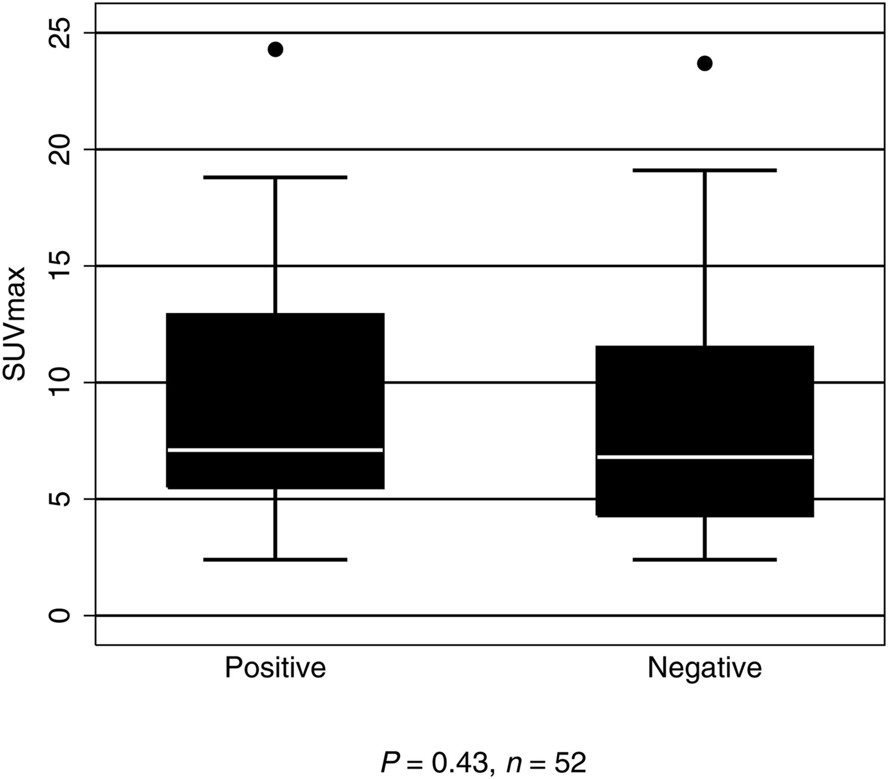
SUVmax, by HER2 status.

No difference was seen between HER2+ and HER2− cases with regards to tumor histology (81% and 57% intestinal type, respectively; P = 0.11), tumor differentiation (47% and 68% poorly differentiated, respectively; P = 0.06), or presence of lymph node metastases (60% and 40% with metastases, respectively; P = 0.23). The pathologic characteristics of the cases studied are summarized in Table 2.

View this table:
  • View inline
  • View popup
TABLE 2

Tumor Pathologic Characteristics

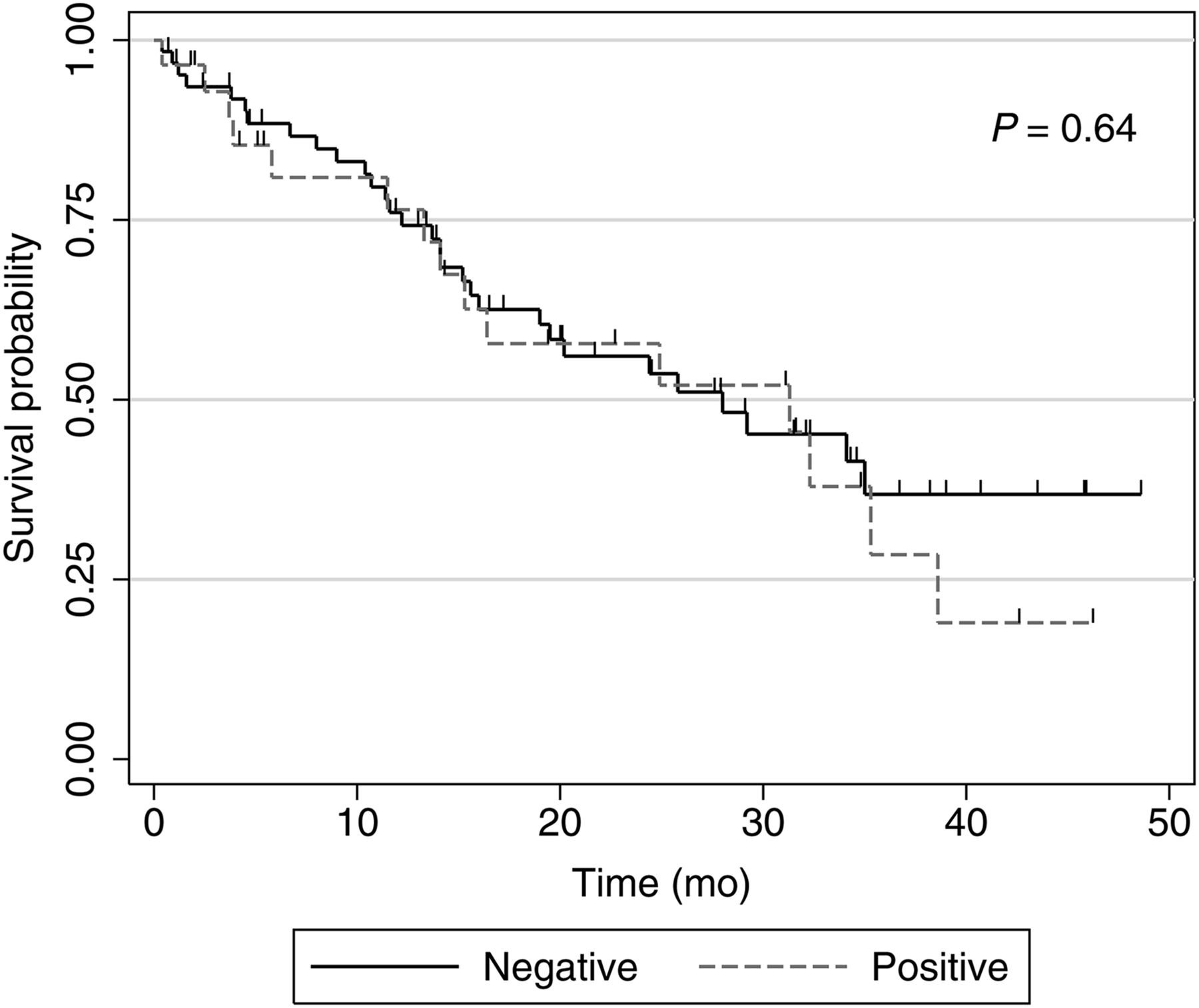
The Kaplan–Meier analysis did not show any survival difference between patients with HER2+ and HER2− cancers (P = 0.64, Fig. 2).

FIGURE 2.
  • Download figure
  • Open in new tab
  • Download powerpoint
FIGURE 2.

Overall survival, by HER2 status.

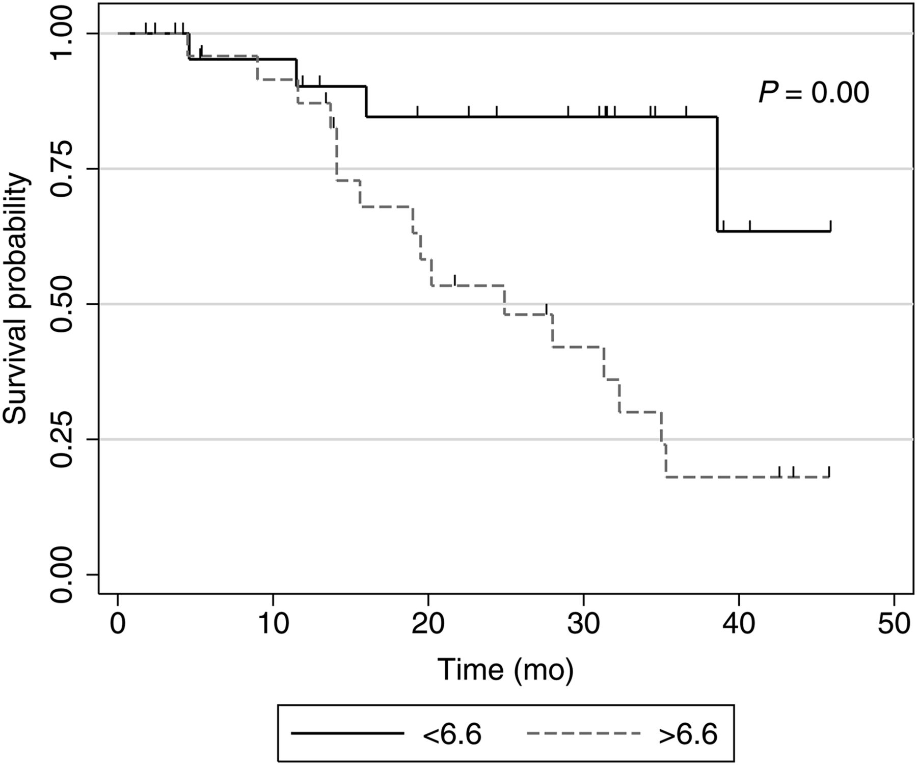
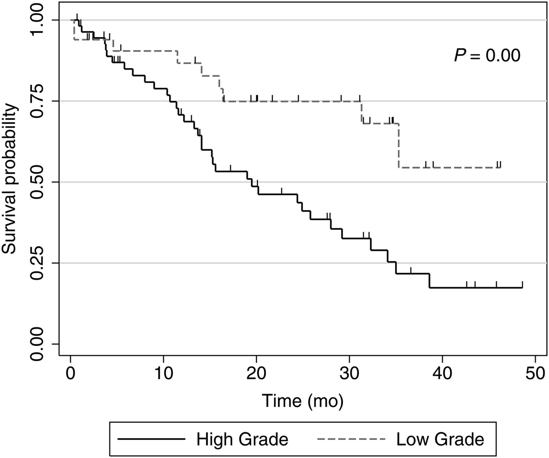
However, there was a statistically significant difference in overall survival between the patients with high tumor metabolic signatures (SUVmax above the median of 6.6) compared with those with metabolic signatures lower than the median SUVmax. The cumulative incidence of death for tumors with high metabolic signatures was 60% compared with 18% in patients with low metabolic signature (P = 0.00), during the study period (Fig. 3). The mean SUVmax of patients who survived during the study period was 7.6 (±5.4) mo versus 10.0 (±4.2) mo among patients who died. Additionally, Kaplan–Meier analysis demonstrated a significant association of survival with tumor differentiation, with poorly differentiated tumors more likely to be associated with death (61% cumulative death incidence vs. 28% in moderately differentiated, P = 0.02, Fig. 4). This was also statistically significant when the tumors were grouped into high-grade tumors (poorly differentiated and anaplastic tumors) and low-grade tumors (moderately and well-differentiated tumors). Patients with high-grade tumors showing an SUVmax above 6.6 had a mean survival of 19.0 mo, whereas those with low-grade tumors and SUVmax below 6.6 had a mean survival of 26.9 mo (sample too small to establish statistical significance). Tumor differentiation and SUVmax were found not to be independent predictors of survival by multivariate analysis. Two tumors in the cohort were well differentiated, and their mean SUVmax was 2.4 (neither was above 6.6).

FIGURE 3.
  • Download figure
  • Open in new tab
  • Download powerpoint
FIGURE 3.

Overall survival, by SUVmax (above and below median SUVmax of 6.6).

FIGURE 4.
  • Download figure
  • Open in new tab
  • Download powerpoint
FIGURE 4.

Overall survival, by tumor differentiation.

DISCUSSION

At present, treatment decisions for GC/GEJC follow pathologic assessment of the tissue specimen. In an era of expanding use of efficient noninvasive diagnostic methods, an imaging biomarker used to predict HER2 overexpression in tumors would be a welcome tool, particularly if it could contribute toward treatment decisions. This is of particular importance because trastuzumab is increasingly being used in a neoadjuvant role for HER2+ GC/GEJC based on the level of HER2 expression (11,12). The criteria for HER2 positivity require only a small proportion of cells (10% in resections, and only 5 clustered cells in biopsies) to be positive, and heterogeneity of HER2 expression is well recognized, which can lead to false-negative results in small biopsies (3,13). Trastuzumab is expensive and its use is not without risk and is known to potentially cause significant cardiac side effects. Thus, finding noninvasive measures that may help predict response to HER2-directed therapies that also overcome sampling issues inherent in small biopsies can be of clinical value (14).

Chen et al. (15) recently identified a relationship between HER2 positivity and decreased SUVmax in gastric cancers of all anatomic subtypes; however, to our knowledge this is the first study comparing the radiologic SUVmax from patients with HER2+ and HER2− GC/GEJC matched by age, sex, tumor size, and histology. There was no significant difference in SUVmax between the 2 groups, and hence the metabolic signature cannot be used to predict HER2 expression status of GC/GEJC on initial staging. However, there was a significant association of high SUVmax with worse survival, with patients with SUVmax above 6.6 showing a mean survival of 12.2 mo. In addition, the mean SUVmax of all patients who died was higher (10.0 vs. 7.7), and the cumulative likelihood of death with an SUVmax more than 6.6 was 60% over 4 y. Taken together, these findings suggest that the metabolic signature (SUVmax) is a useful indicator of biologic aggressiveness and survival. This finding is not entirely surprising, having been replicated previously in gastric carcinoma (16–18). When adding the variable of tumor differentiation (identified at biopsy), cases with an SUVmax above 6.6 and poor differentiation (high grade) showed a shorter interval to death than low-grade tumors, although this trend was not significant.

The strength of this study rests on its direct comparison between the radiologic and pathologic parameters in 1 cohort using prognosis as the outcome. Moreover, we found no difference in tumor size, histologic type, or lymph node metastasis among HER2+ and HER2− cases, all of which are parameters that have been shown to demonstrate differential 18F-FDG uptake (17,19–21). Our study does not address the pathobiologic link between HER2 overexpression and tumor metabolic activity. If HER2 overexpression is indeed associated with increased gastric tumor metabolic activity, as has been shown previously in the breast (22), then the lack of a difference between HER2 groups in SUVmax could reflect tumor heterogeneity. If true, this reflects a significant weakness in the current HER2 testing on limited biopsy samples. Alternatively, gastric HER2+ overexpression may have no correlation with increased glycolysis or glucose transporter expression, pathobiologic factors that have been shown to be independently linked to higher 18F-FDG uptake in the GC/GEJ (19–21,23). Additional studies examining the relationship of HER2 and mechanisms of metabolism in gastric cancer are required. An additional caveat is that the metabolic signature of tumors can show variability after treatment as noted in a previous study, and this may also be a valuable tool in assessing the molecular genotype (24), survival, or treatment strategies at the initial staging or restaging phase.

Our study did not show any difference in several clinicopathologic parameters including patient age, sex, or tumor size with regards to HER2+ and HER2− status of the patients. These findings are similar to many previous studies, but other, discrepant findings have also been reported in the literature (13,25–30). Recent meta-analyses also show conflicting conclusions regarding effect of HER2 expression on patient survival (8,13). Indeed, the correlation of HER2 expression in gastric carcinoma with various clinicopathologic parameters and survival remains controversial. Chief among cited reasons for these discrepancies is variability in pathologic HER2 assessment methodologies (29). The immunohistochemistry and fluorescence in situ hybridization methods used in our laboratory have been validated and reproduced, and are currently considered standard of care (5). HER2 overexpression clearly characterizes a subset of GC/GEJC that benefits from targeted therapy, but the clinical and pathologic consequences of this finding still remain largely unclear.

Our study has some limitations. The cohort, particularly the HER2+ patients, is small for a comparison study. This can be explained in part because the prevalence of HER2+ gastric cases in our hands is relatively low compared with the others reported in the literature (25,31). This may be due to several preanalytical variables, such as the antibody used for immunohistochemistry. Although, the performance of the Dako HercepTest A0485 antibody seems comparable with the Ventana 4B5 (32), a recent study that directly compared results of these antibodies showed a slightly better performance with the Ventana system (33). For the purposes of this investigation, increased specificity allows us to be confident that our HER2+ cases are truly HER2-overexpressing at the molecular/genetic level. Tumor heterogeneity is well recognized in HER2 expression in GC/GEJC, and cases that are negative on biopsies may show different results on resection specimens, and the chances of positivity is increased when multiple blocks are used. In this study, we tested either the biopsy or the resection, whichever specimen was available before therapy, and only 1 block was tested in each case.

CONCLUSION

We present the first study comparing tumor SUVmax and HER2 status in age- and sex-matched patients with GC/GEJC. We did not find a significant difference in SUVmax between HER2+ and HER2− cases. HER2 status did not predict overall survival; however, patients with higher metabolic signatures (irrespective of HER2 status) showed overall decreased survival. This effect is amplified when considering tumors, which—in addition to showing high metabolic signature—are also poorly differentiated. These findings elucidate important aspects of the pathobiology of GC/GEJC—higher metabolic signature (SUVmax) and poor differentiation are more indicative of clinical aggressiveness than HER2+ status. Further investigation using larger cohorts is required to replicate these findings and identify the most effective treatment strategies based on radiologic-pathologic correlation.

DISCLOSURE

No potential conflict of interest relevant to this article was reported.

Footnotes

  • Published online Oct. 27, 2016.

REFERENCES

  1. 1.↵
    2012 Stomach cancer statistics. World Cancer Research Fund International website. http://www.wcrf.org/int/cancer-facts-figures/data-specific-cancers/stomach-cancer-statistics. Accessed October 24, 2016.
    Google Scholar
  2. 2.↵
    1. Siegel R,
    2. Naishadham D,
    3. Jemal A,
    4. et al
    . Cancer statistics, 2014. CA Cancer J Clin. 2014;64:9–29.
    OpenUrlCrossRefPubMedGoogle Scholar
  3. 3.↵
    1. Bang YJ,
    2. Van Cutsem E,
    3. Feyereislova A,
    4. et al
    . Trastuzumab in combination with chemotherapy versus chemotherapy alone for treatment of HER2-positive advanced gastric or gastro-oesophageal junction cancer (ToGA): a phase 3, open-label, randomised controlled trial. Lancet. 2010;376:687–697.
    OpenUrlCrossRefPubMedGoogle Scholar
  4. 4.↵
    1. Gunturu KS,
    2. Woo Y,
    3. Beaubier N,
    4. Remotti HE,
    5. Saif MW
    . Gastric cancer and trastuzumab: first biologic therapy in gastric cancer. Ther Adv Med Oncol. 2013;5:143–151.
    OpenUrlAbstract/FREE Full TextGoogle Scholar
  5. 5.↵
    1. Rüschoff J,
    2. Hanna W,
    3. Bilous M,
    4. et al
    . HER2 testing in gastric cancer: a practical approach. Mod Pathol. 2012;25:637–650.
    OpenUrlCrossRefPubMedGoogle Scholar
  6. 6.↵
    1. Fletcher JW,
    2. Djulbegovic B,
    3. Soares HP,
    4. et al
    . Recommendations on the use of 18F-FDG PET in oncology. J Nucl Med. 2008;49:480–508.
    OpenUrlAbstract/FREE Full TextGoogle Scholar
  7. 7.↵
    1. Ananiev J,
    2. Gulubova M,
    3. Manolova I,
    4. Tchernev G
    . Prognostic significance of HER2/neu expression in gastric cancer. Wien Klin Wochenschr. 2011;123:450–454.
    OpenUrlPubMedGoogle Scholar
  8. 8.↵
    1. Liang JW,
    2. Zhang JJ,
    3. Zhang T,
    4. Zheng ZC
    . Clinicopathological and prognostic significance of HER2 overexpression in gastric cancer: a meta-analysis of the literature. Tumour Biol. 2014;35:4849–4858.
    OpenUrlCrossRefPubMedGoogle Scholar
  9. 9.↵
    1. Im S-A,
    2. Lee KE,
    3. Nam E,
    4. et al
    . Potential prognostic significance of p185(HER2) overexpression with loss of PTEN expression in gastric carcinomas. Tumori. 2005;91:513–521.
    OpenUrlPubMedGoogle Scholar
  10. 10.↵
    1. Hofmann M,
    2. Stoss O,
    3. Shi D,
    4. et al
    . Assessment of a HER2 scoring system for gastric cancer: Results from a validation study. Histopathology. 2008;52:797–805.
    OpenUrlCrossRefPubMedGoogle Scholar
  11. 11.↵
    1. Sbitti Y,
    2. Essaidi I,
    3. Debbagh A,
    4. et al
    . Is there any advantage to combined trastuzumab and chemotherapy in perioperative setting her 2neu positive localized gastric adenocarcinoma? World J Surg Oncol. 2011;9:112.
    OpenUrlPubMedGoogle Scholar
  12. 12.↵
    1. Wang J,
    2. Saukel GW,
    3. Garberoglio CA,
    4. Srikureja W,
    5. Hsueh C-T
    . Pathological complete response after neoadjuvant chemotherapy with trastuzumab-containing regimen in gastric cancer: a case report. J Hematol Oncol. 2010;3:31.
    OpenUrlPubMedGoogle Scholar
  13. 13.↵
    1. Gu J,
    2. Zheng L,
    3. Wang Y,
    4. Zhu M,
    5. Wang Q,
    6. Li X
    . Prognostic significance of HER2 expression based on trastuzumab for gastric cancer (ToGA) criteria in gastric cancer: an updated meta-analysis. Tumour Biol. 2014;35:5315–5321.
    OpenUrlGoogle Scholar
  14. 14.↵
    1. Hudis CA
    . Trastuzumab: mechanism of action and use in clinical practice. N Engl J Med. 2007;357:39–51.
    OpenUrlCrossRefPubMedGoogle Scholar
  15. 15.↵
    1. Chen R,
    2. Zhou X,
    3. Liu J,
    4. et al
    . Relationship between 18F-FDG PET/CT findings and HER2 expression in gastric cancer. J Nucl Med. 2016;57:1040–1044.
    OpenUrlAbstract/FREE Full TextGoogle Scholar
  16. 16.↵
    1. Mochiki E,
    2. Kuwano H,
    3. Katoh H,
    4. Asao T,
    5. Oriuchi N,
    6. Endo K
    . Evaluation of 18F-2-deoxy-2-fluoro-D-glucose positron emission tomography for gastric cancer. World J Surg. 2004;28:247–253.
    OpenUrlCrossRefPubMedGoogle Scholar
  17. 17.↵
    1. Wu C-X,
    2. Zhu Z-H
    . Diagnosis and evaluation of gastric cancer by positron emission tomography. World J Gastroenterol. 2014;20:4574–4585.
    OpenUrlGoogle Scholar
  18. 18.↵
    1. Lee JW,
    2. Lee SM,
    3. Lee MS,
    4. Shin HC
    . Role of 18F-FDG PET/CT in the prediction of gastric cancer recurrence after curative surgical resection. Eur J Nucl Med Mol Imaging. 2012;39:1425–1434.
    OpenUrlCrossRefPubMedGoogle Scholar
  19. 19.↵
    1. Ott K,
    2. Fink U,
    3. Becker K,
    4. et al
    . Prediction of response to preoperative chemotherapy in gastric carcinoma by metabolic imaging: results of a prospective trial. J Clin Oncol. 2003;21:4604–4610.
    OpenUrlAbstract/FREE Full TextGoogle Scholar
  20. 20.
    1. Stahl A,
    2. Ott K,
    3. Weber WA,
    4. et al
    . FDG PET imaging of locally advanced gastric carcinomas: correlation with endoscopic and histopathological findings. Eur J Nucl Med Mol Imaging. 2003;30:288–295.
    OpenUrlCrossRefPubMedGoogle Scholar
  21. 21.↵
    1. Wei B,
    2. Chen L,
    3. Li J
    . Expression of glucose transporter 1 in gastric carcinoma and metastatic lymph nodes and its association with prognosis. Zhonghua Wei Chang Wai Ke Za Zhi. 2009;12:277–280.
    OpenUrlPubMedGoogle Scholar
  22. 22.↵
    1. Choi J,
    2. Jung WH,
    3. Koo JS
    . Metabolism-related proteins are differentially expressed according to the molecular subtype of invasive breast cancer defined by surrogate immunohistochemistry. Pathobiology. 2013;80:41–52.
    OpenUrlCrossRefPubMedGoogle Scholar
  23. 23.↵
    1. Kawamura T,
    2. Kusakabe T,
    3. Sugino T,
    4. et al
    . Expression of glucose transporter-1 in human gastric carcinoma: association with tumor aggressiveness, metastasis, and patient survival. Cancer. 2001;92:634–641.
    OpenUrlCrossRefPubMedGoogle Scholar
  24. 24.↵
    1. Djekidel M
    . The metabolic signature of tumors as an imaging biomarker in staging, restaging and therapy response on FDG PET. Omi J Radiol. 2013;2:3.
    OpenUrlGoogle Scholar
  25. 25.↵
    1. Shan L,
    2. Ying J,
    3. Lu N
    . HER2 expression and relevant clinicopathological features in gastric and gastroesophageal junction adenocarcinoma in a Chinese population. Diagn Pathol. 2013;8:76.
    OpenUrlGoogle Scholar
  26. 26.
    1. Dang H-Z,
    2. Yu Y,
    3. Jiao S-C
    . Prognosis of HER2 over-expressing gastric cancer patients with liver metastasis. World J Gastroenterol. 2012;18:2402–2407.
    OpenUrlPubMedGoogle Scholar
  27. 27.
    1. Jørgensen JT,
    2. Hersom M
    . HER2 as a prognostic marker in gastric cancer: a systematic analysis of data from the literature. J Cancer. 2012;3:137–144.
    OpenUrlCrossRefPubMedGoogle Scholar
  28. 28.
    1. Janjigian YY,
    2. Werner D,
    3. Pauligk C,
    4. et al
    . Prognosis of metastatic gastric and gastroesophageal junction cancer by HER2 status: a European and USA International collaborative analysis. Ann Oncol. 2012;23:2656–2662.
    OpenUrlAbstract/FREE Full TextGoogle Scholar
  29. 29.↵
    1. Gravalos C,
    2. Jimeno A
    . HER2 in gastric cancer: a new prognostic factor and a novel therapeutic target. Ann Oncol. 2008;19:1523–1529.
    OpenUrlAbstract/FREE Full TextGoogle Scholar
  30. 30.↵
    1. Tang D,
    2. Yj Liu C,
    3. Shen D,
    4. et al
    . Assessment and prognostic analysis of egFr, her2, and her3 protein expression in surgically resected gastric adenocarcinomas. Onco Targets Ther. 2014;8:7–14.
    OpenUrlGoogle Scholar
  31. 31.↵
    1. Yoshida H,
    2. Yamamoto N,
    3. Taniguchi H,
    4. et al
    . Comparison of HER2 status between surgically resected specimens and matched biopsy specimens of gastric intestinal-type adenocarcinoma. Virchows Arch. 2014;465:145–154.
    OpenUrlGoogle Scholar
  32. 32.↵
    1. Radu OM,
    2. Foxwell T,
    3. Cieply K,
    4. et al
    . HER2 amplification in gastroesophageal adenocarcinoma correlation of two antibodies using gastric cancer scoring criteria, H score, and digital image analysis with fluorescence in situ hybridization. Am J Clin Pathol. 2012;137:583–594.
    OpenUrlAbstract/FREE Full TextGoogle Scholar
  33. 33.↵
    1. Valtorta E,
    2. Martino C,
    3. Sartore-Bianchi A,
    4. et al
    . Assessment of a HER2 scoring system for colorectal cancer: results from a validation study. Mod Pathol. 2015;28:1481–1491.
    OpenUrlCrossRefPubMedGoogle Scholar
  • Received for publication July 31, 2016.
  • Accepted for publication October 14, 2016.
PreviousNext
Back to top

In this issue

Journal of Nuclear Medicine Technology: 44 (4)
Journal of Nuclear Medicine Technology
Vol. 44, Issue 4
December 1, 2016
  • Table of Contents
  • About the Cover
  • Index by author
Print
Download PDF
Article Alerts
Email Article
Citation Tools
Share
Metabolic Signature on 18F-FDG PET/CT, HER2 Status, and Survival in Gastric Adenocarcinomas
Romulo Celli, Monica Colunga, Natalie Patel, Mehdi Djekidel, Dhanpat Jain
Journal of Nuclear Medicine Technology Dec 2016, 44 (4) 234-238; DOI: 10.2967/jnmt.116.181479
Twitter logo Facebook logo LinkedIn logo Mendeley logo
  • Tweet Widget
Bookmark this article

Jump to section

  • Article
    • Abstract
    • MATERIALS AND METHODS
    • RESULTS
    • DISCUSSION
    • CONCLUSION
    • DISCLOSURE
    • Footnotes
    • REFERENCES
  • Figures & Data
  • Info & Metrics
  • PDF

Related Articles

  • No related articles found.
  • PubMed
  • Google Scholar

Cited By...

  • No citing articles found.
  • Google Scholar

More in this TOC Section

  • Single- Versus Dual-Time-Point Imaging for Transthyretin Cardiac Amyloid Using 99mTc-Pyrophosphate
  • Effectiveness of Data-Driven Gating FDG PET/CT for Abdominal Region
  • Does Arthrography Improve Accuracy of SPECT/CT for Diagnosis of Aseptic Loosening in Patients with Painful Knee Arthroplasty: A Systematic Review and Metaanalysis
Show more Imaging

Similar Articles

  • Added Value of CT Attenuation Correction and Prone Positioning in Improving Breast and Subdiaphragmatic Attenuation in Myocardial Perfusion Imaging
  • Clinical PET imaging with the C-PET camera.
  • Breast Imaging Devices for Nuclear Medicine
  • Single-Photon Emission Computed Tomography in the Year 2001: Instrumentation and Quality Control
  • Abstract Section
See more

Keywords

  • Metabolic signature
  • Gastric carcinoma
  • HER2
  • 18F-FDG PET/CT
SNMMI

© 2025 SNMMI

Powered by HighWire
Alerts for this Article
Sign In to Email Alerts with your Email Address
Email this Article

Thank you for your interest in spreading the word on Journal of Nuclear Medicine Technology.

NOTE: We only request your email address so that the person you are recommending the page to knows that you wanted them to see it, and that it is not junk mail. We do not capture any email address.

Enter multiple addresses on separate lines or separate them with commas.
Metabolic Signature on 18F-FDG PET/CT, HER2 Status, and Survival in Gastric Adenocarcinomas
(Your Name) has sent you a message from Journal of Nuclear Medicine Technology
(Your Name) thought you would like to see the Journal of Nuclear Medicine Technology web site.
Citation Tools
Metabolic Signature on 18F-FDG PET/CT, HER2 Status, and Survival in Gastric Adenocarcinomas
Romulo Celli, Monica Colunga, Natalie Patel, Mehdi Djekidel, Dhanpat Jain
Journal of Nuclear Medicine Technology Dec 2016, 44 (4) 234-238; DOI: 10.2967/jnmt.116.181479

Citation Manager Formats

  • BibTeX
  • Bookends
  • EasyBib
  • EndNote (tagged)
  • EndNote 8 (xml)
  • Medlars
  • Mendeley
  • Papers
  • RefWorks Tagged
  • Ref Manager
  • RIS
  • Zotero

We use cookies on this site to enhance your user experience

By clicking any link on this page you are giving your consent for us to set cookies.