
Main menu

  • Home
  • Content
    • Current
      • JNMT Supplement
    • Ahead of print
    • Past Issues
    • Continuing Education
    • JNMT Podcast
    • SNMMI Annual Meeting Abstracts
  • Subscriptions
    • Subscribers
    • Rates
    • Journal Claims
    • Institutional and Non-member
  • Authors
    • Submit to JNMT
    • Information for Authors
    • Assignment of Copyright
    • AQARA Requirements
  • Info
    • Reviewers
    • Permissions
    • Advertisers
    • Corporate & Special Sales
  • About
    • About Us
    • Editorial Board
    • Contact Information
  • More
    • Alerts
    • Feedback
    • Help
    • SNMMI Journals
  • SNMMI
    • JNMT
    • JNM
    • SNMMI Journals
    • SNMMI

User menu

  • Subscribe
  • My alerts
  • Log in
  • My Cart

Search

  • Advanced search
Journal of Nuclear Medicine Technology
  • SNMMI
    • JNMT
    • JNM
    • SNMMI Journals
    • SNMMI
  • Subscribe
  • My alerts
  • Log in
  • My Cart
Journal of Nuclear Medicine Technology

Advanced Search

  • Home
  • Content
    • Current
    • Ahead of print
    • Past Issues
    • Continuing Education
    • JNMT Podcast
    • SNMMI Annual Meeting Abstracts
  • Subscriptions
    • Subscribers
    • Rates
    • Journal Claims
    • Institutional and Non-member
  • Authors
    • Submit to JNMT
    • Information for Authors
    • Assignment of Copyright
    • AQARA Requirements
  • Info
    • Reviewers
    • Permissions
    • Advertisers
    • Corporate & Special Sales
  • About
    • About Us
    • Editorial Board
    • Contact Information
  • More
    • Alerts
    • Feedback
    • Help
    • SNMMI Journals
  • Watch or Listen to JNMT Podcast
  • Visit SNMMI on Facebook
  • Join SNMMI on LinkedIn
  • Follow SNMMI on Twitter
  • Subscribe to JNMT RSS feeds
Review ArticleTeaching Case Studies

18F-FDG PET/CT Prediction of an Aggressive Clinical Course for Dermatofibrosarcoma Protuberans

Sandip Basu and Fahim Goliwale
Journal of Nuclear Medicine Technology June 2016, 44 (2) 88-89; DOI: https://doi.org/10.2967/jnmt.115.163212
Sandip Basu
1Radiation Medicine Centre, Bhabha Atomic Research Centre, Tata Memorial Hospital Annexe, Mumbai, India; and
  • Find this author on Google Scholar
  • Find this author on PubMed
  • Search for this author on this site
Fahim Goliwale
2Solapur Cancer Center, Solapur, India
  • Find this author on Google Scholar
  • Find this author on PubMed
  • Search for this author on this site
  • Article
  • Figures & Data
  • Info & Metrics
  • PDF
Loading

Abstract

The ability to assess tumor biology is a benefit of molecular imaging with 18F-FDG PET/CT, which performs better than anatomic imaging in evaluating malignancies. We present an unusual case of fatal dermatofibrosarcoma protuberans, a usually indolent entity for which high-grade 18F-FDG uptake was predictive of an aggressive clinical course unabated by tyrosine kinase inhibitor imatinib mesylate, to which the patient showed a poor response.

  • FDG PET-CT
  • dermatofibrosarcoma protuberans
  • tumor biology

Uptake of 18F-FDG is a useful index of tumor biology and heterogeneity in several types of malignancy (1–3). Although the potential of this application of 18F-FDG in PET/CT imaging has been postulated and published in multiple studies, its routine use in clinics continues to evolve. Here, we discuss a case in which a malignancy that typically is relatively indolent showed high 18F-FDG uptake, which was subsequently found to be commensurate with increased metastatic potential and an aggressive disease course.

CASE REPORT

The patient was a 63-y-old man with left lumbar dermatofibrosarcoma protuberans, for which he had undergone a second surgery about 1 y previously because of local recurrence. In view of the recurrence, he had received local radiotherapy. Recently, he had presented with a left axillary mass for which 18F-FDG PET/CT was performed to assess his whole-body disease status (Fig. 1). Multiple 18F-FDG–avid well-defined lesions were seen bilaterally in the lung parenchyma (the largest, in the left lower lobe, measuring 4.2 × 2.8 cm and having an SUVmax of 14.38). Also noted was a large, well-defined soft-tissue mass in the left axilla (4.9 × 4.1 cm; SUVmax, 13.31), a subcutaneous nodule in the right anterior abdomen (2 × 2 cm; SUVmax, 5.82), and a sclerotic lesion in the right iliac bone (SUVmax, 7.40), all of which showed high 18F-FDG uptake. The patient was started on imatinib mesylate tyrosine, a kinase inhibitor approved for unresectable or metastatic dermatofibrosarcoma protuberans. Early PET/CT monitoring of the treatment was planned, but the patient’s condition deteriorated rapidly and he died about 1 mo after beginning the therapy.

FIGURE 1.
  • Download figure
  • Open in new tab
  • Download powerpoint
FIGURE 1.

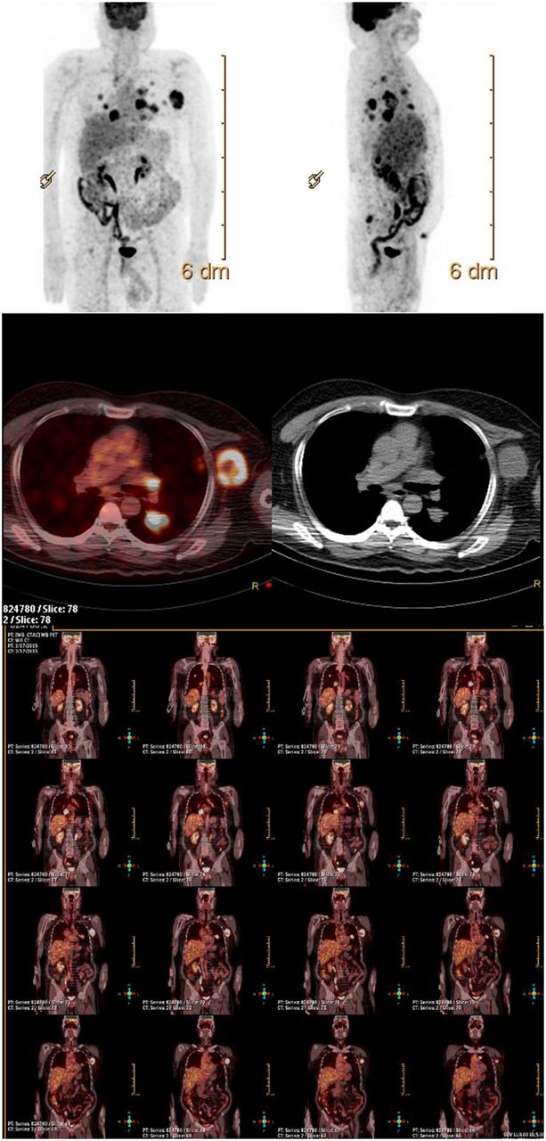
Maximum-intensity-projection (top), transaxial (middle), and coronal (bottom) 18F-FDG PET/CT images showing high uptake in lung, soft-tissue, subcutaneous, and iliac lesions.

DISCUSSION

Dermatofibrosarcoma protuberans is usually a slow-growing tumor that behaves in a benign fashion. Although it can recur locally, it rarely metastasizes (incidence < 5%) and typically has a relatively good prognosis (4,5). Here, we have illustrated the imaging features of 18F-FDG PET/CT in an unusual case of dermatofibrosarcoma protuberans in which high-grade 18F-FDG uptake correctly predicted an aggressive course. The ability to depict tumor biology is a benefit of 18F-FDG PET/CT imaging (1–3), which performs better than anatomic imaging in evaluating malignancies. There have been a couple of reports (6,7) on the role of 18F-FDG PET/CT in detecting distant metastasis of dermatofibrosarcoma protuberans, highlighting the fact that metastasis of this entity, though rare, is possible. Two reports (8,9) have also illustrated a possible role for 18F-FDG PET/CT in monitoring the response of dermatofibrosarcoma protuberans to treatment with imatinib mesylate. However, to our knowledge there has been no specific literature on its role in predicting aggressive biology in individuals showing high uptake at the diseased sites.

CONCLUSION

As illustrated by this case, the ability to predict tumor biology is an important advantage of 18F-FDG PET/CT imaging that has the potential to form the basis of personalized medicine in oncology.

DISCLOSURE

No potential conflict of interest relevant to this article was reported.

Footnotes

  • Published online Sep. 3, 2015.

REFERENCES

  1. 1.↵
    1. Jadvar H,
    2. Alavi A,
    3. Gambhir SS
    . 18F-FDG uptake in lung, breast, and colon cancers: molecular biology correlates and disease characterization. J Nucl Med. 2009;50:1820–1827.
    OpenUrlAbstract/FREE Full Text
  2. 2.
    1. Kwee TC,
    2. Basu S,
    3. Saboury B,
    4. Ambrosini V,
    5. Torigian DA,
    6. Alavi A
    . A new dimension of FDG-PET interpretation: assessment of tumor biology. Eur J Nucl Med Mol Imaging. 2011;38:1158–1170.
    OpenUrlCrossRefPubMed
  3. 3.↵
    1. Basu S,
    2. Sirohi B,
    3. Shrikhande SV
    . Dual tracer imaging approach in assessing tumor biology and heterogeneity in neuroendocrine tumors: its correlation with tumor proliferation index and possible multifaceted implications for personalized clinical management decisions, with focus on PRRT. Eur J Nucl Med Mol Imaging. 2014;41:1492–1496.
    OpenUrlCrossRefPubMed
  4. 4.↵
    1. Mendenhall WM,
    2. Zlotecki RA,
    3. Scarborough MT
    . Dermatofibrosarcoma protuberans. Cancer. 2004;101:2503–2508.
    OpenUrlCrossRefPubMed
  5. 5.↵
    1. Rutkowski P,
    2. Debiec-Rychter M
    . Current treatment options for dermatofibrosarcoma protuberans. Expert Rev Anticancer Ther. 2015;15:901–909.
    OpenUrlCrossRefPubMed
  6. 6.↵
    1. Suman S,
    2. Sharma P,
    3. Jain TK,
    4. Sahoo MK,
    5. Bal C,
    6. Kumar R
    . Recurrent dermatofibrosarcoma protuberans with pulmonary metastases presenting twelve years after initial diagnosis: 18F-FDG PET/CT imaging findings. Clin Nucl Med. 2014;39:77–78.
    OpenUrlCrossRefPubMed
  7. 7.↵
    1. Al-Tamimi A,
    2. Zaheer S,
    3. Pierce CK,
    4. Osmany S,
    5. Sittampalam K
    . Recurrent dermatofibrosarcoma protuberans of the shoulder with rare distant abdominal metastasis detected by fluorodeoxyglucose-positron emission tomography/computed tomography (FDG-PET/CT). Sultan Qaboos Univ Med J. 2012;12:371–374.
    OpenUrlPubMed
  8. 8.↵
    1. Rubin BP,
    2. Schuetze SM,
    3. Eary JF,
    4. et al
    . Molecular targeting of platelet-derived growth factor B by imatinib mesylate in a patient with metastatic dermatofibrosarcoma protuberans. J Clin Oncol. 2002;20:3586–3591.
    OpenUrlAbstract/FREE Full Text
  9. 9.↵
    1. Mizutani K,
    2. Tamada Y,
    3. Hara K,
    4. Tsuzuki T,
    5. Saeki H,
    6. Tamaki K
    . Imatinib mesylate inhibits the growth of metastatic lung lesions in a patient with dermatofibrosarcoma protuberance. Br J Dermatol. 2004;151:235–237.
    OpenUrlCrossRefPubMed
  • Received for publication July 2, 2015.
  • Accepted for publication July 24, 2015.
PreviousNext
Back to top

In this issue

Journal of Nuclear Medicine Technology: 44 (2)
Journal of Nuclear Medicine Technology
Vol. 44, Issue 2
June 1, 2016
  • Table of Contents
  • About the Cover
  • Index by author
Print
Download PDF
Article Alerts
Sign In to Email Alerts with your Email Address
Email Article

Thank you for your interest in spreading the word on Journal of Nuclear Medicine Technology.

NOTE: We only request your email address so that the person you are recommending the page to knows that you wanted them to see it, and that it is not junk mail. We do not capture any email address.

Enter multiple addresses on separate lines or separate them with commas.
18F-FDG PET/CT Prediction of an Aggressive Clinical Course for Dermatofibrosarcoma Protuberans
(Your Name) has sent you a message from Journal of Nuclear Medicine Technology
(Your Name) thought you would like to see the Journal of Nuclear Medicine Technology web site.
Citation Tools
18F-FDG PET/CT Prediction of an Aggressive Clinical Course for Dermatofibrosarcoma Protuberans
Sandip Basu, Fahim Goliwale
Journal of Nuclear Medicine Technology Jun 2016, 44 (2) 88-89; DOI: 10.2967/jnmt.115.163212

Citation Manager Formats

  • BibTeX
  • Bookends
  • EasyBib
  • EndNote (tagged)
  • EndNote 8 (xml)
  • Medlars
  • Mendeley
  • Papers
  • RefWorks Tagged
  • Ref Manager
  • RIS
  • Zotero
Share
18F-FDG PET/CT Prediction of an Aggressive Clinical Course for Dermatofibrosarcoma Protuberans
Sandip Basu, Fahim Goliwale
Journal of Nuclear Medicine Technology Jun 2016, 44 (2) 88-89; DOI: 10.2967/jnmt.115.163212
Twitter logo Facebook logo LinkedIn logo Mendeley logo
  • Tweet Widget
  • Facebook Like
  • Google Plus One
Bookmark this article

Jump to section

  • Article
    • Abstract
    • CASE REPORT
    • DISCUSSION
    • CONCLUSION
    • DISCLOSURE
    • Footnotes
    • REFERENCES
  • Figures & Data
  • Info & Metrics
  • PDF

Related Articles

  • No related articles found.
  • PubMed
  • Google Scholar

Cited By...

  • No citing articles found.
  • Google Scholar

More in this TOC Section

  • High-Sensitivity Troponin Elevation in a Young Woman with Typical Chest Pain: The Heart of the Matter
  • Pulmonary Adenocarcinoma Revealed by Parathyroid Scintigraphy: An Incidental Case to Remember
  • Prominent Right Ventricular Tracer Uptake: A Harbinger of Multivessel Coronary Artery Disease
Show more Teaching Case Studies

Similar Articles

Keywords

  • FDG PET-CT
  • dermatofibrosarcoma protuberans
  • Tumor Biology
SNMMI

© 2025 SNMMI

Powered by HighWire