
Main menu

  • Home
  • Content
    • Current
      • JNMT Supplement
    • Ahead of print
    • Past Issues
    • Continuing Education
    • JNMT Podcast
    • SNMMI Annual Meeting Abstracts
  • Subscriptions
    • Subscribers
    • Rates
    • Journal Claims
    • Institutional and Non-member
  • Authors
    • Submit to JNMT
    • Information for Authors
    • Assignment of Copyright
    • AQARA Requirements
  • Info
    • Reviewers
    • Permissions
    • Advertisers
    • Corporate & Special Sales
  • About
    • About Us
    • Editorial Board
    • Contact Information
  • More
    • Alerts
    • Feedback
    • Help
    • SNMMI Journals
  • SNMMI
    • JNMT
    • JNM
    • SNMMI Journals
    • SNMMI

User menu

  • Subscribe
  • My alerts
  • Log in
  • My Cart

Search

  • Advanced search
Journal of Nuclear Medicine Technology
  • SNMMI
    • JNMT
    • JNM
    • SNMMI Journals
    • SNMMI
  • Subscribe
  • My alerts
  • Log in
  • My Cart
Journal of Nuclear Medicine Technology

Advanced Search

  • Home
  • Content
    • Current
    • Ahead of print
    • Past Issues
    • Continuing Education
    • JNMT Podcast
    • SNMMI Annual Meeting Abstracts
  • Subscriptions
    • Subscribers
    • Rates
    • Journal Claims
    • Institutional and Non-member
  • Authors
    • Submit to JNMT
    • Information for Authors
    • Assignment of Copyright
    • AQARA Requirements
  • Info
    • Reviewers
    • Permissions
    • Advertisers
    • Corporate & Special Sales
  • About
    • About Us
    • Editorial Board
    • Contact Information
  • More
    • Alerts
    • Feedback
    • Help
    • SNMMI Journals
  • Watch or Listen to JNMT Podcast
  • Visit SNMMI on Facebook
  • Join SNMMI on LinkedIn
  • Follow SNMMI on Twitter
  • Subscribe to JNMT RSS feeds
Research ArticleImaging

Development of a 2-Layer Double-Pump Dynamic Cardiac Phantom

Hara Narihiro, Onoguchi Masahisa, Hojyo Osamu, Kawaguchi Hiroyuki, Murai Masakazu and Matsushima Noriko
Journal of Nuclear Medicine Technology March 2016, 44 (1) 31-35; DOI: https://doi.org/10.2967/jnmt.115.168252
Hara Narihiro
1Sumitomo Hospital, Radiological Technology, Nakanoshima, Kita-ku, Osaka, Japan; and
2Kanazawa University, Graduate School of Medical Sciences, Kodatsuno, Kanazawa, Japan
  • Find this author on Google Scholar
  • Find this author on PubMed
  • Search for this author on this site
Onoguchi Masahisa
2Kanazawa University, Graduate School of Medical Sciences, Kodatsuno, Kanazawa, Japan
  • Find this author on Google Scholar
  • Find this author on PubMed
  • Search for this author on this site
Hojyo Osamu
1Sumitomo Hospital, Radiological Technology, Nakanoshima, Kita-ku, Osaka, Japan; and
  • Find this author on Google Scholar
  • Find this author on PubMed
  • Search for this author on this site
Kawaguchi Hiroyuki
1Sumitomo Hospital, Radiological Technology, Nakanoshima, Kita-ku, Osaka, Japan; and
  • Find this author on Google Scholar
  • Find this author on PubMed
  • Search for this author on this site
Murai Masakazu
1Sumitomo Hospital, Radiological Technology, Nakanoshima, Kita-ku, Osaka, Japan; and
  • Find this author on Google Scholar
  • Find this author on PubMed
  • Search for this author on this site
Matsushima Noriko
1Sumitomo Hospital, Radiological Technology, Nakanoshima, Kita-ku, Osaka, Japan; and
  • Find this author on Google Scholar
  • Find this author on PubMed
  • Search for this author on this site
  • Article
  • Figures & Data
  • Info & Metrics
  • PDF
Loading

Abstract

The conventional dynamic cardiac phantom used in the field of nuclear medicine has a structure for which the size of the external side of the heart (the outer membrane substituting the myocardial layer) is fixed and only the inner side (the inner membrane substituting the ventricle part) moves anteroposteriorly. Therefore, its usefulness in technical evaluation is limited. Hence, we developed a new dynamic cardiac phantom in which the outer and inner membranes freely move. Methods: Using a SPECT/CT system, we performed validation by filling the myocardial layer of the dynamic cardiac phantom with solution and the ventricle part with contrast medium. We evaluated myocardial wall motions of 3 segments (basal, mid, and apical) by setting the stroke ratios at 20:20 and 10:10 (ventricle-to-myocardial layer ratio). Results: The myocardial wall motions (mean ± SD) at the stroke ratio of 20:20 were 7.50 ± 0.44, 11.15 ± 0.56, and 9.90 ± 0.24 mm in the basal, mid, and apical segments, respectively. The wall motions (mean ± SD) at the stroke ratio of 10:10 were 3.82 ± 0.43, 5.63 ± 0.39, and 4.53 ± 0.10 mm, respectively. Conclusion: In our dynamic cardiac phantom, different movements could be induced in the myocardial wall by freely changing the stroke ratio. These results suggest that the use of this phantom can realize technical evaluation that presumes various clinical conditions.

  • dynamic cardiac phantom
  • myocardial wall movement
  • gated myocardial SPECT
  • SPECT/CT

Nuclear medicine examination using gated myocardial SPECT can provide image information of high clinical usefulness as a functional diagnostic method (1–5) and much evidence for the diagnosis of ischemic heart disease (6–8), assessment of severity (9,10), determination of treatment efficacy (11,12), prediction of prognosis, and stratification of risk (13–15). To secure and establish evidence, technical evaluation is needed as a basic study. However, the conventional dynamic cardiac phantom used for technical evaluation of gated myocardial SPECT (16) has a structure for which the size of the external side of the heart (the outer membrane substituting the myocardial layer) is fixed and only the inner side (the inner membrane substituting the ventricle) moves anteroposteriorly (Fig. 1A). Therefore, detailed technical evaluation corresponding to actual clinical conditions cannot be performed. To perform technical evaluation toward clinical application, we developed a new dynamic cardiac phantom in which the outer and inner membranes move freely (Figs. 1B and 2A). The myocardial layer comprises outer and inner membranes made of special rubber, and heart movement is reproduced using 2 cylinder pumps (Fig. 2B). The outlet (discharge outlet, 15) for solutions supplied to the area of the ventricle and myocardial layer (e.g., radioactive and contrast agents) has a special structure (Fig. 2C). The contents of the ventricle and myocardial layer can be changed by setting the stroke ratio of the cylinder pumps freely (Fig. 2D).

FIGURE 1.
  • Download figure
  • Open in new tab
  • Download powerpoint
FIGURE 1.

(A) Schematic diagram of conventional dynamic cardiac phantom for nuclear medicine. (B) Schematic diagram of new dynamic cardiac phantom for nuclear medicine.

FIGURE 2.
  • Download figure
  • Open in new tab
  • Download powerpoint
FIGURE 2.

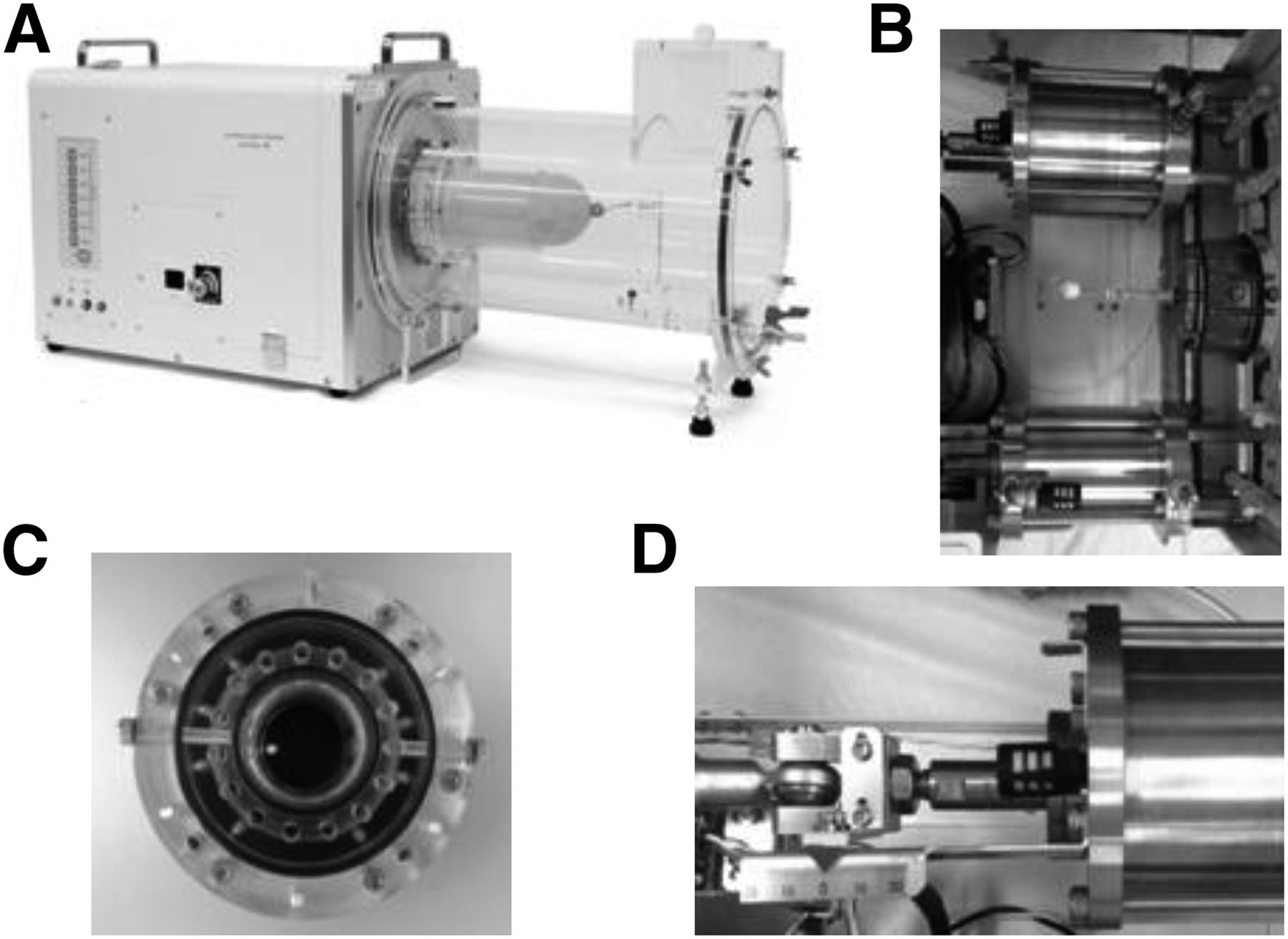
(A) Our new dynamic cardiac phantom. (B) Appearance of 2 cylinder pumps. (C) Appearance of outlet (front). (D) Scale of stroke.

MATERIALS AND METHODS

Setting of Phantom

The movable parts of the myocardial layer and ventricle in the new dynamic cardiac phantom were filled with a mixed solution of radioactive agent (755.00 mL of distilled water and 0.74 MBq of 99mTC solution per mL) and contrast agent (982.00 mL of distilled water and 370 mg of iodine concentration per mL), respectively. Table 1 shows the actual measurement values calculated from the structure of the new dynamic cardiac phantom (end-diastolic volume [EDV], end-systolic volume [ESV], and left ventricular ejection fraction [LVEF]).

View this table:
  • View inline
  • View popup
TABLE 1

Actual Measurement Values and SPECT Analysis Values (EDV, ESV, and LVEF) at Different Stroke Ratios

At the time of SPECT data acquisition, the heart rate was fixed at 60 bpm, and 11 patterns of stroke ratio (ventricle-to-myocardial layer ratio) were set (20:20, 20:10, 20:5, 15:15, 15:10, 15:5, 10:10, 10:5, 10:20, 10:15, and 5:10).

Gated SPECT Data Acquisition and Image Reconstruction

A SPECT/CT system (Precedence 16; Philips) was used. The conditions for gated SPECT data acquisition were data acquisition time at 30 s/step (360°, 64 steps), R-R interval at 16, use of a vertex general-purpose collimator, and matrix sizes of 128 × 128 (pixel size, 3.19 mm) and 64 × 64 (6.39 mm) (concurrent imaging: different acquisition data can be stored at 1 imaging).

Gated myocardial perfusion image reconstruction was performed using filtered backprojection (cutoff of 0.19 cycles/cm and order of 9 for a matrix size of 128 × 128) and a Butterworth filter for the preprocessing filter (cutoff of 0.37 cycles/cm and order of 9 for a matrix size of 64 × 64). A ramp filter was used as the reconstruction filter, without attenuation correction and scatter correction.

Analysis

Results of the cardiac function analysis of the SPECT data (EDV, ESV, LVEF, myocardial wall motion, and changes in wall thickness) were processed using EBW-NM4.5 (Philips). The Statistical Package for Social Sciences software (IBM) was used for the statistical analysis.

RESULTS

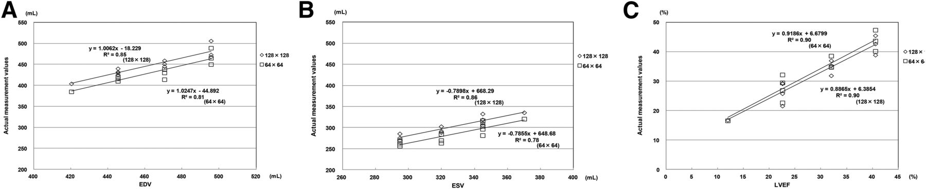
Table 1 shows the actual measurement values obtained using our new dynamic cardiac phantom and the EDV, ESV, and LVEF analyzed from the SPECT images. Figure 3 shows the results of the statistical analysis. In the comparison between the actual measurement values and the SPECT analysis values, high correlations were observed for a matrix size of 128 (EDV, r = 0.92; ESV, r = 0.93; LVEF, r = 0.95) and a matrix size of 64 (EDV, r = 0.90; ESV, r = 0.88; LVEF, r = 0.95).

FIGURE 3.
  • Download figure
  • Open in new tab
  • Download powerpoint
FIGURE 3.

Results of statistical analysis of actual measurement values and SPECT analysis values. (A) EDV. (B) ESV. (C) LVEF.

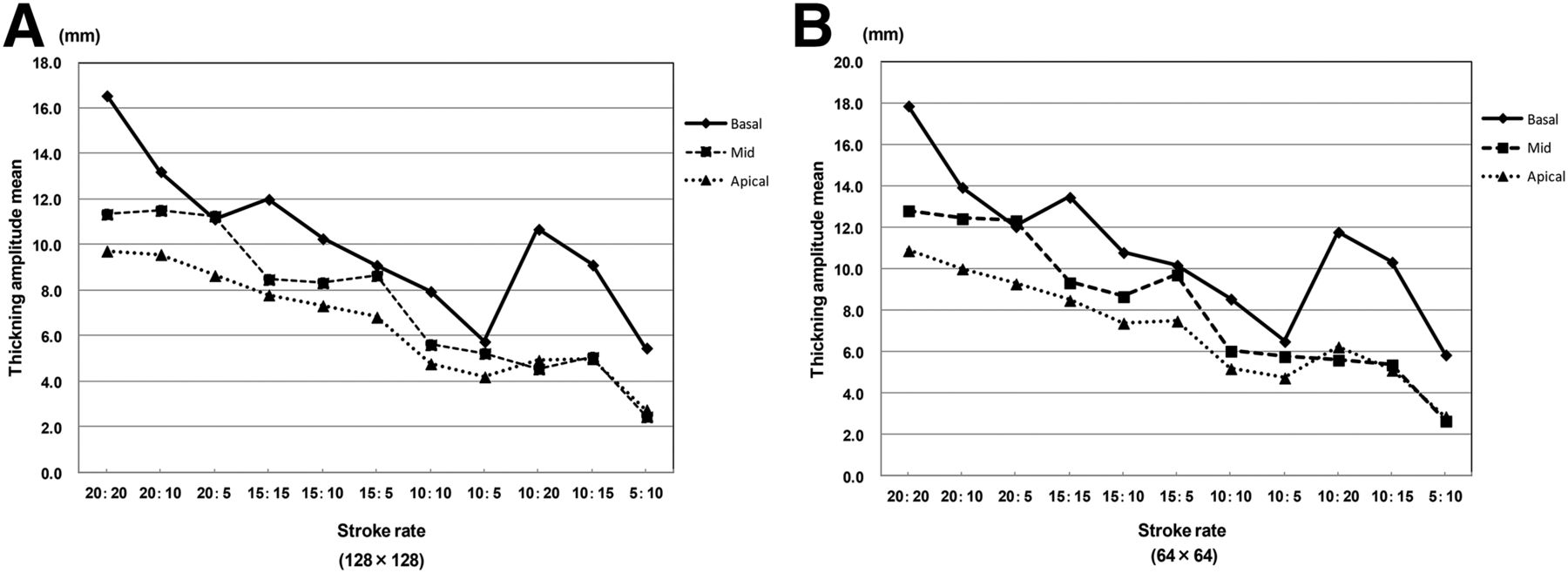
Figure 4 shows the results of the myocardial wall motion at each stroke ratio. The wall motions at each stroke ratio were within the ranges of 2.13–7.50, 3.32–11.10, and 3.00–9.90 mm in the basal, mid, and apical segments, respectively, for a matrix size of 128, and 2.10–7.87, 3.23–11.27, and 3.05–10.25 mm, respectively, for a matrix size of 64 (Fig. 4).

FIGURE 4.
  • Download figure
  • Open in new tab
  • Download powerpoint
FIGURE 4.

Results of stroke ratios and wall motion. (A) Matrix size, 128 × 128. (B) Matrix size, 64 × 64.

Figure 5 shows the results of the changes in wall thickness at each stroke ratio. The maximum and minimum changes in wall thickness at each stroke ratio (mean ± SD) for a matrix size of 128 were 16.55 ± 1.56 and 5.47 ± 0.64 mm, 11.35 ± 3.29 and 2.45 ± 0.85 mm, and 9.73 ± 2.91 and 2.75 ± 1.48 mm in the basal, mid, and apical segments, respectively, and those for a matrix size of 64 were 17.90 ± 1.66 and 5.87 ± 0.65 mm, 12.83 ± 3.49 and 2.67 ± 1.03 mm, and 10.90 ± 3.14 and 2.88 ± 1.45 mm, respectively (Fig. 5).

FIGURE 5.
  • Download figure
  • Open in new tab
  • Download powerpoint
FIGURE 5.

Results of stroke ratios and changes in myocardial wall thickness. (A) Matrix size, 128 × 128. (B) Matrix size, 64 × 64.

DISCUSSION

The usefulness of gated myocardial SPECT in the quantitative evaluation of transient left ventricular dysfunction and myocardial blood flow impairment has been reported, and it has improved the accuracy of evaluations of severity (9,10), risk, and prognosis (13–15). In addition, gated myocardial SPECT has been reported to be useful for the evaluation of EDV, ESV, LVEF, stroke volume/cardiac index, myocardial wall motion, and changes in myocardial wall thickness, which are critical in risk and prognosis assessment in coronary artery diseases (1,2)

Published reports have stated that application of the exercise or drug-loading method by using gated myocardial SPECT can quantitatively evaluate markers of poor prognosis, transient reduction in LVEF after loading, and myocardial wall motion abnormality (3,4). These reports suggest the need of further improvement of the technical accuracy of nuclear cardiology examinations.

Recent advances have led to the development of nuclear cardiology devices mounted with a semiconductor detector, and technical evaluation was performed on these devices (17–19). However, the phantoms used in the previous studies were static cardiac phantoms (17) and cardiac SPECT phantoms (19). With these phantoms, detailed technical evaluation with gated myocardial SPECT cannot be performed. We developed a new dynamic cardiac phantom because the evaluation of myocardial wall motion is considered necessary, in addition to the evaluation of EDV, ESV, and LVEF, to perform technical evaluation with high clinical applicability in the future.

Detailed cardiac function analysis cannot be performed with static cardiac phantoms used for technical evaluation in nuclear cardiology examinations (20–22) or the conventional dynamic cardiac phantom developed by Kubo et al. (16), in which cardiac function analytic values (EDV, ESV, and LVEF) are fixed values calculated from the forms of the phantoms. However, the actual measurement values at the different stroke ratios of our new dynamic cardiac phantom showed high correlation with SPECT analysis values, suggesting that detailed validation of cardiac function analysis can be performed by setting stroke ratios appropriate for clinical application (Table 1; Fig. 5).

The dynamic myocardial phantoms reported by Jacco et al. (23) and Luca et al. (24) have 2 discharge outlets for solutions supplied to the myocardial layer, and the same pump is used to supply solutions to the ventricle part and myocardial layer. Although EDV, ESV, and LVEF were evaluated, no detailed evaluation was performed such as that for myocardial wall motion. In our new dynamic cardiac phantom, the solution supply system to the myocardial layer has a special structure (Fig. 2C; discharge outlet, 15). It has independent supply pumps to the ventricle and myocardial layer (Fig. 2B), and stroke ratios can be set freely (Fig. 2D). In addition to the evaluation of EDV, ESV, and LVEF at different stroke ratios (Fig. 3), various myocardial wall motions can be set through the combination of stroke ratios of the cylinder pumps of the ventricle and myocardial layer (Fig. 4). Among the stroke ratios validated, we could confirm free changes in myocardial wall thickness based on the differences between 20:10 and 10:20 (2.54, 6.97, and 4.63 mm in the basal, mid, and apical segments, respectively), between 15:10 and 10:15 (1.15, 3.26, and 2.33 mm, respectively), and between 10:5 and 5:10 (0.28, 2.78, and 1.45 mm, respectively (Fig. 5).

CONCLUSION

This study demonstrates that our new dynamic cardiac phantom can be used for technical evaluation that presumes various clinical conditions. Technical validation for SPECT image processing and attenuation correction using this phantom is needed in the future.

DISCLOSURE

No potential conflict of interest relevant to this article was reported.

Acknowledgments

We thank Munetomo Sato (Fuyo Corporation) and the staff of the Department of Pharmacy of Sumitomo Hospital for their cooperation in this study.

Footnotes

  • Published online Jan. 14, 2016.

REFERENCES

  1. 1.↵
    1. Nakajima K,
    2. Kusuoka H,
    3. Nishimura S,
    4. et al
    . Normal limits of ejection fraction and volumes determined by gated SPECT in clinically normal patients without cardiac events: a study based on the J-ACCESS database. Eur J Nucl Med Mol Imaging. 2007;34:1088–1096.
    OpenUrlCrossRefPubMed
  2. 2.↵
    1. Nakajima K,
    2. Nishimura T
    . Inter-institution preference-based variability of ejection fraction and volumes using quantitative gated SPECT with 99mTc-tetrofosmin: a multicentre study involving 106 hospitals. Eur J Nucl Med Mol Imaging. 2006;33:127–133.
    OpenUrlCrossRefPubMed
  3. 3.↵
    1. Kapetanopoulos,
    2. Ahlberg AW,
    3. Taub CC,
    4. et al
    . Regional wall-motion abnormalities on post-stress electrocardiographic-gated technetium-99m sestamibi single-photon emission computed tomography imaging predict cardiac events. J Nucl Cardiol. 2007;14:810–817.
    OpenUrlCrossRefPubMed
  4. 4.↵
    1. Sharir T
    . Role of regional myocardial dysfunction by gated myocardial perfusion SPECT in the prognostic evaluation of patients with coronary artery disease. J Nucl Cardiol. 2005;12:5–8.
    OpenUrlCrossRefPubMed
  5. 5.↵
    1. Schaefer WM,
    2. Lipke CS,
    3. Dirk Standke,
    4. et al
    . Quantification of left ventricular volumes and ejection fraction from gated 99mTc-MIBI SPECT: MRI validation and comparison of the Emory cardiac tool box with QGS and 4D-MSPECT. J Nucl Med. 2005;46:1256–1263.
    OpenUrlAbstract/FREE Full Text
  6. 6.↵
    1. Kuwabara Y,
    2. Watanabe S,
    3. Nakaya J,
    4. et al
    . Functional evaluation of myocardial viability by 99mTc tetrofosmin gated SPECT: a quantitative comparison with 18F fluorodeoxyglucose positron emission CT (18F FDG PET). Ann Nucl Med. 1999;13:135–140.
    OpenUrlCrossRefPubMed
  7. 7.
    1. Levine MG,
    2. McGill CC,
    3. Ahlberg AW,
    4. et al
    . Functional assessment with electrocardiographic gated single-photon emission computed tomography improves the ability of technetium-99m sestamibi myocardial perfusion imaging to predict myocardial viability in patients undergoing revascularization. Am J Cardiol. 1999;83:1–5.
    OpenUrlCrossRefPubMed
  8. 8.↵
    1. Pagnanelli RA,
    2. Hanson MW,
    3. Turkington T,
    4. Coleman RE,
    5. Borges-Neto S
    . Gated 99mTc-tetrofosmin and 18F-FDG studies: a comparison of single-acquisition and separate-acquisition protocols. J Nucl Med Technol. 2002;30:175–178.
    OpenUrlAbstract/FREE Full Text
  9. 9.↵
    1. Slomka PJ,
    2. Nishina H,
    3. Berman DS,
    4. et al
    . Automated quantification of myocardial perfusion SPECT using simplified normal limits. J Nucl Cardiol. 2005;12:66–77.
    OpenUrlCrossRefPubMed
  10. 10.↵
    1. Berman DS,
    2. Kang X,
    3. Gransar H,
    4. et al
    . Quantitative assessment of myocardial perfusion abnormality on SPECT myocardial perfusion imaging is more reproducible than expert visual analysis. J Nucl Cardiol. 2009;16:45–53.
    OpenUrlCrossRefPubMed
  11. 11.↵
    1. Nakajima K,
    2. Taki J,
    3. Higuchi T,
    4. et al
    . Gated SPET quantification of small hearts: mathematical simulation and clinical application. Eur J Nucl Med. 2000;27:1372–1379.
    OpenUrlCrossRefPubMed
  12. 12.↵
    1. Emre A,
    2. Ersek B,
    3. Gürsürer M,
    4. et al
    . Angiographic and scintigraphic (perfusion and electrocardiogram-gated SPECT) correlates of clinical presentation in unstable angina. Clin Cardiol. 2000;23:495–500.
    OpenUrlCrossRefPubMed
  13. 13.↵
    1. Matsumoto N,
    2. Sato Y,
    3. Suzuki Y,
    4. et al
    . Incremental prognostic value of cardiac function assessed by ECG-gated myocardial perfusion SPECT for the prediction of future acute coronary syndrome. Circ J. 2008;72:2035–2039.
    OpenUrlCrossRefPubMed
  14. 14.
    1. Petix NR,
    2. Sestini S,
    3. Coppola A,
    4. et al
    . Prognostic value of combined perfusion and function by stress technetium-99m sestamibi gated SPECT myocardial perfusion imaging in patients with suspected or known coronary artery disease. Am J Cardiol. 2005;95:1351–1357.
    OpenUrlCrossRefPubMed
  15. 15.↵
    1. Travin MI,
    2. Heller GV,
    3. Johnson LL,
    4. et al
    . The prognostic value of ECG-gated SPECT imaging in patients undergoing stress Tc-99m sestamibi myocardial perfusion imaging. J Nucl Cardiol. 2004;11:253–262.
    OpenUrlCrossRefPubMed
  16. 16.↵
    1. Kubo N,
    2. Mabuchi M,
    3. Katoh C,
    4. et al
    . Validation of left ventricular function from gated single photon computed emission tomography by using a scintillator-photodiode camera: a dynamic myocardial phantom study. Nucl Med Commun. 2002;23:639–643.
    OpenUrlCrossRefPubMed
  17. 17.↵
    1. Bocher M,
    2. Blevis IM,
    3. Tsukerman L,
    4. et al
    . A fast cardiac gamma camera with dynamic SPECT capabilities: design, system validation and future potential. Eur J Nucl Med Mol Imaging. 2010;37:1887–1902.
    OpenUrlCrossRefPubMed
  18. 18.
    1. Kacperski K,
    2. Erlandsson K,
    3. Haim SB,
    4. et al
    . Iterative deconvolution of simultaneous 99mTc and 201Tl projection data measured on a CdZnTe based cardiac SPECT scanner. Phys Med Biol. 2011;56:1397–1414.
    OpenUrlCrossRefPubMed
  19. 19.↵
    1. Takahashi Y,
    2. Miyagawa M,
    3. Nishiyama Y,
    4. et al
    . Performance of a semiconductor SPECT system: comparison with a conventional Anger-type SPECT instrument. Ann Nucl Med. 2013;27:11–16.
    OpenUrlCrossRefPubMed
  20. 20.↵
    1. Beach RD,
    2. Depold H,
    3. Boening G,
    4. et al
    . An adaptive approach to decomposing patient-motion tracking data acquired during cardiac SPECT imaging. IEEE Trans Nucl Sci. 2007;54:130–139.
    OpenUrlCrossRefPubMed
  21. 21.
    1. Knesaurek K,
    2. Machac J
    . Comparison of 18F SPECT with PET in myocardial imaging: a realistic thorax cardiac phantom study. BMC Nucl Med. 2006;6:5–13.
    OpenUrlCrossRefPubMed
  22. 22.↵
    1. Difilippo FP
    . Design and performance of a multi-pinhole collimation device for small animal imaging with clinical SPECT and SPECT-CT scanners. Phys Med Biol. 2008;53:4185–4201.
    OpenUrlCrossRefPubMed
  23. 23.↵
    1. Visser JJ,
    2. Sokole EB,
    3. Verberne HJ,
    4. et al
    . A realistic 3-D gated cardiac phantom for quality control of gated myocardial perfusion SPET: the Amsterdam gated (AGATE) cardiac phantom. Eur J Nucl Med Mol Imaging. 2004;31:222–228.
    OpenUrlCrossRefPubMed
  24. 24.↵
    1. Ceriani L,
    2. Ruberto T,
    3. Delaloye AB,
    4. Prior JO,
    5. Giovanella L
    . Three-dimensional ordered-subset expectation maximization iterative protocol for evaluation of left ventricular volumes and function by quantitative gated SPECT: a dynamic phantom study. J Nucl Med Technol. 2010;38:18–23.
    OpenUrlAbstract/FREE Full Text
  • Received for publication October 16, 2015.
  • Accepted for publication December 28, 2015.
PreviousNext
Back to top

In this issue

Journal of Nuclear Medicine Technology: 44 (1)
Journal of Nuclear Medicine Technology
Vol. 44, Issue 1
March 1, 2016
  • Table of Contents
  • About the Cover
  • Index by author
Print
Download PDF
Article Alerts
Sign In to Email Alerts with your Email Address
Email Article

Thank you for your interest in spreading the word on Journal of Nuclear Medicine Technology.

NOTE: We only request your email address so that the person you are recommending the page to knows that you wanted them to see it, and that it is not junk mail. We do not capture any email address.

Enter multiple addresses on separate lines or separate them with commas.
Development of a 2-Layer Double-Pump Dynamic Cardiac Phantom
(Your Name) has sent you a message from Journal of Nuclear Medicine Technology
(Your Name) thought you would like to see the Journal of Nuclear Medicine Technology web site.
Citation Tools
Development of a 2-Layer Double-Pump Dynamic Cardiac Phantom
Hara Narihiro, Onoguchi Masahisa, Hojyo Osamu, Kawaguchi Hiroyuki, Murai Masakazu, Matsushima Noriko
Journal of Nuclear Medicine Technology Mar 2016, 44 (1) 31-35; DOI: 10.2967/jnmt.115.168252

Citation Manager Formats

  • BibTeX
  • Bookends
  • EasyBib
  • EndNote (tagged)
  • EndNote 8 (xml)
  • Medlars
  • Mendeley
  • Papers
  • RefWorks Tagged
  • Ref Manager
  • RIS
  • Zotero
Share
Development of a 2-Layer Double-Pump Dynamic Cardiac Phantom
Hara Narihiro, Onoguchi Masahisa, Hojyo Osamu, Kawaguchi Hiroyuki, Murai Masakazu, Matsushima Noriko
Journal of Nuclear Medicine Technology Mar 2016, 44 (1) 31-35; DOI: 10.2967/jnmt.115.168252
Twitter logo Facebook logo LinkedIn logo Mendeley logo
  • Tweet Widget
  • Facebook Like
  • Google Plus One
Bookmark this article

Jump to section

  • Article
    • Abstract
    • MATERIALS AND METHODS
    • RESULTS
    • DISCUSSION
    • CONCLUSION
    • DISCLOSURE
    • Acknowledgments
    • Footnotes
    • REFERENCES
  • Figures & Data
  • Info & Metrics
  • PDF

Related Articles

  • No related articles found.
  • PubMed
  • Google Scholar

Cited By...

  • Study of Attenuation Correction Using a Cardiac Dynamic Phantom: Synchronized Time-Phase-Gated Attenuation Correction Method
  • Google Scholar

More in this TOC Section

  • Early 10-Minute Postinjection [18F]F-FAPI-42 uEXPLORER Total-Body PET/CT Scanning Protocol for Staging Lung Cancer Using HYPER Iterative Reconstruction
  • Single- Versus Dual-Time-Point Imaging for Transthyretin Cardiac Amyloid Using 99mTc-Pyrophosphate
  • Does Arthrography Improve Accuracy of SPECT/CT for Diagnosis of Aseptic Loosening in Patients with Painful Knee Arthroplasty: A Systematic Review and Metaanalysis
Show more Imaging

Similar Articles

Keywords

  • Dynamic cardiac phantom
  • Myocardial wall movement
  • Gated myocardial SPECT
  • SPECT/CT
SNMMI

© 2025 SNMMI

Powered by HighWire