
Main menu

  • Home
  • Content
    • Current
      • JNMT Supplement
    • Ahead of print
    • Past Issues
    • Continuing Education
    • JNMT Podcast
    • SNMMI Annual Meeting Abstracts
  • Subscriptions
    • Subscribers
    • Rates
    • Journal Claims
    • Institutional and Non-member
  • Authors
    • Submit to JNMT
    • Information for Authors
    • Assignment of Copyright
    • AQARA Requirements
  • Info
    • Reviewers
    • Permissions
    • Advertisers
    • Corporate & Special Sales
  • About
    • About Us
    • Editorial Board
    • Contact Information
  • More
    • Alerts
    • Feedback
    • Help
    • SNMMI Journals
  • SNMMI
    • JNMT
    • JNM
    • SNMMI Journals
    • SNMMI

User menu

  • Subscribe
  • My alerts
  • Log in
  • My Cart

Search

  • Advanced search
Journal of Nuclear Medicine Technology
  • SNMMI
    • JNMT
    • JNM
    • SNMMI Journals
    • SNMMI
  • Subscribe
  • My alerts
  • Log in
  • My Cart
Journal of Nuclear Medicine Technology

Advanced Search

  • Home
  • Content
    • Current
    • Ahead of print
    • Past Issues
    • Continuing Education
    • JNMT Podcast
    • SNMMI Annual Meeting Abstracts
  • Subscriptions
    • Subscribers
    • Rates
    • Journal Claims
    • Institutional and Non-member
  • Authors
    • Submit to JNMT
    • Information for Authors
    • Assignment of Copyright
    • AQARA Requirements
  • Info
    • Reviewers
    • Permissions
    • Advertisers
    • Corporate & Special Sales
  • About
    • About Us
    • Editorial Board
    • Contact Information
  • More
    • Alerts
    • Feedback
    • Help
    • SNMMI Journals
  • Watch or Listen to JNMT Podcast
  • Visit SNMMI on Facebook
  • Join SNMMI on LinkedIn
  • Follow SNMMI on Twitter
  • Subscribe to JNMT RSS feeds
Research ArticleImaging

Tailoring 99mTc-Macroaggregated Albumin Administration to Optimize Patient Dose Reduction

Sarah L. Gales, Sharn K. Peters, Nouria Salehi and Nathan Better
Journal of Nuclear Medicine Technology September 2015, 43 (3) 220-221; DOI: https://doi.org/10.2967/jnmt.114.151811
Sarah L. Gales
1Department of Nuclear Medicine, St. F.X. Cabrini Hospital, Malvern, Victoria, Australia; and
  • Find this author on Google Scholar
  • Find this author on PubMed
  • Search for this author on this site
Sharn K. Peters
1Department of Nuclear Medicine, St. F.X. Cabrini Hospital, Malvern, Victoria, Australia; and
  • Find this author on Google Scholar
  • Find this author on PubMed
  • Search for this author on this site
Nouria Salehi
2Department of Nuclear Medicine, Royal Melbourne Hospital, Parkville, Victoria, Australia
  • Find this author on Google Scholar
  • Find this author on PubMed
  • Search for this author on this site
Nathan Better
1Department of Nuclear Medicine, St. F.X. Cabrini Hospital, Malvern, Victoria, Australia; and
2Department of Nuclear Medicine, Royal Melbourne Hospital, Parkville, Victoria, Australia
  • Find this author on Google Scholar
  • Find this author on PubMed
  • Search for this author on this site
  • Article
  • Figures & Data
  • Info & Metrics
  • PDF
Loading

Abstract

In the ever-changing field of nuclear medicine, best-practice considerations cannot simply go unchallenged for months and years, with the need to minimize radiation exposure to patients highlighted in “as low as reasonably achievable” principles. The Australian Radiation Protection and Nuclear Safety Agency reports that the dose for 99mTc-macroaggregated albumin (99mTc-MAA) administered should be 180–200 MBq. An objective of imaging in pulmonary embolism, or indeed any diagnostic procedure involving radiation, is to minimize radiation exposure without sacrificing image quality and diagnostic accuracy. The amount of radiation involved must be considered together with imaging protocols. Our aim was to reduce the amount of 99mTc-MAA administered without compromising the diagnostic quality of the scan. Methods: To achieve a ventilation-to-perfusion ratio of 1:4, we ventilated the patient as per standard protocol and then placed intravenous access into the patient. For the perfusion component, 180–200 MBq were prepared in a 2-mL injection. Aliquots of 0.5 mL of 99mTc-MAA were administered every 30 s followed by a 5-mL saline flush until the required ventilation-to-perfusion ratio was achieved. Results: With this protocol, the average administered dose was 105 ± 20.7 MBq (vs. 180 ± 5.3 MBq, P < 0.0001). Conclusion: By individually tailoring the administered dose, diagnostic quality is maintained while achieving a significant dose reduction.

  • MAA
  • dose reduction
  • VQ scan

In the ever-changing field of nuclear medicine, best-practice considerations cannot simply go unchallenged for months and years. The need to minimize radiation exposure to patients is highlighted in “as low as reasonably achievable” principles. The Australian Radiation Protection and Nuclear Safety Agency reports that the dose for 99mTc-macroaggregated albumin (99mTc-MAA) administered is 180–200 MBq (1). The Society of Nuclear Medicine and Molecular Imaging reported the dose to be 40–150 MBq (2). When describing the use of aerosols in ventilation–perfusion (VQ) imaging, the society states that “Because both agents are labeled with 99mTc, it is extremely important that the counting rate of the second study is at least 3 to 4 times the counting rate of the first study” (2,3).

One objective of imaging in pulmonary embolus detection or indeed any diagnostic procedure involving radiation is to minimize radiation exposure without sacrificing image quality and diagnostic accuracy (4). The amount of radiation involved must be considered together with imaging protocols. We aimed to reduce the amount of 99mTc-MAA administered without compromising the diagnostic quality of the scan.

MATERIALS AND METHODS

In a VQ lung scan at St. F.X. Cabrini Hospital, the patient inhales 99mTc-Technegas for the ventilation component: 1,000−1,500 cps as detected on the γ camera, or 700 cps over the right lung using a Geiger counter measurement. The dose administered equates to approximately 40 MBq (3). For the perfusion component, we administer 180–200 MBq of Technescan LyoMAA (Mallinckrodt) intravenously. The detected 99mTc-MAA counting rate must be at least 4 times greater than the ventilation component (2).

Our routine acquisition is SPECT with low-energy high-resolution collimators, 48 views over 180° using 2 detectors. The matrix is 128 × 128, and the zoom is 1.23. The ventilation imaging is performed at 12 s/stop, and the perfusion imaging at 10 s/stop (5–8).

A control sample of 10 patients underwent lung scanning using the standard 180–200 MBq (average, 185 MBq ± 5.27 [SD]) Technescan LyoMAA dose. A VQ ratio was then calculated for each patient.

Our trial patients were ventilated per the previously discussed standard protocol, and intravenous access was placed with the patient under the camera. For the perfusion component, 180–200 MBq of Technescan LyoMAA were prepared in a 2-mL injection with normal saline. Aliquots of 0.5 mL followed by a 5-mL saline flush were administered every 30 s, and counts were performed over the right lung until a VQ ratio of at least 1:4 was achieved. The residual dose in the syringe was measured to calculate the administered dose. Intravenous cannulas were also measured. It was found that the amount left in the cannula was negligible.

All patients presenting to our department for a VQ study during the period of the trial were included in the analysis; no exclusions were required.

Statistics were analyzed using a Student t distribution test.

Approval for this study was sought and obtained from the Ethics Committee at St. F.X. Cabrini Hospital.

RESULTS

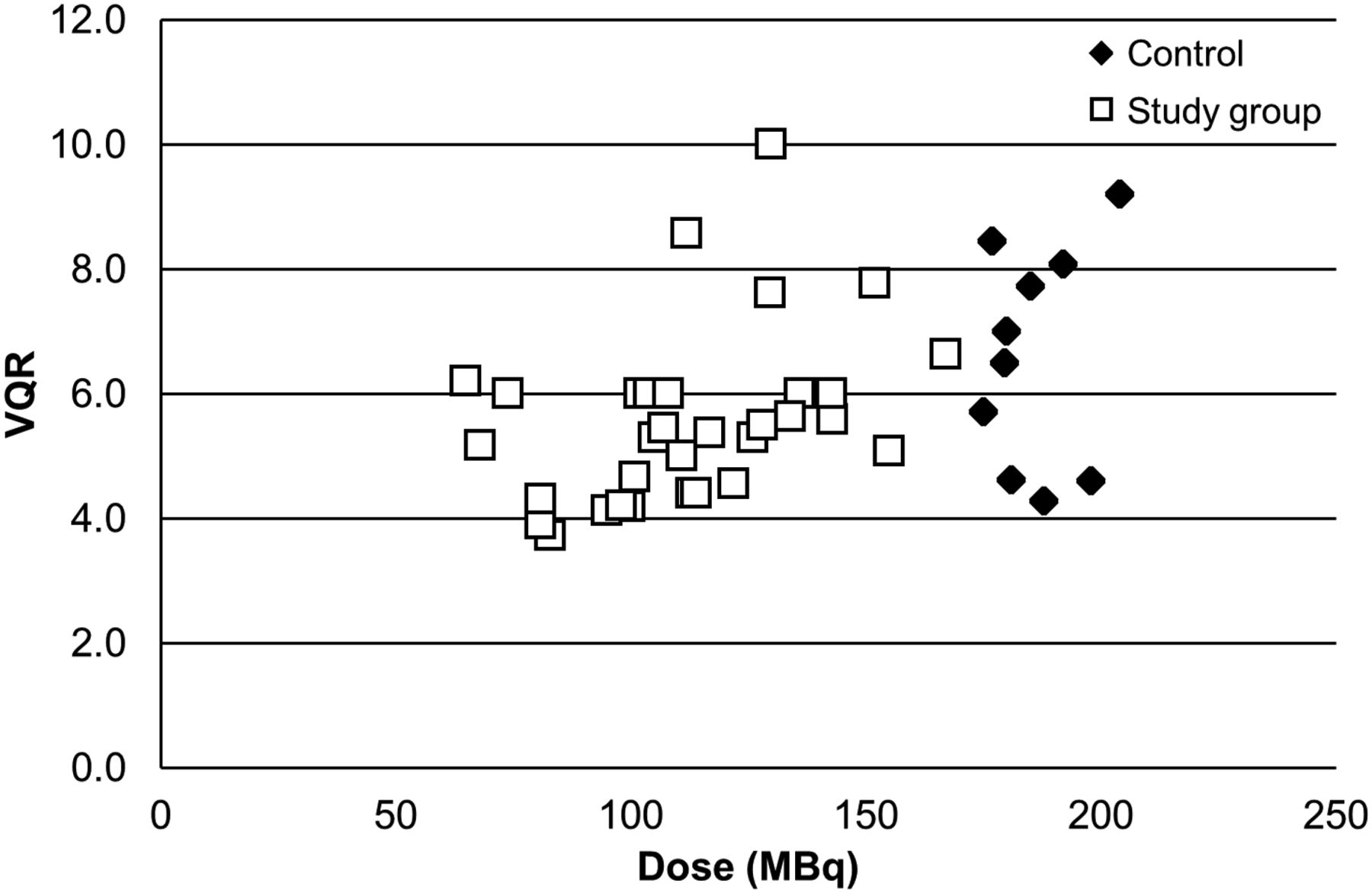
The average administered dose was 105 ± 20.7 MBq for our trial group of 18 patients and 180 ± 5.3 MBq for our controls (P < 0.0001) (Fig. 1).

FIGURE 1.
  • Download figure
  • Open in new tab
  • Download powerpoint
FIGURE 1.

Dose vs. VQ ratio (VQR). Administered perfusion dose of 99mTc-MAA is compared between control group (n = 10) and trial group (n = 18). Although VQ ratio was greater in control patients, value still remained above 4 in trial group while allowing significant reduction in administered dose from 180 ± 5.3 to 105 ± 20.7 MBq (P < 0.0001).

As expected, the VQ ratio was significantly higher in the control group than in the trial group: 1:6.6 ± 1.7 versus 1:4.7 ± 0.7 (P < 0.001). However, the ratio in the trial patients was still over the required 1:4.

DISCUSSION

Perfusion scintigraphy is accomplished by microembolization with radiolabeled particles injected into a peripheral vein. The commercially used particles are MAA, which are labeled with 99mTc. They are 15–100 μm and lodge in the pulmonary capillaries and precapillary arterioles (9,10).

The particle distribution accurately defines regional lung perfusion. An important factor is the number of particles injected. A minimum of 60,000 particles is required to obtain a uniform distribution of activity reflecting regional perfusion. Normally, about 400,000 labeled particles are injected. Bearing in mind that there are over 280 billion pulmonary capillaries and 300 million precapillary arterioles, the administration of up to 400,000 particles will obstruct only a small fraction of pulmonary vessels (11). A reduction in the number of particles administered to between 100,000 and 200,000 is recommended in patients with known pulmonary hypertension, right-to-left heart shunt, or a single transplanted lung. In infants and children, the number of particles may be further reduced in accordance with weight (10–12). To reduce radiation exposure, the dose can be reduced in pregnant women (9–11). Current guidelines have also emphasized the importance of dose minimization in all patient subgroups (9). A diagnostic scan can still be obtained with this reduction in dose. It is this process that prompted the research project to reduce the standard recommended dose.

Our results demonstrate the ability to reduce the dose and not compromise scan quality by following this administration protocol (Fig. 1).

This protocol can tailor the dose to each patient to obtain perfusion images while significantly reducing dose. All scans were clinically indicated and were interpreted by the nuclear medicine physician; each scan in both groups was of excellent diagnostic quality. The sample size was small and a larger series would be useful, but the dose reduction was statistically significant.

The pattern of lung perfusion may be heterogeneous in certain subgroups (e.g., chronic obstructive pulmonary disease), and the ratio was often lower in these subgroups than in patients without such diseases. Whether this specifically affects use of the technique for dose reduction in these subgroups warrants further research.

CONCLUSION

Our novel protocol allows the perfusion dose to be individually tailored to the patient. In this way, a diagnostic scan can be achieved at a significantly reduced dose, thereby decreasing patient and staff exposure alike.

DISCLOSURE

No potential conflict of interest relevant to this article was reported.

Acknowledgments

We thank the staff in the Nuclear Medicine Department at St. F.X. Cabrini Hospital for helping to collate the data.

Footnotes

  • Published online Aug. 13, 2015.

REFERENCES

  1. 1.↵
    National Diagnostic Reference Level Fact Sheet. Australian Radiation Protection and Nuclear Safety Agency website. http://www.arpansa.gov.au/services/ndrl/ndrlfactsheet.cfm. Accessed July 13, 2015.
  2. 2.↵
    1. Parker JA,
    2. Coleman ER,
    3. Erin G,
    4. et al
    . SNM Practice Guideline for Lung Scintigraphy 4.0. J Nucl Med Technol. 2012;40:57–65.
    OpenUrlFREE Full Text
  3. 3.↵
    1. Burch WM,
    2. Sullivan PJ,
    3. Lomas FE,
    4. Evans VA,
    5. McLaren CJ,
    6. Arnot RN
    . Lung ventilation studies with technetium-99m Pseudogas. J Nucl Med. 1986;27:842–846.
    OpenUrlAbstract/FREE Full Text
  4. 4.↵
    1. Albert C,
    2. Sylviane P,
    3. Sokole EB,
    4. et al
    . Best practice in nuclear medicine: part 2—a technologist’s guide. European Association of Nuclear Medicine website. http://www.eanm.org/publications/tech_guidelines/docs/tech_best_practice2.pdf. Accessed July 13, 2015.
  5. 5.↵
    1. Murray IPC,
    2. Ell PJ
    . Nuclear Medicine in Clinical Diagnosis and Treatment. 2nd ed. London, U.K.: Churchill Livingstone; 1998:37–50.
  6. 6.
    1. Shackett P
    . Nuclear Medicine Technology, Procedures and Quick Reference. Lippincott, Williams, and Wilkins, USA, 2000:154–170.
  7. 7.
    Parker A, Coleman RE, Hilson AJW, Royal HD, Siegel BA. Society of Nuclear Medicine Procedure Guideline for Lung Scintigraphy, version 3.0, approved February 7, 2004. Society of Nuclear Medicine and Molecular Imaging website. Accessed July 13, 2015.
  8. 8.↵
    Sharp PF, Gemmell HG, Murray A, eds. Practical Nuclear Medicine. 3rd ed. New York, NY: Springer; 2005.
  9. 9.↵
    1. Bajc M,
    2. Neilly JB,
    3. Miniati M,
    4. Schuemichen C,
    5. Meignan M,
    6. Jonson B
    . EANM guidelines for ventilation/perfusion scintigraphy, part 1. Pulmonary imaging with ventilation/perfusion single photon emission tomography. Eur J Nucl Med Mol Imaging. 2009;36:1356–1370.
    OpenUrlCrossRefPubMed
  10. 10.↵
    Saha GB. Fundamentals of Nuclear Pharmacy. 4th ed. New York, NY: Springer; 1998.
  11. 11.↵
    1. Heck LL,
    2. Duley JW Jr.
    . Statistical considerations in lung imaging with 99mTc albumin particles. Radiology. 1974;113:675–679.
    OpenUrlCrossRefPubMed
  12. 12.↵
    1. Ciofetta G,
    2. Piepsz A,
    3. Roca I,
    4. et al
    . Guidelines for lung scintigraphy in children. Eur J Nucl Med Mol Imaging. 2007;34:1518–1526.
    OpenUrlCrossRefPubMed
  • Received for publication May 12, 2015.
  • Accepted for publication July 1, 2015.
PreviousNext
Back to top

In this issue

Journal of Nuclear Medicine Technology: 43 (3)
Journal of Nuclear Medicine Technology
Vol. 43, Issue 3
September 1, 2015
  • Table of Contents
  • About the Cover
  • Index by author
Print
Download PDF
Article Alerts
Sign In to Email Alerts with your Email Address
Email Article

Thank you for your interest in spreading the word on Journal of Nuclear Medicine Technology.

NOTE: We only request your email address so that the person you are recommending the page to knows that you wanted them to see it, and that it is not junk mail. We do not capture any email address.

Enter multiple addresses on separate lines or separate them with commas.
Tailoring 99mTc-Macroaggregated Albumin Administration to Optimize Patient Dose Reduction
(Your Name) has sent you a message from Journal of Nuclear Medicine Technology
(Your Name) thought you would like to see the Journal of Nuclear Medicine Technology web site.
Citation Tools
Tailoring 99mTc-Macroaggregated Albumin Administration to Optimize Patient Dose Reduction
Sarah L. Gales, Sharn K. Peters, Nouria Salehi, Nathan Better
Journal of Nuclear Medicine Technology Sep 2015, 43 (3) 220-221; DOI: 10.2967/jnmt.114.151811

Citation Manager Formats

  • BibTeX
  • Bookends
  • EasyBib
  • EndNote (tagged)
  • EndNote 8 (xml)
  • Medlars
  • Mendeley
  • Papers
  • RefWorks Tagged
  • Ref Manager
  • RIS
  • Zotero
Share
Tailoring 99mTc-Macroaggregated Albumin Administration to Optimize Patient Dose Reduction
Sarah L. Gales, Sharn K. Peters, Nouria Salehi, Nathan Better
Journal of Nuclear Medicine Technology Sep 2015, 43 (3) 220-221; DOI: 10.2967/jnmt.114.151811
Twitter logo Facebook logo LinkedIn logo Mendeley logo
  • Tweet Widget
  • Facebook Like
  • Google Plus One
Bookmark this article

Jump to section

  • Article
    • Abstract
    • MATERIALS AND METHODS
    • RESULTS
    • DISCUSSION
    • CONCLUSION
    • DISCLOSURE
    • Acknowledgments
    • Footnotes
    • REFERENCES
  • Figures & Data
  • Info & Metrics
  • PDF

Related Articles

  • No related articles found.
  • PubMed
  • Google Scholar

Cited By...

  • No citing articles found.
  • Google Scholar

More in this TOC Section

  • Early 10-Minute Postinjection [18F]F-FAPI-42 uEXPLORER Total-Body PET/CT Scanning Protocol for Staging Lung Cancer Using HYPER Iterative Reconstruction
  • Single- Versus Dual-Time-Point Imaging for Transthyretin Cardiac Amyloid Using 99mTc-Pyrophosphate
  • Does Arthrography Improve Accuracy of SPECT/CT for Diagnosis of Aseptic Loosening in Patients with Painful Knee Arthroplasty: A Systematic Review and Metaanalysis
Show more Imaging

Similar Articles

Keywords

  • MAA
  • Dose reduction
  • VQ scan
SNMMI

© 2025 SNMMI

Powered by HighWire