
Main menu

  • Home
  • Content
    • Current
      • JNMT Supplement
    • Ahead of print
    • Past Issues
    • Continuing Education
    • JNMT Podcast
    • SNMMI Annual Meeting Abstracts
  • Subscriptions
    • Subscribers
    • Rates
    • Journal Claims
    • Institutional and Non-member
  • Authors
    • Submit to JNMT
    • Information for Authors
    • Assignment of Copyright
    • AQARA Requirements
  • Info
    • Reviewers
    • Permissions
    • Advertisers
    • Corporate & Special Sales
  • About
    • About Us
    • Editorial Board
    • Contact Information
  • More
    • Alerts
    • Feedback
    • Help
    • SNMMI Journals
  • SNMMI
    • JNMT
    • JNM
    • SNMMI Journals
    • SNMMI

User menu

  • Subscribe
  • My alerts
  • Log in
  • My Cart

Search

  • Advanced search
Journal of Nuclear Medicine Technology
  • SNMMI
    • JNMT
    • JNM
    • SNMMI Journals
    • SNMMI
  • Subscribe
  • My alerts
  • Log in
  • My Cart
Journal of Nuclear Medicine Technology

Advanced Search

  • Home
  • Content
    • Current
    • Ahead of print
    • Past Issues
    • Continuing Education
    • JNMT Podcast
    • SNMMI Annual Meeting Abstracts
  • Subscriptions
    • Subscribers
    • Rates
    • Journal Claims
    • Institutional and Non-member
  • Authors
    • Submit to JNMT
    • Information for Authors
    • Assignment of Copyright
    • AQARA Requirements
  • Info
    • Reviewers
    • Permissions
    • Advertisers
    • Corporate & Special Sales
  • About
    • About Us
    • Editorial Board
    • Contact Information
  • More
    • Alerts
    • Feedback
    • Help
    • SNMMI Journals
  • Watch or Listen to JNMT Podcast
  • Visit SNMMI on Facebook
  • Join SNMMI on LinkedIn
  • Follow SNMMI on Twitter
  • Subscribe to JNMT RSS feeds
Review ArticleTeaching Case Studies

Lumbosacral Transitional Vertebra Diagnosed on 99mTc-Methylene Diphosphonate SPECT/CT

Mohammad A. Kassir, Zaid Al-faham, Nicholas Abel and Helena R. Balon
Journal of Nuclear Medicine Technology June 2015, 43 (2) 137-138; DOI: https://doi.org/10.2967/jnmt.114.146639
Mohammad A. Kassir
Department of Diagnostic Radiology and Molecular Imaging, Oakland University William Beaumont School of Medicine and Beaumont Health System, Royal Oak, Michigan
  • Find this author on Google Scholar
  • Find this author on PubMed
  • Search for this author on this site
Zaid Al-faham
Department of Diagnostic Radiology and Molecular Imaging, Oakland University William Beaumont School of Medicine and Beaumont Health System, Royal Oak, Michigan
  • Find this author on Google Scholar
  • Find this author on PubMed
  • Search for this author on this site
Nicholas Abel
Department of Diagnostic Radiology and Molecular Imaging, Oakland University William Beaumont School of Medicine and Beaumont Health System, Royal Oak, Michigan
  • Find this author on Google Scholar
  • Find this author on PubMed
  • Search for this author on this site
Helena R. Balon
Department of Diagnostic Radiology and Molecular Imaging, Oakland University William Beaumont School of Medicine and Beaumont Health System, Royal Oak, Michigan
  • Find this author on Google Scholar
  • Find this author on PubMed
  • Search for this author on this site
  • Article
  • Figures & Data
  • Info & Metrics
  • PDF
Loading

Abstract

Lumbosacral transitional vertebra (LSTV) is a congenital anomaly of the lumbosacral junction. The association between back pain and LSTV is controversial; however, in our patient the symptoms localized to a hemisacralized left transverse process of L5. LSTV should be included in the differential diagnosis in young patients with lower back pain, and scintigraphic imaging should be considered for diagnostic purposes.

  • bone
  • SPECT
  • SPECT/CT
  • Bertolloti syndrome
  • lumbosacral transitional vertebra

Lumbosacral transitional vertebra (LSTV) is a congenital malformation of the lumbosacral spine that can present as lumbarization of the first sacral vertebra or sacralization of the lowest lumbar vertebra. Association of LSTV with lower back pain is known as Bertolotti syndrome and is thought to be related to increased stress at the lumbosacral articulation. LSTV is usually diagnosed on lumbar radiography, CT, or MR imaging. 99mTc-methylene diphosphonate SPECT bone scanning can be of additional value, as it helps determine whether there is stress at the articulation between the transitional vertebra and the sacrum.

CASE REPORT

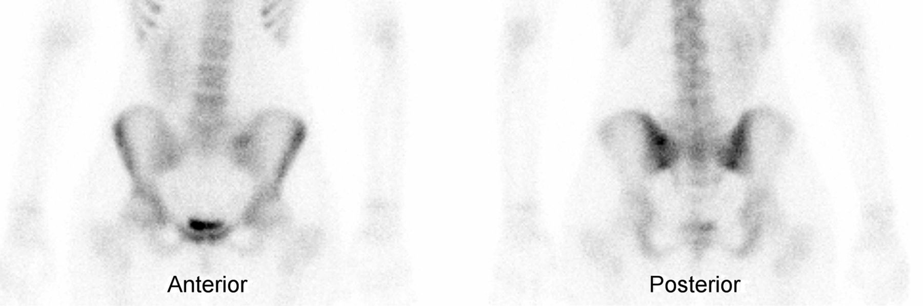
A 16-y-old female presented with lower back pain for 8 mo. Her only reported medical history was left renal agenesis. She was a cheerleader but denied history of recent trauma. A bone scan was requested for suspected pars interarticularis defect. Whole-body bone scintigraphy was performed approximately 3 h after intravenous injection of 762 MBq (20.6 mCi) of 99mTc-methylene diphosphonate. Planar images showed a focus of increased radiotracer activity in the left lumbosacral area overlying the left sacroiliac joint (Fig. 1). Because this pattern was considered atypical for pars interarticularis fracture, we obtained another planar image with a marker placed over the area of pain and additional SPECT with low-dose CT imaging of the lumbosacral spine, instead of our routine protocol for pars interarticularis defect (SPECT without CT). The SPECT/CT images localized the focus of increased radiotracer activity to a hemisacralized left transverse process of L5 (Fig. 2). The CT appearance was consistent with a type IIa LSTV.

FIGURE 1.
  • Download figure
  • Open in new tab
  • Download powerpoint
FIGURE 1.

Anterior and posterior spot images of postvoid pelvis show focus of increased radiotracer activity in left lumbosacral area overlying left sacroiliac joint. Marker used to denote site of pain correlated closely to focus of increased radiotracer intensity (not shown).

FIGURE 2.
  • Download figure
  • Open in new tab
  • Download powerpoint
FIGURE 2.

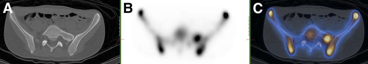
SPECT (A), low-dose CT (B), and hybrid SPECT/CT (C) images of lower lumbar spine and pelvis reveal hemisacralized left transverse process at level L5. Hybrid SPECT/CT image shows that focus of increased radiotracer activity corresponds to hemisacralized left transverse process of L5.

DISCUSSION

The lumbosacral junction often exhibits a transitional vertebra with sacralization of the lower-most lumbar segment or lumbarization of superior-most sacral segment. The Castellvi system is most commonly used for morphologic classification of LSTV (1). It defines types I–IV, with type I characterized by an enlarged (>19 mm) transverse process, type II representing vertebra with an incompletely fused broad transverse process forming a diarthrodial joint with the sacrum, and type III having complete fusion of the transverse process with the sacrum. Under each type, unilateral findings are denoted as subtype a, and bilateral findings are denoted b. Type IV consists of a unilateral complete fusion with contralateral incomplete fusion (type III on one side and type II on the other).

LSTV can be diagnosed using several imaging modalities. Historically, an anteroposterior radiograph of the lumbosacral junction angled 30° cranially, known as a Ferguson radiograph, was the best study for evaluating transitional vertebrae (2). However, with the widespread use of CT and MR imaging, LSTV is now usually incidentally discovered on CT or MR imaging done for workup of back pain or for other reasons. Low-back pain associated with a LSTV may arise from the level above the transition, the contralateral facet when unilateral, or the anomalous articulation when present (3). In addition to standard radiologic modalities, nuclear medicine studies can be useful in the workup of LSTV.

SPECT can play an important role in the evaluation of patients with LSTV and lower back pain. Although the presence of a transitional vertebra can be established with plain film radiography, CT, or MR imaging, demonstration of stress at the transverse–sacral articulation is best accomplished using bone scanning with SPECT (4). The finding of intense radiotracer uptake at the transverse–sacral articulation in patients with LSTV seems to correlate with lower back pain symptoms. This is because the increase in radiotracer uptake will identify sites of increased osseous metabolic activity at the LSTV articulation site (5). These findings suggest that SPECT imaging can be a good tool for determining whether lower back pain in patients with LSTV is caused by the transitional vertebra or attributable to other causes. We believe hybrid SPECT/CT imaging improves the diagnostic capabilities of the bone scan by localizing the increased osseous activity more accurately.

CONCLUSION

LSTV is a congenital malformation of the lumbosacral spine. It is mostly asymptomatic; however, it can present with lower back pain. Bertolotti syndrome should always be included in the differential diagnosis of lower back pain in young patients. LSTV can be diagnosed using several imaging modalities including radiography, MR imaging, and CT. This case shows that a whole-body bone scan with hybrid SPECT/CT imaging of the area of pain can be a tool to help diagnose LSTV.

DISCLOSURE

No potential conflict of interest relevant to this article was reported.

Footnotes

  • Published online Feb. 5, 2015.

REFERENCES

  1. 1. ↵
    1. Castellvi AE ,
    2. Goldstein LA ,
    3. Chan DP
    . Lumbosacral transitional vertebrae and their relationship with lumbar extradural defects. Spine (Phila Pa 1976). 1984;9:493–495.
  2. 2. ↵
    1. Hughes RJ ,
    2. Saifuddin A
    . Imaging of lumbosacral transitional vertebrae. Clin Radiol. 2004;59:984–991.
    OpenUrlCrossRefPubMed
  3. 3. ↵
    1. Konin GP ,
    2. Walz DM
    . Lumbosacral transitional vertebrae: classification, imaging findings, and clinical relevance. AJNR. 2010;31:1778–1786.
    OpenUrlAbstract/FREE Full Text
  4. 4. ↵
    1. Connolly LP ,
    2. d'Hemecourt PA ,
    3. Connolly SA ,
    4. Drubach LA ,
    5. Micheli LJ ,
    6. Treves ST
    . Skeletal scintigraphy of young patients with low-back pain and a lumbosacral transitional vertebra. J Nucl Med. 2003;44:909–914.
    OpenUrlAbstract/FREE Full Text
  5. 5. ↵
    1. Pekindil G ,
    2. Sarikaya A ,
    3. Pekindil Y ,
    4. Gültekin A ,
    5. Kokino S
    . Lumbosacral transitional vertebra articulation: evaluation by planar and SPECT bone scintigraphy. Nucl Med Commun. 2004;25:29–37.
    OpenUrlCrossRefPubMed
  • Received for publication August 2, 2014.
  • Accepted for publication October 23, 2014.
PreviousNext
Back to top

In this issue

Journal of Nuclear Medicine Technology: 43 (2)
Journal of Nuclear Medicine Technology
Vol. 43, Issue 2
June 1, 2015
  • Table of Contents
  • About the Cover
  • Index by author
Print
Download PDF
Article Alerts
Sign In to Email Alerts with your Email Address
Email Article

Thank you for your interest in spreading the word on Journal of Nuclear Medicine Technology.

NOTE: We only request your email address so that the person you are recommending the page to knows that you wanted them to see it, and that it is not junk mail. We do not capture any email address.

Enter multiple addresses on separate lines or separate them with commas.
Lumbosacral Transitional Vertebra Diagnosed on 99mTc-Methylene Diphosphonate SPECT/CT
(Your Name) has sent you a message from Journal of Nuclear Medicine Technology
(Your Name) thought you would like to see the Journal of Nuclear Medicine Technology web site.
Citation Tools
Lumbosacral Transitional Vertebra Diagnosed on 99mTc-Methylene Diphosphonate SPECT/CT
Mohammad A. Kassir, Zaid Al-faham, Nicholas Abel, Helena R. Balon
Journal of Nuclear Medicine Technology Jun 2015, 43 (2) 137-138; DOI: 10.2967/jnmt.114.146639

Citation Manager Formats

  • BibTeX
  • Bookends
  • EasyBib
  • EndNote (tagged)
  • EndNote 8 (xml)
  • Medlars
  • Mendeley
  • Papers
  • RefWorks Tagged
  • Ref Manager
  • RIS
  • Zotero
Share
Lumbosacral Transitional Vertebra Diagnosed on 99mTc-Methylene Diphosphonate SPECT/CT
Mohammad A. Kassir, Zaid Al-faham, Nicholas Abel, Helena R. Balon
Journal of Nuclear Medicine Technology Jun 2015, 43 (2) 137-138; DOI: 10.2967/jnmt.114.146639
Twitter logo Facebook logo LinkedIn logo Mendeley logo
  • Tweet Widget
  • Facebook Like
  • Google Plus One
Bookmark this article

Jump to section

  • Article
    • Abstract
    • CASE REPORT
    • DISCUSSION
    • CONCLUSION
    • DISCLOSURE
    • Footnotes
    • REFERENCES
  • Figures & Data
  • Info & Metrics
  • PDF

Related Articles

  • No related articles found.
  • PubMed
  • Google Scholar

Cited By...

  • No citing articles found.
  • Google Scholar

More in this TOC Section

  • High-Sensitivity Troponin Elevation in a Young Woman with Typical Chest Pain: The Heart of the Matter
  • Pulmonary Adenocarcinoma Revealed by Parathyroid Scintigraphy: An Incidental Case to Remember
  • Prominent Right Ventricular Tracer Uptake: A Harbinger of Multivessel Coronary Artery Disease
Show more Teaching Case Studies

Similar Articles

Keywords

  • bone
  • SPECT
  • SPECT/CT
  • Bertolloti Syndrome
  • Lumbosacral Transitional Vertebra
SNMMI

© 2025 SNMMI

Powered by HighWire