
Main menu

  • Home
  • Content
    • Current
      • JNMT Supplement
    • Ahead of print
    • Past Issues
    • Continuing Education
    • JNMT Podcast
    • SNMMI Annual Meeting Abstracts
  • Subscriptions
    • Subscribers
    • Rates
    • Journal Claims
    • Institutional and Non-member
  • Authors
    • Submit to JNMT
    • Information for Authors
    • Assignment of Copyright
    • AQARA Requirements
  • Info
    • Reviewers
    • Permissions
    • Advertisers
    • Corporate & Special Sales
  • About
    • About Us
    • Editorial Board
    • Contact Information
  • More
    • Alerts
    • Feedback
    • Help
    • SNMMI Journals
  • SNMMI
    • JNMT
    • JNM
    • SNMMI Journals
    • SNMMI

User menu

  • Subscribe
  • My alerts
  • Log in
  • Log out
  • My Cart

Search

  • Advanced search
Journal of Nuclear Medicine Technology
  • SNMMI
    • JNMT
    • JNM
    • SNMMI Journals
    • SNMMI
  • Subscribe
  • My alerts
  • Log in
  • Log out
  • My Cart
Journal of Nuclear Medicine Technology

Advanced Search

  • Home
  • Content
    • Current
    • Ahead of print
    • Past Issues
    • Continuing Education
    • JNMT Podcast
    • SNMMI Annual Meeting Abstracts
  • Subscriptions
    • Subscribers
    • Rates
    • Journal Claims
    • Institutional and Non-member
  • Authors
    • Submit to JNMT
    • Information for Authors
    • Assignment of Copyright
    • AQARA Requirements
  • Info
    • Reviewers
    • Permissions
    • Advertisers
    • Corporate & Special Sales
  • About
    • About Us
    • Editorial Board
    • Contact Information
  • More
    • Alerts
    • Feedback
    • Help
    • SNMMI Journals
  • Watch or Listen to JNMT Podcast
  • Visit SNMMI on Facebook
  • Join SNMMI on LinkedIn
  • Follow SNMMI on Twitter
  • Subscribe to JNMT RSS feeds
Review ArticleTEACHING FILE

13-Year-Old Boy with a 3-Day History of Melena

Katie Traylor, Laura Gonzalez-Krellwitz and Helena Balon
Journal of Nuclear Medicine Technology June 2012, 40 (2) 119-121; DOI: https://doi.org/10.2967/jnmt.112.106310
Katie Traylor
  • Find this author on Google Scholar
  • Find this author on PubMed
  • Search for this author on this site
Laura Gonzalez-Krellwitz
  • Find this author on Google Scholar
  • Find this author on PubMed
  • Search for this author on this site
Helena Balon
  • Find this author on Google Scholar
  • Find this author on PubMed
  • Search for this author on this site
  • Article
  • Figures & Data
  • Info & Metrics
  • PDF
Loading

A 13-y-old boy presented to the emergency center with a 3-d history of melena. On admission the patient was anemic (hemoglobin = 9.3 g/dL, hematocrit = 26.2%). The patient was injected with 334 MBq (9 mCi) of 99mTc-pertechnetate. Anterior, posterior, and lateral views of the abdomen were acquired at 15, 40, and 60 min after injection.

QUESTION 1

On examining all the images, what is the ordering physician evaluating? Please select the best answer.

  • A. Gastrointestinal bleeding.

  • B. Bleeding from diverticulitis.

  • C. Meckel diverticulum.

  • D. Appendicitis.

FIGURE 1.
  • Download figure
  • Open in new tab
  • Download powerpoint
FIGURE 1.

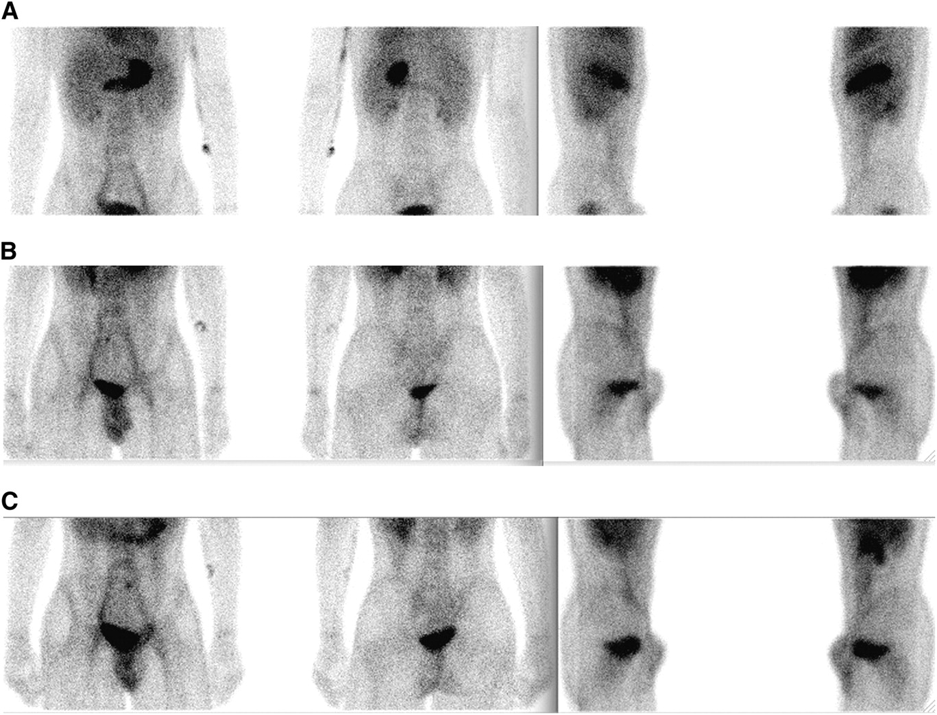
(A) Anterior, posterior, and lateral projections at 15 min after injection; (B) anterior, posterior, and lateral projections at 40 min after injection; and (C) anterior, posterior, and lateral projections at 60 min after injection.

QUESTION 2

Which of the following would cause a false-negative finding on a Meckel scan?

  • A. Ulcerative colitis.

  • B. Intestinal obstruction.

  • C. Laxative abuse.

  • D. Irritable bowel in the right lower quadrant.

FIGURE 2.
  • Download figure
  • Open in new tab
  • Download powerpoint
FIGURE 2.

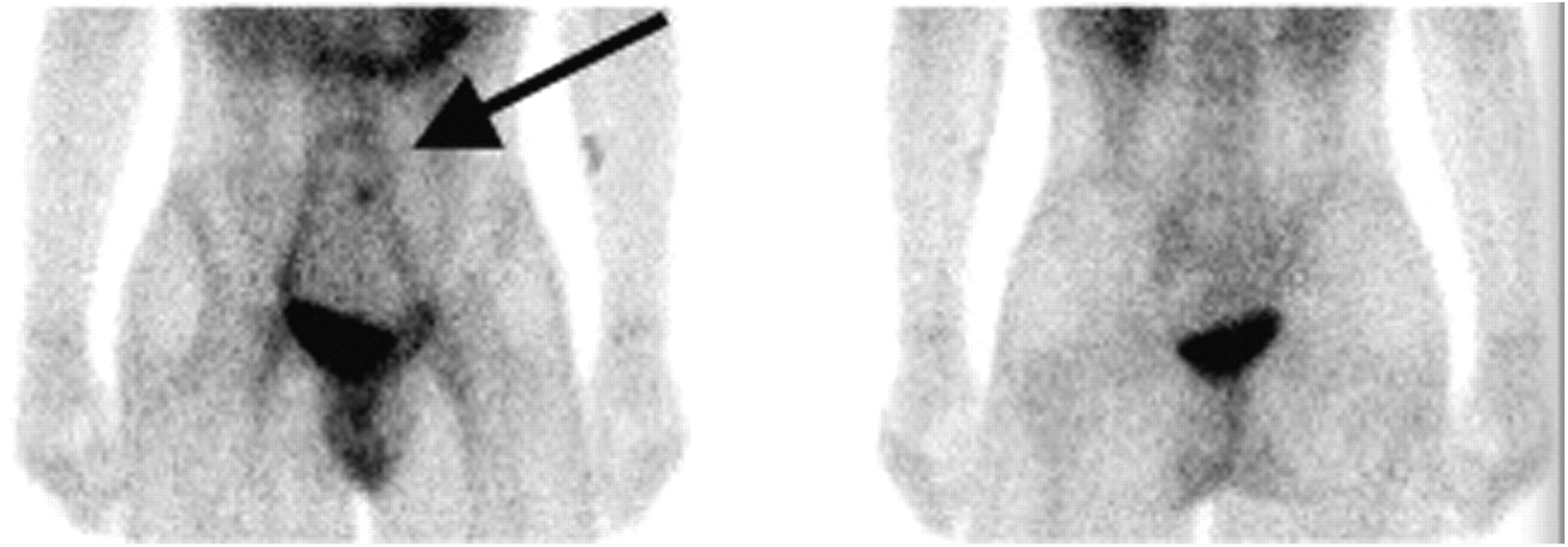
Anterior and posterior projections at 60 min after injection. Arrow illustrates focus of ectopic gastric mucosa in Meckel diverticulum.

QUESTION 3

What pharmacologic interventions can be used to improve visualization of the Meckel diverticulum by decreasing bowel peristalsis?

  • A. 20 mg of cimetidine per kilogram per day for 2 d before study.

  • B. 50 μg of glucagon per kilogram administered intravenously 5–10 min before the study.

  • C. 6 μg/kg of pentagastrin 5–10 min before the study.

  • D. 0.07 mL of saturated solution of potassium iodide given to the patient before injection and continued 3 d after the study.

CASE DISCUSSION

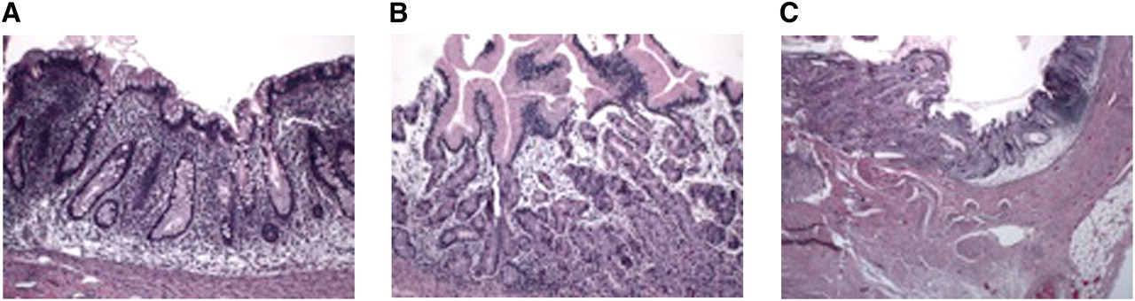
Meckel diverticulum is the most common congenital abnormality and arises because of persistence of the omphalomesenteric duct (vitelline duct), which normally should obliterate by the fifth week of gestation. It affects approximately 2% of the population, is usually symptomatic in children less than 2 y old, is located within 61 cm (2 ft) of the ileocecal valve, and usually has a length of 5 cm (2 in). Meckel diverticula contain ectopic mucosa in 50% of the cases, which could include gastric, pancreatic, or colonic tissue (1–3). The diverticulum causes complications in 20% of the patients, such as gastrointestinal bleeding due to ulceration, acute diverticulitis, intestinal obstruction due to intussusception, or chronic abdominal pain (4,5). The normal gastric mucosa contains not only parietal, chief, and G cells but also columnar mucin-secreting cells—the cell type that concentrates pertechnetate. Detection of ectopic gastric mucosa is the basis for the Meckel scan (4). Figure 3 illustrates the histology of colonic and gastric mucosa and compares it with the histology of ectopic gastric mucosa from our patient’s Meckel diverticulum.

FIGURE 3.
  • Download figure
  • Open in new tab
  • Download powerpoint
FIGURE 3.

(A) Colonic mucosa, (B) gastric mucosa in stomach, and (C) transition point from colonic mucosa to ectopic gastric mucosa in our patient. Left side of slide illustrates ectopic gastric mucosa, with transition to colonic mucosa on right side of slide.

The pertechnetate scan is more than 85% sensitive, more than 95% specific, and 83%–88% accurate in identifying ectopic gastric mucosa. 99mTc-pertechnetate is concentrated by mucoid cells of the gastric mucosa, and its excretion is not dependent on the presence of parietal cells (3,5). The sensitivity of this scan decreases after adolescence because patients who are asymptomatic throughout childhood are less likely to have ectopic gastric mucosa (1,3). A typical positive scan should have a small focus of increased radiotracer accumulation, usually within the right lower quadrant, which appears at the same time as or shortly after visualization of the stomach; however, a small Meckel diverticulum may appear later than the stomach. The intensity of radiotracer activity should increase with time parallel to that of the stomach (6). It is important to realize that a Meckel diverticulum that does not contain gastric mucosa will not be identified by this scan.

Acknowledgments

The histology photos were prepared by Laura Gonzalez-Krellwitz, from the William Beaumont Hospital Department of Pathology.

Footnotes

  • Published online May 8, 2012.

  • ↵* For the answers, see page 20A

REFERENCES

  1. 1.↵
    1. Howarth DM
    . The role of nuclear medicine in the detection of acute gastrointestinal bleeding. Semin Nucl Med. 2006;36:133–146.
    OpenUrlCrossRefPubMed
  2. 2.
    1. Seth A,
    2. Seth J
    . Axial torsion as a rare and unusual complication of a Meckel’s diverticulum: a case report and review of the literature. J Med Case Reports. 2011;5:118.
    OpenUrlCrossRefPubMed
  3. 3.↵
    1. Malik AA,
    2. Shams-ul-Bari,
    3. Wani KA,
    4. Khaja AR
    . Meckel’s diverticulum: revisited. Saudi J Gastroenterol. 2010;16:3–7.
    OpenUrlCrossRefPubMed
  4. 4.↵
    1. Kiratli PO,
    2. Aksoy T,
    3. Bozkurt MF,
    4. Orhan D
    . Detection of ectopic gastric mucosa using 99mTc-pertechnetate: review of the literature. Ann Nucl Med. 2009;23:97–105.
    OpenUrlCrossRefPubMed
  5. 5.↵
    1. Rossi P,
    2. Gourtsoyiannis N,
    3. Bezzi M,
    4. et al
    . Meckel’s diverticulum: imaging diagnosis. AJR. 1996;166:567–573.
    OpenUrlPubMed
  6. 6.↵
    1. Mariani G,
    2. Pauwels EK,
    3. AlSharif A,
    4. et al
    . Radionuclide evaluation of the lower gastrointestinal tract. J Nucl Med. 2008;49:776–787.
    OpenUrlAbstract/FREE Full Text
PreviousNext
Back to top

In this issue

Journal of Nuclear Medicine Technology: 40 (2)
Journal of Nuclear Medicine Technology
Vol. 40, Issue 2
June 1, 2012
  • Table of Contents
  • About the Cover
  • Index by author
Print
Download PDF
Article Alerts
Sign In to Email Alerts with your Email Address
Email Article

Thank you for your interest in spreading the word on Journal of Nuclear Medicine Technology.

NOTE: We only request your email address so that the person you are recommending the page to knows that you wanted them to see it, and that it is not junk mail. We do not capture any email address.

Enter multiple addresses on separate lines or separate them with commas.
13-Year-Old Boy with a 3-Day History of Melena
(Your Name) has sent you a message from Journal of Nuclear Medicine Technology
(Your Name) thought you would like to see the Journal of Nuclear Medicine Technology web site.
Citation Tools
13-Year-Old Boy with a 3-Day History of Melena
Katie Traylor, Laura Gonzalez-Krellwitz, Helena Balon
Journal of Nuclear Medicine Technology Jun 2012, 40 (2) 119-121; DOI: 10.2967/jnmt.112.106310

Citation Manager Formats

  • BibTeX
  • Bookends
  • EasyBib
  • EndNote (tagged)
  • EndNote 8 (xml)
  • Medlars
  • Mendeley
  • Papers
  • RefWorks Tagged
  • Ref Manager
  • RIS
  • Zotero
Share
13-Year-Old Boy with a 3-Day History of Melena
Katie Traylor, Laura Gonzalez-Krellwitz, Helena Balon
Journal of Nuclear Medicine Technology Jun 2012, 40 (2) 119-121; DOI: 10.2967/jnmt.112.106310
Twitter logo Facebook logo LinkedIn logo Mendeley logo
  • Tweet Widget
  • Facebook Like
  • Google Plus One
Bookmark this article

Jump to section

  • Article
    • QUESTION 1
    • QUESTION 2
    • QUESTION 3
    • CASE DISCUSSION
    • Acknowledgments
    • Footnotes
    • REFERENCES
  • Figures & Data
  • Info & Metrics
  • PDF

Related Articles

  • No related articles found.
  • PubMed
  • Google Scholar

Cited By...

  • No citing articles found.
  • Google Scholar

More in this TOC Section

  • Brain SPECT Imaging with Acetazolamide Challenge
  • Scintigraphic Evaluation of a Suspected Cavernous Hemangioma
  • Evaluation of Pleural-Based Nodules in a 52-Year-Old Woman with a History of Metastatic Breast Cancer
Show more Teaching File

Similar Articles

SNMMI

© 2025 SNMMI

Powered by HighWire