

FIGURE 17.
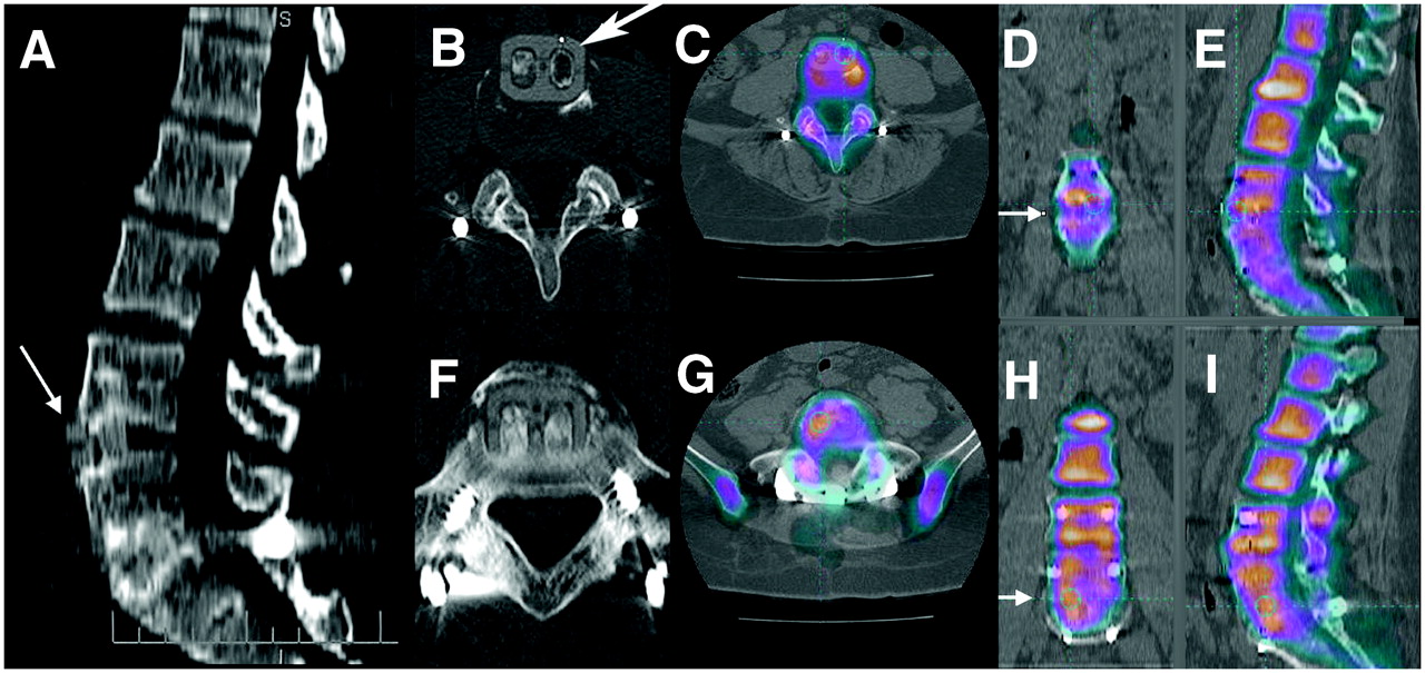
Failed front–back lumbar fusion: patient fell after recent surgery. (A) CT sagittal reformatted image shows that L4–L5 anterior interbody cassette has subluxed forward (arrow). L5–S1 cassette is in place. (B and C) Transaxial CT image of slipped cassette at L4–L5 (arrow) and fused PET bone/CT image of area of devitalized bone. (D and E) 18F and CT fusion in coronal and sagittal planes confirms devitalization of bone grafts at L4–L5. No activity crosses space between L4 and L5 (arrow in D). (F and G) CT and CT fusion of L5–S1. (H and I) Images showing that vital bone links L5 and S1 (arrow in H), unlike absence of activity across endplates at L4–L5. Improved resolution of 18F PET bone scanning allows interrogation of separate channels of cassettes.
In this issue


Related Articles
Cited By...
- Bone-Targeted Imaging and Radionuclide Therapy in Prostate Cancer
- 18F-Fluoride PET in the Assessment of Malignant Bone Disease
- The Value of Observer Performance Studies in Dose Optimization: A Focus on Free-Response Receiver Operating Characteristic Methods
- Validation of a Paper Chromatographic Methodology as an Alternative for Determination of the Radiochemical Purity of Na18F
- Software Fusion: An Option Never Fully Explored