


Abstract
Objective:
Quantitative analysis of 99mTc-pertechnetate salivary gland scintigraphy has been used in the evaluation of salivary gland function, but so far no one method can be considered optimal for this task. In this study, a semiquantitative method providing 2 functional parameters for objective assessment of salivary gland function by scintillation camera imaging was tested.
Methods:
Twenty-one patients referred for 99mTc-pertechnetate thyroid scanning were studied. Two patients with salivary complaints were also included. Dynamic imaging of the anterior head using a scintillation camera was started after a bolus intravenous injection of 185 MBq (5 mCi) 99mTc-pertechnetate at 1 frame per 30 s for 30 min. At 15 min after injection, diluted lemon juice was administered orally. Analysis of the dynamic study included time–activity curves of 4 salivary glands (right and left parotid and right and left submandibular). Two parameters of function were defined: uptake rate, taken as the value of the initial slope of the time–activity curve, and washout fraction, which was the relative mobilizable radioactivity from each salivary gland after ingestion of the sialogogue. A parametric image of the washout fraction was also generated.
Results:
The images showed gradual uptake in the parotid and submandibular glands. Washout was noted immediately after ingestion of the lemon juice. The pattern of the time–activity curve in all glands showed an early fast-rising part followed by a slow-rising component to nearly a plateau within 6–10 min after injection. The mean value of the uptake rate parameter was 0.10 ± 0.09 cps/s. There was no significant difference between the parotid and submandibular glands or the right and left sides. Uptake in the parotid gland was 1.5–2 times that in the submandibular gland. The washout fraction was 1.40 ± 1.60 for the parotid glands and 0.77 ± 0.41 for the submandibular glands (P = 0.005).
Conclusion:
The quantitative analysis method including the uptake rate and the washout fraction parameters would enable objective assessment of salivary function and provide a reproducible means for follow-up of functional impairment in certain diseases.
Salivary gland dysfunction is encountered in various pathologic processes presenting as a dry mouth or, rarely, overproduction of saliva (1). The diagnosis of salivary gland disease relies mainly on the clinical presentation and in certain conditions on more specialized investigations (2). Of the few tests currently available for investigation of salivary gland function, sodium pertechnetate scintigraphy is a noninvasive one (3). This technique provides images of the parotid and submandibular glands. In addition, physiologic intervention by administration of a sialogogue such as lemon juice provides information on the patency of the salivary ducts and on the overall functional integrity of the system.
Salivary scintigraphy has been useful for investigation of multiple diseases affecting the salivary glands. In Sjögren’s syndrome, pertechnetate imaging can gauge the severity of salivary gland involvement, which may not be accurately reflected by the symptoms (i.e., xerostomia and other features of the sicca syndrome) (4,5). In other clinical situations, such as iatrogenic irradiation of the salivary glands for therapy of head and neck tumors or radioiodine treatment of thyroid cancer, salivary scintigraphy helps assess functional damage and monitor recovery in these patients (6,7).
Recently, salivary scintigraphy has been refined toward providing quantitative information on changes in gland function after parenchymal insult, whether inflammatory or radiation induced. Most attempts have used data derived from the kinetics of uptake and clearance of radioactivity in the major salivary glands (8–11). In addition, grading and comparison of uptake in the salivary gland in successive scans have been used to objectively and reproducibly follow changes, thus indicating progression or improvement of the pathologic condition (7,12).
In this report, we present a semiquantitative method based on analysis of the kinetics of uptake of pertechnetate in the salivary glands for accurate and reproducible assessment of function. Two functional parameters are introduced. The first reflects the uptake rate of the radiotracer by the individual salivary gland. The second is the washout fraction from each salivary gland, given in terms of the relative amount of mobilizable saliva from the gland after stimulation with a sialogogue (lemon juice). It is expected that such quantitation will improve diagnostic accuracy and allow for objective follow-up of pathologic conditions of the salivary glands.
MATERIALS AND METHODS
Subjects
Twenty-one patients referred over a 6-mo period for 99mTc-pertechnetate thyroid scanning were included in this study (mean age, 34 y; female-to-male ratio, 2.5:1). The data on individual patient age and sex are shown in Table 1. The patients did not complain of salivary gland abnormalities but were referred for thyroid scanning for various reasons, including thyroid nodules and hyper- or hypothyroidism. All patients gave informed consent to participate in the study, which did not alter their future management. The approval of the local ethics committee was also obtained.
Age, Sex, Uptake Rate, and Washout Fraction of Patients Studied
In addition, 2 patients who had been referred because of suspected abnormalities of the salivary glands were studied. The first patient, a 40-y-old man, complained of atypical pain behind the jaw, and in the second, a 45-y-old man, Sjögren’s syndrome had been diagnosed.
Imaging Technique
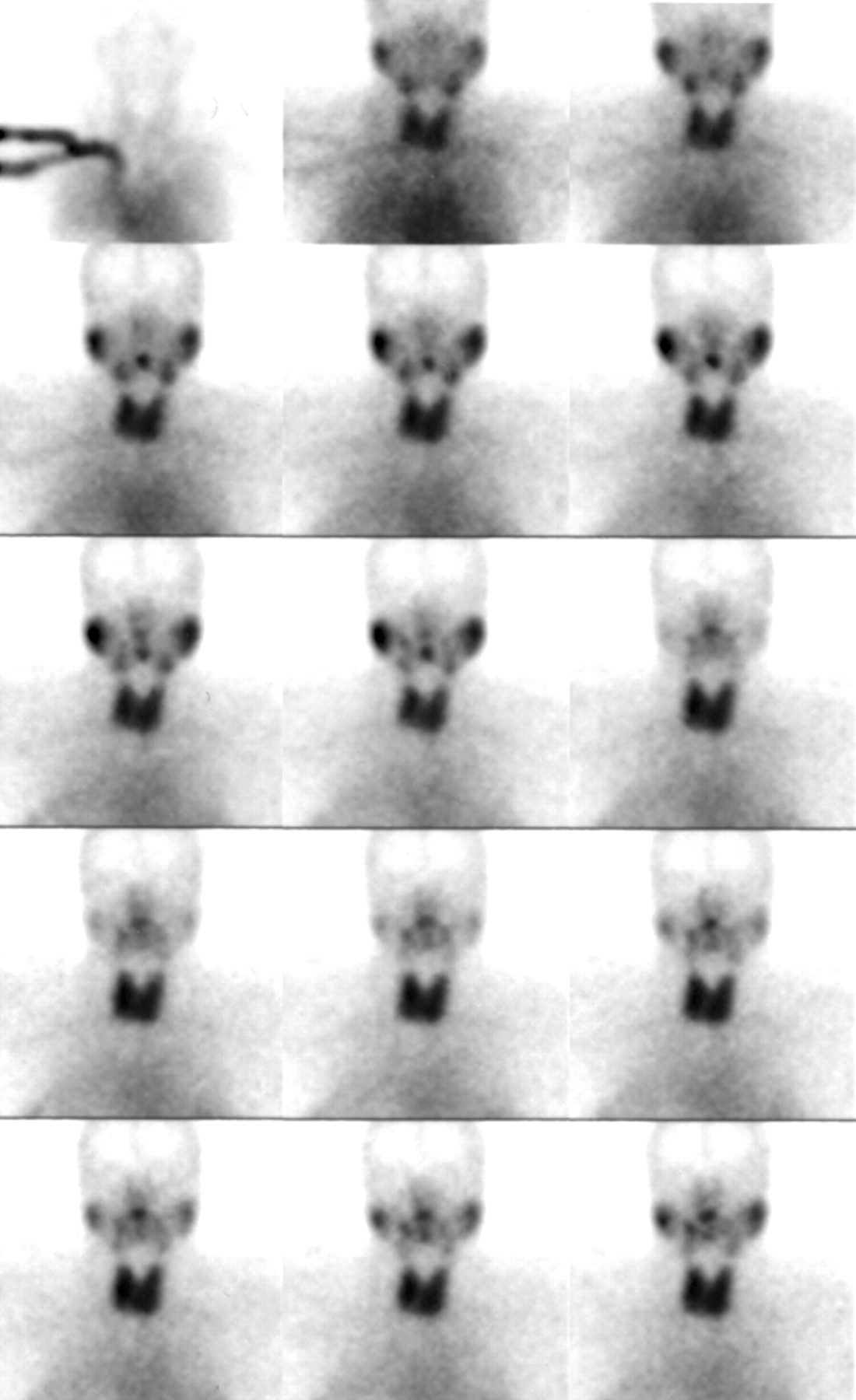
Imaging was performed using a mobile small-field-of-view scintillation camera (Starcam 300; General Electric Medical Systems) equipped with a low-energy all-purpose parallel-hole collimator. The patient was supine, and the camera was positioned for an anterior head-and-neck projection. Dynamic imaging was performed in a 64 × 64 pixel matrix at 30 s per frame starting immediately after a bolus intravenous injection of 185 MBq (5 mCi) 99mTc-sodium pertechnetate. Imaging continued for 30 min after injection. At 15 min after injection, each patient drank the freshly squeezed juice of half a lemon in water, using a straw and without moving, while imaging was continued. The study was reviewed to check for patient motion and for suitability for further analysis (Fig. 1). Afterward, standard imaging of the thyroid proceeded as set out in our protocol manual, using the same camera, to address the question for which the patient was referred.

Reframed dynamic images (1 min per frame) show parotid and submandibular glands. Washout after lemon juice is seen halfway through study (3rd frame of 3rd row).
Analysis
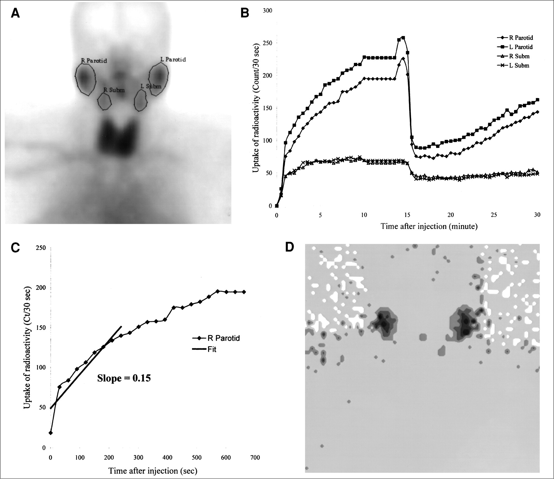
Regions of interest were drawn on the dynamic images of the parotid glands (right and left) and submandibular glands (right and left) (Fig. 2). Time–activity curves were generated for each region. These were analyzed as follows: First, the initial slope of the time–activity curve for each salivary gland was calculated from a linear fit of the rapidly rising part of the curve 0–240 s after injection of the radiotracer. This value was considered the uptake rate parameter of the salivary gland and was expressed as count rate per second (cps/s). Second, the response of the salivary gland to lemon juice was noted on the time–activity curves as a sharp decline of the activity in the gland with subsequent slow buildup. The washout fraction in each salivary gland was defined as the amount of cleared radioactivity from the gland divided by the postclearance activity (nonmobilizable uptake). A parametric image of the washout fraction in each gland was generated by subtraction of the image immediately after washout from the image at peak activity, followed by division of the result by the former image (peak − post/post).

(A) Regions of interest drawn around individual salivary glands. (B) Time–activity curves of parotid and submandibular glands. (C) Linear curve fit for right parotid curve from 0 to 240 s. (D) Parametric washout fraction image. Ct = count; Subm = submandibular.
The parotid and submandibular glands and the right and left sides were compared for these parameters using the paired t test. A P value < 0.05 was considered statistically significant.
RESULTS
The time–activity curves of the individual salivary glands showed an early fast-rising uptake followed by a slow-rising component to nearly a plateau in all glands (Fig. 2).
In Table 1, the results of the calculated parameters of salivary gland function (i.e., uptake rate and washout fraction for the parotid and submandibular glands) are exhibited.
The average value for uptake rate was 0.10 ± 0.09 cps/s. The uptake rates were similar for all salivary glands and did not significantly differ between parotid and submandibular glands or right and left sides. The P values for the comparisons using the paired t test were as follows: P = 0.17 for parotid versus submandibular, P = 0.28 for right versus left parotid, and P = 0.81 for right versus left submandibular.
The washout fraction showed similar values for the right and left parotid glands (P = 0.21) and for the right and left submandibular glands (P = 0.09). However, a significant difference was noted between the parotid and submandibular glands (P = 0.005), with the parotid glands exhibiting a higher washout fraction (1.40 ± 1.60) than the submandibular glands (0.77 ± 0.41). A parametric image of the washout fraction of the salivary glands is shown in Figure 2.
Of the 2 patients with suspected salivary gland pathology, the first showed normal scan findings and parameters within the range of the controls. In contrast, the second showed a markedly depressed washout fraction (parotid, 0.4; submandibular, 0.15), whereas the uptake rate (0.07 in this patient) remained within the average range shown above.
DISCUSSION
In this study, a semiquantitative method for analysis of 99mTc-sodium pertechnetate salivary scintigraphy was used to assess the function of, as well as image, the parotid and submandibular glands. Semiquantitation was performed using a standard dynamic imaging protocol that included administration of a sialogogue halfway through the dynamic study. Two parameters of function were calculated, namely uptake rate and washout fraction, providing information that could be useful to categorize the effects of insults to the salivary glands.
To establish the method of quantitation and provide guidelines for its use, including expected values and normal range, studies of 21 healthy volunteers were performed. These subjects were selected from a pool of patients referred for thyroid scintigraphy who did not have a history or clinical evidence of salivary gland dysfunction. The possibility that thyroid disease could affect the results of the quantitation method was considered minimal, since only relative quantitation was performed and thyroid uptake would usually be a small fraction of the total injected dose (0.7%–3.0%) (13).
The uptake rate parameter was derived from the early rising part of the individual salivary gland time–activity curve. It represented a simple measure of how well the gland could extract the radiotracer from the circulation, thus avoiding the need for absolute quantitation of uptake and ignoring the subsequent fate of the radiotracer in the gland (dealt with using the second parameter). The linear fit of the time–activity curves from 0 to 240 s was chosen because progressive accumulation, approximating a straight line, was seen in the salivary glands during that time. A diseased salivary gland would be expected to show a slowly rising curve with a resulting decreased slope, compared with normal.
The washout fraction parameter would be most useful to track subtle changes in salivary gland function, providing a marker for follow-up with time. The parameter calculation was based on the assumption that once the plateau of the time–activity curves was reached (usually between 6 and 10 min after injection), radioactivity in the salivary gland was located in 2 separate pools: a mobilizable one in the excreted saliva and a nonmobilizable one remaining in the parenchymal cells. The 2 pools were separated using intervention with a sialogogue at 15 min after injection. The ratio of the mobilizable radioactivity to the remaining activity gave a measure of the specific function, denoted as the washout fraction, of the individual salivary gland. Our observation that the washout fraction of the parotid glands was higher than that of the submandibular glands agreed with other reports in the literature and might have had to do with the type of saliva produced (parotid, serous, vs. submandibular, mucous) (14). This parameter helped us recognize and confirm the disturbance in the second patient and provided a baseline for future follow-up.
A review of previous reports on quantitation of salivary gland function, comparing the 2 parameters introduced in this study with others (3,4,10,12,15,16), showed that most functional indices were derived from individual salivary gland time–activity curves generated from a dynamic study similar to that outlined here. In certain cases, the functional indicators were somewhat difficult to define and would vary with the amount of injected radioactivity, requiring absolute quantitation of the uptake for reproducibility (12). The semiquantitative reporting of the uptake and clearance data in the salivary time–activity curve (i.e., the slope of the curve and the ratios of counts), as presented in this study, would make the data easier to reproduce using different imaging equipment or amounts of administered radioactivity.
The clinical experience with the 2 patients suspected of salivary gland disease in this study encourages the use of the semiquantitative method when salivary function impairment is suspected, the rationale being that function is assessed from 2 different aspects, for changes occurring in disease that are related to separate impairment of blood flow and secretory stimulation (8). The application of the semiquantitative method to a larger number of patients with salivary dysfunction is required, however, to establish the usefulness of the method in clinical practice.
CONCLUSION
Salivary scintigraphy using the described semiquantitative method including the uptake rate and washout fraction parameters would provide an objective means of diagnosis and a reproducible tool for follow-up of salivary function impairment.
Acknowledgments
Part of this research was presented orally, with the same title, at the technologist session of the 48th annual meeting of the Society of Nuclear Medicine, Toronto, Ontario, Canada, June 2001.
Footnotes
For correspondence or reprints contact: Issa Loutfi, MD, PhD, Department of Nuclear Medicine, Faculty of Medicine, Kuwait University, P.O. Box 24923, Safat 13110, Kuwait.
E-mail: loutfi{at}hsc.kuniv.edu.kw
REFERENCES


Jump to section
Related Articles
Cited By...
- Assessment of Salivary Gland Dysfunction after Radioiodine Therapy for Thyroid Carcinoma Using Non-Contrast-Enhanced CT: The Significance of Changes in Volume and Attenuation of the Glands
- Infrequently Performed Studies in Nuclear Medicine: Part 2
- Application of 99mTc-Pertechnetate Scintigraphy to Microvascular Autologous Transplantation of the Submandibular Gland in Patients with Severe Keratoconjunctivitis Sicca
- Does Lemon Candy Decrease Salivary Gland Damage After Radioiodine Therapy for Thyroid Cancer?