
Main menu

  • Home
  • Content
    • Current
      • JNMT Supplement
    • Ahead of print
    • Past Issues
    • Continuing Education
    • JNMT Podcast
    • SNMMI Annual Meeting Abstracts
  • Subscriptions
    • Subscribers
    • Rates
    • Journal Claims
    • Institutional and Non-member
  • Authors
    • Submit to JNMT
    • Information for Authors
    • Assignment of Copyright
    • AQARA Requirements
  • Info
    • Reviewers
    • Permissions
    • Advertisers
    • Corporate & Special Sales
  • About
    • About Us
    • Editorial Board
    • Contact Information
  • More
    • Alerts
    • Feedback
    • Help
    • SNMMI Journals
  • SNMMI
    • JNMT
    • JNM
    • SNMMI Journals
    • SNMMI

User menu

  • Subscribe
  • My alerts
  • Log in
  • My Cart

Search

  • Advanced search
Journal of Nuclear Medicine Technology
  • SNMMI
    • JNMT
    • JNM
    • SNMMI Journals
    • SNMMI
  • Subscribe
  • My alerts
  • Log in
  • My Cart
Journal of Nuclear Medicine Technology

Advanced Search

  • Home
  • Content
    • Current
    • Ahead of print
    • Past Issues
    • Continuing Education
    • JNMT Podcast
    • SNMMI Annual Meeting Abstracts
  • Subscriptions
    • Subscribers
    • Rates
    • Journal Claims
    • Institutional and Non-member
  • Authors
    • Submit to JNMT
    • Information for Authors
    • Assignment of Copyright
    • AQARA Requirements
  • Info
    • Reviewers
    • Permissions
    • Advertisers
    • Corporate & Special Sales
  • About
    • About Us
    • Editorial Board
    • Contact Information
  • More
    • Alerts
    • Feedback
    • Help
    • SNMMI Journals
  • Watch or Listen to JNMT Podcast
  • Visit SNMMI on Facebook
  • Join SNMMI on LinkedIn
  • Follow SNMMI on Twitter
  • Subscribe to JNMT RSS feeds
OtherIMAGING

Visualization in the Ipsilateral Lymph Nodes Secondary to Extravasation of a Bone-Imaging Agent in the Left Hand: A Case Report

Wei-Jen Shih, Judy Collins and Vickie Kiefer
Journal of Nuclear Medicine Technology September 2001, 29 (3) 154-155;
Wei-Jen Shih
  • Find this author on Google Scholar
  • Find this author on PubMed
  • Search for this author on this site
Judy Collins
  • Find this author on Google Scholar
  • Find this author on PubMed
  • Search for this author on this site
Vickie Kiefer
  • Find this author on Google Scholar
  • Find this author on PubMed
  • Search for this author on this site
  • Article
  • Figures & Data
  • Info & Metrics
  • PDF
Loading

Abstract

Axillary or elbow lymph node visualization after subcutaneous infiltration of the bone-imaging agent on a routine bone scintigraphy has been reported. The prostate cancer patient in this case report underwent bone scintigraphy; in 3-h bone images, the lymph nodes in the wrist, elbow, and axillary regions were simultaneously visualized. This was caused by extravasation of the intravenous injection of bone-imaging agent in the dorsal part of the patient’s hand.

  • bone scintigraphy
  • lymph nodes
  • extravasation
  • bone-imaging agent

A 69-y-old man with prostate cancer was referred for bone scintigraphy because of an increasing serum prostate-specific antigen level. A total-body image was acquired 3 h after intravenous injection of 762.2 MBq (20.6 mCi) 99mTc-HMDP. This image showed a large area of intense uptake in the dorsal portion of the left hand corresponding to the area of injection and infiltration of the radiopharmaceutical. A small area of intense uptake was seen in the left wrist, an area of substantially less activity was seen in the elbow, and an area of faint uptake was seen in the left axilla (Fig. 1). Multiple anterior, posterior, and left posterior oblique images of the chest and forearm showed areas of radiopharmaceutical uptake that incrementally decreased in size and intensity proceeding from the wrist and through the elbow and axilla (Fig. 2).

FIGURE 1.
  • Download figure
  • Open in new tab
  • Download powerpoint
FIGURE 1.

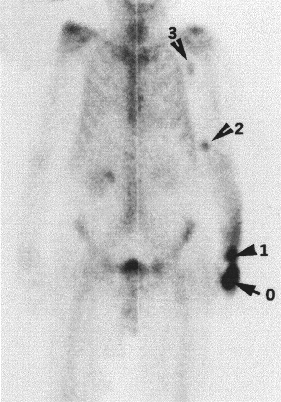
99mTc-HMDP scan shows large area of intense uptake in dorsal portion of left hand (0), a relatively smaller area of intense uptake in left wrist (1), area of substantial decrease in radioactivity in elbow (2), and area of rather faint radioactivity in left axilla (3).

FIGURE 2.
  • Download figure
  • Open in new tab
  • Download powerpoint
FIGURE 2.

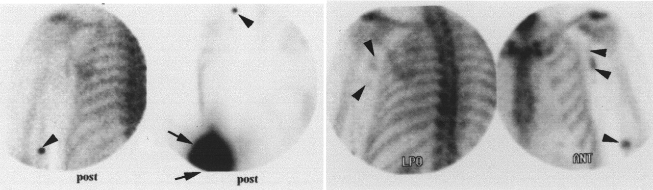
Multiple posterior (first 2 images from left), left posterior oblique (LPO, second image from right), and anterior (far right) images of chest and forearm show lymph nodes in elbow and axilla (arrowhead) and left wrist (arrow). Lymphatic channel (2 arrowheads) is also faintly visualized in anterior image.

DISCUSSION

Incidental axillary or elbow lymph node visualization after subcutaneous infiltration of the bone-imaging agent into the antecubital or wrist region has been reported (1–5). Visualization of lymph nodes in the elbow region resulting from the extravasation of bone-imaging agent in the wrist area has also been described (6). Our patient’s injection site for the bone-imaging agent was located on the distal dorsal area of the left hand, near the dorsal portion of the little and ring fingers. As per routine imaging, the bone scintigraphy was acquired 3 h after radiopharmaceutical injection. The extravasation appeared to be a substantially large amount of the total injected dose. This was shown by the intense uptake in the large area in the dorsal aspect of the left hand even 3 h after administration. Also note that the time required to acquire the total-body image (Fig. 1) and spot images (Fig. 2) was unusually prolonged. Over 3 h, gradual transportation of bone-imaging agent in the cephalic direction occurred, resulting in an incremental decrease in radioactive uptake in the lymph nodes in the distal to proximal direction.

Footnotes

  • For correspondence or reprints contact: Dr. Wei-Jen Shih, Nuclear Medicine Service, Lexington VA Medical Center, Lexington, KY 40511; Phone: 849-381-5928; Fax: 849-381-5934.

REFERENCES

  1. ↵
    Ongseng F, Goldfarb CR, Finestone H. Axillary lymph node uptake of 99mTc MDP. J Nucl Med 1995;36:1797.–1799.
    OpenUrlAbstract/FREE Full Text
  2. Chatterton BE, Vannitamby M, Cook DJ. Lymph node visualisation: an unusual artifact in the 99mTc pyrophosphate bone scan. Eur J Nucl Med 1980;5:187.–188.
    OpenUrlPubMed
  3. Penney HF, Styles CB. Fortuitous lymph node visualization after interstitial injection of 99mTc-MDP. Clin Nucl Med 1982;7:84.–85.
    OpenUrlPubMed
  4. Wallis JW, Fisher S, Wahl RL. 99mTc MDP uptake by lymph nodes following tracer infiltration: clinical and laboratory evaluation. Nucl Med Commun 1987:8:357–363.
  5. ↵
    Dogan AS, Rezai K. Incidental lymph node visualization on bone scan due to subcutaneous infiltration of 99mTc MDP. A potential for false-positive interpretation. Clin Nucl Med 1993;18:208.–209.
    OpenUrlPubMed
  6. ↵
    Shih WJ, Wierzbinski B, Magoun S. Lymph node visualization in the elbow region. J Nucl Med 1996;37:1913.
    OpenUrlFREE Full Text
PreviousNext
Back to top

In this issue

Journal of Nuclear Medicine Technology: 29 (3)
Journal of Nuclear Medicine Technology
Vol. 29, Issue 3
September 1, 2001
  • Table of Contents
  • About the Cover
  • Index by author
Print
Download PDF
Article Alerts
Sign In to Email Alerts with your Email Address
Email Article

Thank you for your interest in spreading the word on Journal of Nuclear Medicine Technology.

NOTE: We only request your email address so that the person you are recommending the page to knows that you wanted them to see it, and that it is not junk mail. We do not capture any email address.

Enter multiple addresses on separate lines or separate them with commas.
Visualization in the Ipsilateral Lymph Nodes Secondary to Extravasation of a Bone-Imaging Agent in the Left Hand: A Case Report
(Your Name) has sent you a message from Journal of Nuclear Medicine Technology
(Your Name) thought you would like to see the Journal of Nuclear Medicine Technology web site.
Citation Tools
Visualization in the Ipsilateral Lymph Nodes Secondary to Extravasation of a Bone-Imaging Agent in the Left Hand: A Case Report
Wei-Jen Shih, Judy Collins, Vickie Kiefer
Journal of Nuclear Medicine Technology Sep 2001, 29 (3) 154-155;

Citation Manager Formats

  • BibTeX
  • Bookends
  • EasyBib
  • EndNote (tagged)
  • EndNote 8 (xml)
  • Medlars
  • Mendeley
  • Papers
  • RefWorks Tagged
  • Ref Manager
  • RIS
  • Zotero
Share
Visualization in the Ipsilateral Lymph Nodes Secondary to Extravasation of a Bone-Imaging Agent in the Left Hand: A Case Report
Wei-Jen Shih, Judy Collins, Vickie Kiefer
Journal of Nuclear Medicine Technology Sep 2001, 29 (3) 154-155;
Twitter logo Facebook logo LinkedIn logo Mendeley logo
  • Tweet Widget
  • Facebook Like
  • Google Plus One
Bookmark this article

Jump to section

  • Article
    • Abstract
    • DISCUSSION
    • Footnotes
    • REFERENCES
  • Figures & Data
  • Info & Metrics
  • PDF

Related Articles

  • No related articles found.
  • PubMed
  • Google Scholar

Cited By...

  • Adverse Clinical Events at the Injection Site Are Exceedingly Rare After Reported Radiopharmaceutical Extravasation in Patients Undergoing 99mTc-MDP Whole-Body Bone Scintigraphy: A 12-Year Experience
  • Inadvertent Intraarterial Injection of 18F-FDG: A Case Report and Literature Review of Hot Forearm and Hot Hand Signs
  • Google Scholar

More in this TOC Section

  • Early 10-Minute Postinjection [18F]F-FAPI-42 uEXPLORER Total-Body PET/CT Scanning Protocol for Staging Lung Cancer Using HYPER Iterative Reconstruction
  • Single- Versus Dual-Time-Point Imaging for Transthyretin Cardiac Amyloid Using 99mTc-Pyrophosphate
  • Does Arthrography Improve Accuracy of SPECT/CT for Diagnosis of Aseptic Loosening in Patients with Painful Knee Arthroplasty: A Systematic Review and Metaanalysis
Show more Imaging

Similar Articles

SNMMI

© 2025 SNMMI

Powered by HighWire